

 PDF
PDF ePub
ePub Citation
 Print
Print


Introduction
Second trimester ultrasonography has become an essential part of antenatal obstetric care. As detailed fetal anatomical surveillances are performed, some organizations recommend guidelines for the second trimester fetal ultrasonography scan [12]. They describe prerequisites, precautions, and checklists in performing it. They also provide management recommendations when abnormalities, especially soft markers, are found. Soft markers are sonographic findings of the fetuses that are considered as normal variants, but possibly associated with increased risk of chromosomal abnormalities [34]. As the risks of chromosomal abnormalities vary greatly among reports [56], the management plan would be very diverse among the centers and each obstetrician. With this background, Korean Society of Ultrasound in Obstetrics and Gynecology (KSUOG) Research Group designed a questionnaire survey on the practice pattern of second trimester ultrasonography to give information and suggestions regarding second trimester fetal anatomic surveillance.
The objectives of this study were to analyze practice patterns and checklists of second trimester ultrasonography, and to investigate management plans when soft markers are found in the second trimester fetal anatomic surveillance among KSUOG members.
Materials and methods
1. Questionnaire development and data collection
To collect the information regarding practice patterns of second trimester ultrasonography and management plans when soft markers are found during fetal anatomic surveillance, internet-based, self-administered questionnaire survey was performed between 26th, August and 4th, October 2014. The members of KSUOG research group developed the questionnaire which was composed of two parts: 1) questions regarding general information about the performance of the second trimester fetal ultrasonography and 2) those regarding management plans with the findings of the soft markers. An e-mail of invitation was sent to a total of 446 KSUOG members, and if the recipient accepted to respond the questionnaire, the data were gathered through Google Drive platform. The questionnaire can be accessed at the following internet address: https://docs.google.com/forms/d/1A4JiVg4NOyWyaNmjTuz06MVPNZgYYlHIHE_Y1RssQa4/viewform.
KSUOG members were also requested for the checklists they were using in second trimester fetal anatomic surveillance. The checklists were sent to one of the author (HSP) through e-mail, facsimile and hardcopy between 29th, July and 3rd, September 2014.
2. Data analysis
In questionnaire survey, respondents were asked to enter basic information such as medical practice settings, age, sex, and year when they were certified as obstetrics and gynecology specialists. Practice patterns of the second trimester ultrasonography collected include gestational age at examination, characteristics of examiners and subjects, whether or not giving the informed consent, and examination time. Soft markers of interest included increased nuchal fold thickness, intracardiac echogenic focus, echogenic bowel, mild pyelectasia defined as kidney pelvis anterior-posterior diameter of 4 to 10 mm, short femur, and choroid plexus cyst. In addition, mild ventriculomegaly defined as lateral ventricular diameter of 10 to 12 mm, and megacisterna magna defined as cisterna magna diameter ≥10 mm were also included in the survey. The definition of short femur was also asked. Finally, respondents were asked to choose three most important soft markers that lead to genetic counseling.
Results
1. Characteristics of the respondents
A total of 101 KSUOG members participated in this survey. The mean age of the respondents was 42.0 years. The male to female ratio of respondents was nearly 1 (51% vs. 49%). About 97% of respondents (98 of 101) were working in the hospitals that run delivery rooms. Three thirds have been practicing as board-certified obstetrics and gynecology specialists less than 14 years.
2. General aspects of second trimester fetal ultrasonography
Eighty-eight (87.1%) respondents were routinely recommending second trimester anatomic surveillances to mothers. As to the question asking when they perform second trimester ultrasonography, 91.1% (92/101) performed the ultrasonography scan between 20+0 and 23+6 weeks of gestation (20+0 to 21+6 weeks, 68/101; 22+0 to 23+6 weeks, 24/101). The mean time needed for the scan ranged from less than ten minutes to an hour with the following order: 20 to 29 minutes (41.6%, 42/101), 10 to 19 minutes (32.7%, 33/101), 30 to 39 minutes (15.8%, 16/101), less than ten minutes (4%, 4/101), 40 to 49 minutes (3%, 3/101), and 50 to 59 minutes (3%, 3/101). Written informed consent was given by only 15.8% (16/101) of the respondents. Many physicians explained the purpose and limit of the ultrasonography with or without documenting it in the medical records (14.9%, 15/101 and 44.6%, 45/101, respectively). Twelve respondents (11.9%) answered that they gave the written instructions to the mothers without explaining or documenting, and another twelve responded that they explained nothing.
3. Practice patterns when the soft markers are found
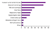
Table 1 shows general practice patterns of respondents when soft markers are found during second trimester ultrasonography. Nearly 60% offered invasive test when multiple soft markers were found and/or advanced maternal age was combined. Similar practice patterns were found in the analysis of the individual soft markers (Table 2). However, when such isolated soft markers as increased nuchal skinfold thickness, and mild ventriculomegaly were found, more than 20% of respondents recommended invasive test for chromosomal analysis. We found a wide, even, conflicting variations in counseling pattern among clinicians over same soft markers. For example, when intracardiac echogenic focus was found, amniocentesis was offered in about 51.5% (to offer invasive test with isolated finding + to offer invasive test if advanced maternal age present + to offer invasive test if accompanied by other soft markers) (Table 2). At the same time, however, in about 37.6% (not documented + none irrespective of any other soft marker accompanied), amniocentesis was not offered, or the finding was ignored. In cases of pyelectasia, megacisterna magna, short femur, and choroid plexus cyst, both conflicting recommendations accounted more than 20% each. As for the definition of the short femur, discrepancy of more than three weeks of gestational references was most frequently accepted. The preferences varied depending on the clinical settings (Table 3). Finally, participants were asked to select three most important things that lead to genetic counseling. These were increased nuchal skinfold thickness, advanced maternal age and mild ventriculomegaly (Fig. 1).
4. Analysis of checklists in second trimester fetal ultrasonography
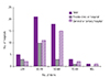
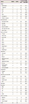
Checklists of 52 hospitals were submitted. As five hospitals used the same checklist, 48 were included in the analysis. The median number of items in the checklists was 46.5 (range, 17 to 109). The number of items in checklists varied according to the hospital practice setting. There was a tendency that more items were listed in the checklists of the general or tertiary hospitals than in those of private clinics or hospitals (Fig. 2). The items were compared with those of checklists recommended by ISUOG and ACOG [12] (Table 4). The numbers of items in the checklists suggested by ISUOG and ACOG are about 43 and 28, respectively. Of the 49 items of checklists recommended by ISUOG and/or ACOG, 28.6% (14/49) were found in less than 50% of the checklists analyzed in this study. Those were skull shape, falx, nose, chin, neck, thorax, heart size, heart axis, gall bladder, abdominal mass, placental location, genitalia, myoma, and ovary.
Discussion
In the first part of this survey, we intended to look into the general practice patterns of the second trimester fetal anatomic surveillance. The practice patterns were relatively consistent. For example, nearly 90% routinely recommended second trimester fetal anatomic surveillance to mothers. Ultrasonography was conducted between 20 and 24 weeks in 91%. Finally, time needed to complete ultrasound exam was between 20 and 40 minutes. However, only 15.8% of respondents gave written informed consents explaining the purposes, processes, and limitations of the ultrasonography. The percentages need to be increased, as other medical societies recommended [12]. There are some discrepancies between what we are doing and the other societies' recommendations. First, gestational age of 18 to 22 weeks is suggested to be best in performing second trimester scan. This is intended to have enough time for possible genetic testing and pregnancy termination, which is prohibited in our country in most cases. Another thing we should point out is that the way to describe the conclusions and further plans of the second trimester ultrasonography. In ISUOG practice guideline, they clearly describe the conclusions such as 1) normal and complete examination, 2) normal but incomplete examination, and 3) abnormal examination. Likewise, the follow-up plans need to be clearly described.
The principal findings of the second part of the questionnaire survey were: 1) many KSUOG members recommended invasive test for chromosomal analysis when multiple soft markers or risk factors including advanced maternal age were found; and 2) practice patterns when they detect soft markers were very diverse, and sometimes conflicting with each other. If the practice patterns over same medical condition are conflicting, it would be very confusing to the pregnant women. For example, when intracardiac echogenic foci and pyelectasia are found in a fetus, about 40% to 50% of doctors would recommend invasive test, but about 30% to 37% might say, "Amniocentesis is not needed" or "I don't think that those soft markers are important" (Table 2). In this regards, it would be very helpful to both doctors and pregnant women if a practice recommendations are provided by a medical society of Korea. As the frequency of soft markers may vary according to the ethnicity like that in intracardiac echogenic foci [78], it is of importance to have our own practice schemes. Although genetic counseling were not offered frequently in the management of choroid plexus cyst, about 22% of members offered invasive test when cyst is bilateral, single large, or multiple, which is not recently recommended [91011].
In the checklists analysis, the number of items varied very much. Some used very detailed checklists but others did not. If we take an example of the spine, some listed only 'spine', but others used more detailed lists like, 'axial, coronal, sagittal view of the spine, spinal defects, lumbosacral mass, etc.' There arise the question of 'How much detailed items should be included in the checklists?' The numbers of items from the recommendation of ISUOG or ACOG are 43 and 28, respectively, which are less than our median number of 46.5 [12]. If detailed explanation about the items can be offered along with the checklist, the number of items can be decreased. That strategy would also be very comprehensive and easy to understand. The wide range of spectrum of the item number suggests the need for standardization of the second trimester ultrasonography. One thing to keep in mind is that cultural difference can be reflected in the practice patterns or checklists. For example, in the checklist made by ISUOG, counting fingers or toes is not considered a part of routine mid-trimester scan, which are considered essential by many pregnant women in our country.
There are 14 items that were included less than 50% of the KSUOG member's checklists submitted. Items such as skull shape, nose, thorax, heart size and axis might be evaluated when head size, cleft lips, and four-chamber view of heart are examined. However, items such as chin, neck, and abdominal mass should not be omitted. Because the presence of myoma and adnexal mass might have been already evaluated during described in the checklists of second trimester ultrasonography in our country.
This is the first survey to investigate the practice patterns of second trimester fetal ultrasonography. This report can contribute to drafting recommendations of the second trimester ultrasonography in Korea, in association with the article about the survey of the first trimester ultrasound, published last year [12]. The main shortcoming of this article is that the number of respondents was small. However, it seems that a response rate of about 22.6% (101/446) is not a poor number.
In summary, the practice patterns of second trimester fetal ultrasonography were relatively consistent, but more informed consents should be given to pregnant women. Many KSUOG members offer invasive test when multiple soft markers are found, or a single soft marker is detected in association with advanced maternal age. However, these practice patterns have very wide range of spectrum, and this is the case with the checklists of the second trimester fetal ultrasonography. It would be desirable to make our own recommendations, or endorse and use other medical society's. This applies to all aspect of the second trimester fetal ultrasonography, and the management of soft markers.


 XML Download