

 PDF
PDF ePub
ePub Citation
 Print
Print


Introduction
Post hysterectomy complications at the vault site such as a bleeding incident can be commonly observed at a short term postoperative period. Other delayed complications often occur as a hematoma, granuloma, keloid, incisional hernia, and or vascular formation at the vault [12]. Many of these complications may be accompanied with bleeding symptoms. However a sudden massive bleeding occurring after more than a year postoperatively is rare.
Here we report a case of sudden vaginal vault bleeding in a post-hysterectomy state patient with no known history of endometriosis, hysterectomy undergone prior 13 months ago, proven to be a rare case of vault endometriosis. Hence, we would like to present how such a rare complication might be misdiagnosed and moreover increase the awareness of such complication of the vault in a post-hysterectomy patient.
Case report
Early on October 2014, a 45-year-old woman was presented at our outpatient gynecology clinic with sudden lower pelvic discomfort and vaginal bleeding symptoms. The patient had a history of hysterectomy 13 months ago due to symptomatic multiple leiomyomas and adenomyosis.
The previous surgery was conducted as a single-port approach laparoscopic-assisted vaginal hysterectomy in July 2013. Surgical findings showed an enlarged uterus of about a 14 weeks gestational age size. Both adnexa were grossly normal in appearance and the peritoneum was clear with no signs of endometriosis. The vaginal vault was sutured vaginally using a 1-0 vicryl. No complications were observed during the postoperative period and the patient was discharged as scheduled. The histology of the uterus was confirmed as adenomyosis with leiomyoma with a secretory phase endometrium. Follow up examinations at 3 and 6 months showed no complications and thus no additional follow-ups were required afterwards.
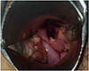
However on October 2014, after more than a year from her last check up, the patient appeared at our clinic complaining of sudden pelvic discomfort and vaginal bleeding symptoms. Pelvic examinations showed no signs of active bleeding. Yet, a dark wine colored papule suggestive of a small hematoma or ulcerative lesion was observed upon the previous operative vaginal vault site (Fig. 1.).
Her vitals were stable and she showed no signs of fever. The pain was tolerable during manipulation of the vault site. The ulcerative lesion was suspected as an old hematoma or granulation formation of the previous hysterectomy vault or possibly due to an unknown malignancy or cancerous change.
For further investigation, a quick excision biopsy using sharp scissors was performed under local anesthesia at the outpatient operation room. Bleeding control was done with sutures and tampon gauzes were inserted intravaginally. Nevertheless, the patient returned to the clinic within a few hours with excessive bleeding. Upon re-examination, active bleeding was visible at the vault site, and additional maneuvers including sutures were performed for hemostasis.
The pathology reports showed normal vaginal tissue with non-specific loss of surface epithelium and subepithelial hemorrhage. The diagnosis was uncertain but any kind of malignancy could be ruled out.
Ten days later, the patient revisited our clinic with another incidence of massive vaginal bleeding with large clots. She looked pale and anemic in general with vital signs showing an increased pulse with a decreased blood pressure at an initial 130/70 to 100/60. The patient also complained of symptoms of dizziness. Her lab results showed a hemoglobin level of 6.9 g/dL and hematocrit of 20.2%. Further attempts to achieve hemostasis with ball electrode cauterization were insufficient. The lesion healed only for a short while and the fragile tissue continued to bleed. Because of the massive bleeding, a clinical diagnosis of a possible vascular malformation or uterine artery pseudoaneurysm was considered.
An emergent exploratory laparoscopic operation was performed to control vault bleeding and to evaluate any possible vascular complication or collateral arterial bleeding. Due to anemic conditions, transfusion with 2 pints of packed red blood cells were necessary prior to the operation.
Upon surgery, other peritoneal structures including both ovaries were grossly normal and showed no signs of endometriosis. The pelvic side vault site was securely sealed with normal peritoneum covering the vault with no dehiscence or any other complication. Under conventional laparoscopy, after dissecting the anterior rectal wall and bladder peritoneum, a full thickness excision of approximately 2 cm in diameter was performed at the vault site and resealed with sutures.
Pathology results of the excised lesion was confirmed to be consistent with endometriosis (Fig. 2.). Hence, our difficult diagnosis of delayed vault site bleeding turned out to be a rare case of vault site iatrogenic endometriosis.
Discussion
Endometriosis is defined as the presence of endometrial tissue lesions or nodules that are similar to the endometrium but are present at sites outside the uterus [3]. Common sites of endometriosis include the ovaries, pelvic viscera and the peritoneum and the presentations can vary from a few minimal lesions on pelvic organs to massive endometric ovary cysts or even extensive adhesions involving subperitoneal spaces, intestinal system, and urinary system [45].
Rare cases of extrapelvic endometriosis may result from vascular or lymphatic dissemination of endometrial cells to many gynecologic and non gynecologic sites. It has been reported that endometriosis of the skin and soft tissue makes up about 3.5% of cases of extrapelvic endometriosis with a majority of such cases occurring in surgical scars following operations of the uterus [567].
Although rare, there have been few reports of vaginal vault endometriosis with patients presenting with irregular or cyclic menstrual bleeding several months or years after hysterectomy [8]. However, those cases had a history of a functional endometriosis at the ovaries with adhesions or a fistulous tract to the vault or even some endometriotic spots left behind near the vault site [9].
Usually in cases of sudden bleeding with a history of prior hysterectomy, pathologic causes such as granulation tissue formation or malignancies such as cervical stump cancer must be excluded [10]. Among the various complications of hysterectomy, here we report an incidence of a patient who developed endometriosis at the vaginal vault with no previous history of the disease.
There have been some reports where endometriosis found on scar tissues with no prior history of the disease. Cases such as scar endometriosis developing after a cesarean section are one of them [11]. However vault endometriosis after hysterectomy is an extremely rare complication and therefore difficult to diagnose.
In our case, the previous hysterectomy had no evidence of endometriosis on other sites. Even the pathologist, although having suspicions for endometriosis, could not give a clear diagnosis due to such a rare occurrence. However after examining the final vault excision specimen and correlating with the symptoms with our patient, the pathologist confirmed both biopsy results as a typical histology for endometriosis.
The possible pathophysiology in our particular case is suspected to be endometrial fragment implantation to the vaginal vault site at the time of surgery. We believe a risk of development into vault site endometriosis could be due to a uterus showing adenomyosis and or maybe due to a secretory phase endometrium. Our patient was on the 25th to 26th day of her menstrual cycle during the previous surgery, where the endometrium thickness was about 9.2 mm and the pathology proved the endometrium to be at secretory phase. Implantation during morcellation of the adenomyotic uterus or the secretory phase endometrium probably had an effect on the living tissues of the vagina, with transplantations to the site through cell adhesion mechanisms and then progression to iatrogenic endometriosis of the vaginal vault [38]. This theory is highly possible although further investigation and studies are required.
Although such complications are extremely rare, iatrogenic vault site endometriosis must be considered in delayed bleeding occurring in a post-hysterectomy patient when other diagnoses have been excluded. Due to the nature of these lesions being highly likely to recur, the principle of treatment as in any other extra pelvic endometriosis, is total surgical excision.

 XML Download