Abstract
Objective
Women with Lynch syndrome have an increased risk of developing colorectal and gynecologic malignancies such as endometrial cancer. Complex hyperplasia has about a 30% risk of developing into endometrial cancer. The aim of this study was to determine the genetic risk for developing endometrial cancer by immunohistochemical staining of premalignant lesions for mutL homolog 1, mutS homolog 2, mutS homolog 6, and postmeiotic segregation increased 2.
Methods
Twenty cases (n=20) were selected from among patients with available sample blocks for analysis. Clinical information was obtained from medical chart review. Immunohistochemical staining was performed for all of the tumor blocks. Staining was scored based on the intensity (intensity score 0-3) .
Results
Among the 20 cases of complex endometrial hyperplasia, 11 (55%) patients showed loss of expression of at least one of the following proteins: mutL homolog 1, mutS homolog 2, mutS homolog 6, or postmeiotic segregation increased 2. Seven (35%) patients were negative for the expression of two or more proteins, and one patient (5%) was negative for the expression of all four proteins.
Endometrial carcinoma is the second most frequent gynecologic malignancy in women with 49,560 cases reported and 8,190 deaths from this disease in the United States in 2013 [1]. Endometrial carcinoma typically arises from atypical complex hyperplasia and develops in estrogen-rich environments, such as those that are present in the cases of obesity, anovulation, and excessive exogenous estrogen [2]. The endometrioid subtype of endometrial adenocarcinoma comprises 80% to 85% of cancers arising from the lining of the endometrium and is frequently preceded by a precursor lesion, such as endometrial hyperplasia [3]. The cumulative 20-year progression risk among women who remain at risk for at least 1 year is 28% for atypical complex hyperplasia; this is 14-fold the risk faced by women with non-atypical endometrial hyperplasia [4].
The detection and appropriate treatment of complex endometrial hyperplasia with atypia are important to prevent endometrial carcinoma. About 5% of endometrial carcinoma cases are thought to be caused by inherited genetic changes [5]. Lynch syndrome or hereditary nonpolyposis colorectal cancer (HNPCC) accounts for 2% to 3% of all endometrial carcinomas [6]. Lynch syndrome is an autosomal dominant disorder known to occur at various extracolonic sites [7].
Most Lynch syndrome cases are caused by germline mutations of DNA mismatch repair genes (mutL homolog 1 [MLH1], mutS homolog 2 [MSH2], mutS homolog 6 [MSH6], and post-meiotic segregation increased 2 [PMS2]) [8,9,10,11,12,13]. These genetic defects in the DNA mismatch repair system result in replication errors in repetitive DNA segments, known as microsatellite instability (MSI), and the absence of expression of mismatch repair proteins in the tumor. Currently, Lynch syndrome is diagnosed based on family history, immunohistochemistry (IHC), MSI, and gene sequencing chromatograms (revised Amsterdam, 1998 [7] or Bethesda, 2002 [14] criteria).
More than 75% of endometrial tumors in HNPCC mutation carriers show MSI [15,16]. Furthermore, HNPCC-related endometrial cancer studies have shown a correlation between MSI and the lack of related protein expression as detected by IHC [17,18,19,20].
Our aim was to investigate the association between premalignant lesions of the endometrium by evaluating MLH1, MSH2, MSH6, and PMS2 expression in patients with complex endometrial hyperplasia by IHC to find appropriate group for further genetic counseling and test.
The study sample was identified through a retrospective search of medical records. Among the patients with pathologically proven complex endometrial hyperplasia who were treated at the Department of Obstetrics and Gynecology, Samsung Changwon Hospital from 2001 to 2012, 20 patients with available blocks were chosen. Their medical records were reviewed for clinical characteristics. After acquiring approval from the institutional review board (IRB) at the Samsung Changwon Hospital (IRB no. 2012-SCMC-028-00), IHC staining of MSH2, MLH1, MSH6, and PMS2 was performed.
After confirmation of the pathologic diagnosis of complex endometrial hyperplasia, the 20 patients were treated with either medical therapy (e.g., progesterone) or surgical treatment such as hysterectomy.
IHC analysis of endometrial hyperplasia specimens was performed to determine the protein expression of MLH1 (Novocastra, Newcastle, UK), MSH2 (Novocastra), MSH6 (Novocastra), and PMS2 (Novocastra). Immunohistochemical staining was performed using the streptavidin-biotin method. Sections (4-µm thick) were deparaffinized with xylene, rehydrated, and incubated with fresh 0.3% hydrogen peroxide in methanol for 30 min at room temperature. Specimens were rehydrated through a graded ethanol series and washed in phosphate-buffered saline (PBS). After blocking treatment, specimens were incubated with monoclonal antibodies at a dilution of 1:200 in PBS containing 1% bovine serum albumin at 4℃ overnight. Sections were washed with PBS and incubated in secondary antibody for 30 minutes at room temperature. The chromogen was a 3.3% to 0.02% solution containing 0.005% H2O2 in 50 mM ammonium acetate-citric acid buffer (pH 6.0). Tissue sections were lightly counterstained with hematoxylin and then examined by light microscopy. There was no detectable staining in the negative controls prepared by omitting the primary antibody.
Staining was evaluated by two independent observers without knowledge of clinical outcomes. One dedicated gynecologic pathologist and one gynecologic oncologist reviewed the slides in a blinded manner and evaluated the immunohistochemical data independently. The staining intensity was scored as follows: 0, no appreciable staining in tumor cells; 1, barely detectable staining in the cytoplasm, nucleus, or membrane compared with stromal elements; 2, readily detectable brown staining distinctly marking the tumor cell cytoplasm, nucleus, or membrane; and 3, dark brown staining in tumor cells completely obscuring the cytoplasm, nucleus, or membrane (Fig. 1). A previous study regarding Lynch syndrome reported that complete loss of expression of these genes in the setting of a positive internal control is interpreted as a positive result [21].
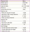
The demographic and clinical characteristics of the 20 patients are described in Table 1. The median age at diagnosis was 49 years (range, 38 to 66 years). The median body mass index of the patients was 24.19, and 35% of the patients had a body mass index above 25. Parity was known in all 20 patients. Of these, two (10%) were nulliparous.
After the dilatation and curettage biopsy (DCB) diagnosis of complex hyperplasia, four (20%) patients received medical treatment (progesterone) for 3 months only, and the follow-up endometrial biopsy revealed no lesion, two (10%) patients received medical treatment and surgical treatment (hysterectomy) with persistent disease, and 11 (55%) underwent surgical treatment only. Three (15%) patients refused treatment after diagnosis (Table 1). Among the surgery types, DCB only was performed in seven patients, hysterectomy after DCB in 12 patients, and hysterectomy only in one patient. The mean medication treatment period for six patients was 24 months. Of the 20 cases of complex hyperplasia, four cases were diagnosed with complex hyperplasia without atypia, and 16 (80%) with complex hyperplasia with atypia after the final pathology.
After surgery as an adjuvant treatment, endometrial cancer was found incidentally by pathological analysis in two of the 16 patients diagnosed with complex hyperplasia with atypia. Surgical staging was performed for these two patients; both had endometrial cancer stage 1, and no additional treatment was applied.
The results of the IHC expression of MLH1, MSH2, MSH6, and PMS2 are summarized in Table 2. Eleven patients (55%) showed loss of expression of at least one of the following proteins: MLH1, MSH2, MSH6, or PMS2. Seven patients (35%) were negative for the expression of two or more proteins, and one patient (5%) was negative for the expression of all four proteins (Table 2).
Approximately 247,000 and 1,600,000 new cancer cases are expected to occur in Korea and the United States [1] in 2013, respectively. In Korea, the expected incidence and mortality due to endometrial cancer are expected to be 2,175 and 262 cases, respectively [22]. Appropriate cancer treatment and early diagnosis and prevention are required to decrease mortality and the economic burden associated with cancer diagnosis.
The present study is the first to determine the correlation between mismatch repair gene (MLH1, MSH2, MSH6, and PMS2) protein expression and complex hyperplasia using IHC in a Korean population. Further genetic counseling, tests, and long-term follow up are needed to prevent endometrial carcinoma in this subgroup of patients.
IHC is an easy and inexpensive test. The sensitivity and specificity of IHC for predicting Lynch syndrome are both near 100% [23]. Complex hyperplasia has a high cumulative progression risk for endometrial carcinoma. The identification of patients with a genetic risk among those with complex hyperplasia based on IHC is important for cancer prevention and early detection. However, IHC is not a confirmative test, and gene sequencing tests are expensive. Furthermore, the evaluation of IHC can vary among pathologists.
A previous study showed that loss of MLH1 expression was correlated with the loss of PMS2 and that the loss of MSH2 expression was associated with the loss of MSH6 expression [24]. In our study, seven patients (35%) were negative for two or more proteins, and one patient (5%) was negative for all four proteins. One MSH2-negative patient was also negative for MSH6. Similarly, two MLH1-negative patients did not express PMS2. Further genetic tests are highly recommended for these three patients.
Lynch syndrome is an autosomal dominant cancer. The lifetime risk of endometrial cancer for women with Lynch syndrome is approximately 40% to 60%, a level that equals or exceeds their risk of colorectal cancer. Prophylactic hysterectomy with bilateral salpingoophorectomy is an effective strategy for preventing endometrial and ovarian cancer in women with Lynch syndrome [25].
Atypical hyperplasia is known to increase the risk of endometrial cancer by about 30%. The cumulative 20-year progression risk among women who remain at risk for at least 1 year is less than 5% for non-atypical endometrial hyperplasia but is 28% for atypical hyperplasia [4].
The expression of MLH1, MSH2, MSH6, and PMS2 proteins is strongly associated with Lynch syndrome [26]. In the present study of 20 cases of complex endometrial hyperplasia, more than half of the patients showed loss of expression of at least one of the above proteins. The limitations of our study are 1) the small number in the study group, 2) not having a confirmative test, and 3) the inability to define the cut-off level of intensity and proportion for further genetic tests. The present study suggests the necessity for further large-scale screening for Lynch syndrome in patients with complex endometrial hyperplasia to facilitate the early diagnosis of malignancy.
Figures and Tables
Fig. 1
Immunohistochemistry examples of the comparative expression of mutL homolog 1 (MLH1), mutS homolog 2 (MSH2), mutS homolog 6 (MSH6), and post-meiotic segregation increased 2 (PMS2) in complex endometrial hyperplasia lesion (PMS2 picture show only mild positive expression).

Acknowledgments
This study was supported by a grant from the Samsung Biomedical Research Institute (SMR112162).
References
1. Siegel R, Naishadham D, Jemal A. Cancer statistics, 2013. CA Cancer J Clin. 2013; 63:11–30.
2. Emons G, Fleckenstein G, Hinney B, Huschmand A, Heyl W. Hormonal interactions in endometrial cancer. Endocr Relat Cancer. 2000; 7:227–242.
3. Sherman ME. Theories of endometrial carcinogenesis: a multidisciplinary approach. Mod Pathol. 2000; 13:295–308.
4. Lacey JV Jr, Sherman ME, Rush BB, Ronnett BM, Ioffe OB, Duggan MA, et al. Absolute risk of endometrial carcinoma during 20-year follow-up among women with endometrial hyperplasia. J Clin Oncol. 2010; 28:788–792.
5. Gruber SB, Thompson WD. A population-based study of endometrial cancer and familial risk in younger women. Cancer and Steroid Hormone Study Group. Cancer Epidemiol Biomarkers Prev. 1996; 5:411–417.
6. Meyer LA, Broaddus RR, Lu KH. Endometrial cancer and Lynch syndrome: clinical and pathologic considerations. Cancer Control. 2009; 16:14–22.
7. Vasen HF, Watson P, Mecklin JP, Lynch HT. New clinical criteria for hereditary nonpolyposis colorectal cancer (HNPCC, Lynch syndrome) proposed by the International Collaborative group on HNPCC. Gastroenterology. 1999; 116:1453–1456.
8. Fishel R, Lescoe MK, Rao MR, Copeland NG, Jenkins NA, Garber J, et al. The human mutator gene homolog MSH2 and its association with hereditary nonpolyposis colon cancer. Cell. 1993; 75:1027–1038.
9. Papadopoulos N, Nicolaides NC, Wei YF, Ruben SM, Carter KC, Rosen CA, et al. Mutation of a mutL homolog in hereditary colon cancer. Science. 1994; 263:1625–1629.
10. Leach FS, Nicolaides NC, Papadopoulos N, Liu B, Jen J, Parsons R, et al. Mutations of a mutS homolog in hereditary nonpolyposis colorectal cancer. Cell. 1993; 75:1215–1225.
11. Bronner CE, Baker SM, Morrison PT, Warren G, Smith LG, Lescoe MK, et al. Mutation in the DNA mismatch repair gene homologue hMLH1 is associated with hereditary non-polyposis colon cancer. Nature. 1994; 368:258–261.
12. Burgart LJ. Testing for defective DNA mismatch repair in colorectal carcinoma: a practical guide. Arch Pathol Lab Med. 2005; 129:1385–1389.
13. Hendriks YM, Jagmohan-Changur S, van der Klift HM, Morreau H, van Puijenbroek M, Tops C, et al. Heterozygous mutations in PMS2 cause hereditary nonpolyposis colorectal carcinoma (Lynch syndrome). Gastroenterology. 2006; 130:312–322.
14. Umar A, Boland CR, Terdiman JP, Syngal S, de la Chapelle A, Ruschoff J, et al. Revised Bethesda Guidelines for hereditary nonpolyposis colorectal cancer (Lynch syndrome) and microsatellite instability. J Natl Cancer Inst. 2004; 96:261–268.
15. Liu B, Parsons R, Papadopoulos N, Nicolaides NC, Lynch HT, Watson P, et al. Analysis of mismatch repair genes in hereditary non-polyposis colorectal cancer patients. Nat Med. 1996; 2:169–174.
16. Risinger JI, Berchuck A, Kohler MF, Watson P, Lynch HT, Boyd J. Genetic instability of microsatellites in endometrial carcinoma. Cancer Res. 1993; 53:5100–5103.
17. Thibodeau SN, French AJ, Roche PC, Cunningham JM, Tester DJ, Lindor NM, et al. Altered expression of hMSH2 and hMLH1 in tumors with microsatellite instability and genetic alterations in mismatch repair genes. Cancer Res. 1996; 56:4836–4840.
18. Dietmaier W, Wallinger S, Bocker T, Kullmann F, Fishel R, Ruschoff J. Diagnostic microsatellite instability: definition and correlation with mismatch repair protein expression. Cancer Res. 1997; 57:4749–4756.
19. Leach FS, Polyak K, Burrell M, Johnson KA, Hill D, Dunlop MG, et al. Expression of the human mismatch repair gene hMSH2 in normal and neoplastic tissues. Cancer Res. 1996; 56:235–240.
20. Marcus VA, Madlensky L, Gryfe R, Kim H, So K, Millar A, et al. Immunohistochemistry for hMLH1 and hMSH2: a practical test for DNA mismatch repair-deficient tumors. Am J Surg Pathol. 1999; 23:1248–1255.
21. Tafe LJ, Riggs ER, Tsongalis GJ. Lynch syndrome presenting as endometrial cancer. Clin Chem. 2014; 60:111–121.
22. Jung KW, Won YJ, Kong HJ, Oh CM, Seo HG, Lee JS. Prediction of cancer incidence and mortality in Korea, 2013. Cancer Res Treat. 2013; 45:15–21.
23. Resnick KE, Hampel H, Fishel R, Cohn DE. Current and emerging trends in Lynch syndrome identification in women with endometrial cancer. Gynecol Oncol. 2009; 114:128–134.
24. Shia J. Immunohistochemistry versus microsatellite instability testing for screening colorectal cancer patients at risk for hereditary nonpolyposis colorectal cancer syndrome. Part I. The utility of immunohistochemistry. J Mol Diagn. 2008; 10:293–300.
25. Schmeler KM, Lynch HT, Chen LM, Munsell MF, Soliman PT, Clark MB, et al. Prophylactic surgery to reduce the risk of gynecologic cancers in the Lynch syndrome. N Engl J Med. 2006; 354:261–269.
26. Shia J, Holck S, Depetris G, Greenson JK, Klimstra DS. Lynch syndrome-associated neoplasms: a discussion on histopathology and immunohistochemistry. Fam Cancer. 2013; 12:241–260.


 PDF
PDF ePub
ePub Citation
 Print
Print


 XML Download