

 PDF
PDF ePub
ePub Citation
 Print
Print


Introduction
Placenta previa is an obstetric complication wherein the placenta invades the lower uterine segment. Placenta percreta is the most serious complication of placenta previa, and is associated with massive obstetric hemorrhage. A study reported previous cesarean delivery as a risk factor for placenta accreta [1]. Several surgical techniques during cesarean delivery and subsequent cesarean hysterectomy for placenta previa are reported; however, to improve maternal outcome, further investigation is needed [2]. We present a case of placenta percreta that involved the anterior wall; this is the rarest and most complicated form of placenta accreta. For management, we made a transverse uterine fundal incision to avoid cutting through the placenta, and then successfully performed a subsequent cesarean hysterectomy. The patient was discharged without any complications. However, postoperative day 48, she experienced a watery discharge and was diagnosed with a vaginal fistula. This is the first case report of placenta percreta complicated with a vaginal fistula after a uterine transverse fundal incision and a subsequent cesarean hysterectomy.
Case report
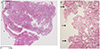
A 36-year-old female (gravida 2 and para 1) was referred to our hospital from a private clinic for placenta previa at 28 weeks of gestation. The patient had a history of a cesarean delivery, and in her current pregnancy, the placenta covered the entire anterior uterine wall and totally embedded within the lower uterine segment. We performed ultrasonography and magnetic resonance imaging, and diagnosed her with placenta previa and placenta accreta without bladder invasion (Fig. 1A). The patient was considered at risk for severe obstetrical complications due to placenta accreta; therefore, a cesarean section was planned at 36 weeks of gestation to avoid emergency surgery. At 35 weeks, she experienced a sudden onset of vaginal bleeding from the placenta and was admitted to our hospital. Total blood loss was estimated to be over 500 mL, and we performed an emergency cesarean section under general anesthesia. Laparotomy revealed large blood vessels and portions of the placenta present throughout the anterior uterine wall (Fig. 1B). Thus, we suspected placenta percreta and made an incision in the transverse uterine fundus to avoid cutting through the placenta. After elevating the uterus outside the abdominal wall, an ultrasonography-guided transverse incision was made at the uterine fundus to avoid rupturing the membrane or producing a bulge on the fetal membrane (Fig. 1C). The incision produced minimal bleeding, and a sleeping male infant with a birth weight of 2,560 g was successfully delivered (Apgar scores of 2 and 6 at 1 and 5 minutes, respectively). The placenta was not spontaneously delivered because of abnormal uterine adherence; therefore, we performed a hysterectomy, which was completed without complications, and closed the abdomen. Operation time was 139 min, and total blood loss was approximately 2,000 mL (including the preoperative bleeding), and bleeding was primarily observed from the placental site. Minimal bleeding was observed from the surgical site. Although abnormal adherence was observed between the anterior uterine wall and the bladder because of placenta percreta, it was difficult to strip the bladder from the uterus; we were finally able to strip the bladder without injury to any part of the bladder. The patient was transfused with 840 mL of red blood cells. The patient was discharged 7 days later without any complications in good condition. Histopathological analysis was revealed the placenta percreta as shown in Fig. 2. Postoperative examination revealed no complications 35 days after surgery. However, postoperative day 48, she experienced a watery discharge and was diagnosed with a vaginal fistula by internal examination and cystoscopy. Her vaginal fistula improved after 20 days and no recurrence occurred after one year after the diagnosis.
Discussion
Several studies have reported an association between the frequency of placenta accreta and the number of cesarean deliveries [1]. In addition to multiple cesarean deliveries, placenta previa is the most serious risk factor for placenta accreta [1]. Because the number of cesarean deliveries continues to increase, it is expected that the prevalence of placenta accreta will also continue to increase.
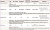
Recent studies have reported that making a transverse uterine fundal incision for managing placenta accreta can effectively avoid cutting through the placenta and subsequently decrease fetal and maternal blood loss [3,4]. We summarized previous English reports of transverse uterine fundal incision for placenta previa involving the entire anterior uterine wall in Table 1 [3,4,5]. These reports concluded that transverse uterine fundal incision is a useful technique for placenta previa covering the entire uterine anterior wall, with no major complications reported. The amount of blood loss from the incision was minimal, and the operating obstetrician could directly observe the placenta through the surgical wound. Most importantly, a transverse uterine fundal incision can prevent the necessity of an incision in placenta accreta, which subsequently circumvents massive bleeding. Therefore, the obstetrician can begin a planned hysterectomy at delivery and minimize bleeding. In contrast, a drawback is the limited data regarding the influence of this procedure on subsequent pregnancies. Therefore, we are currently unable to recommend this procedure for patients who desire subsequent pregnancies.
To manage placenta percreta, a recent study reported that the average blood loss during the surgery is 4,800 mL, which increases the difficulty of the operative technique [6]. In our case, the placenta covered the entire uterine anterior wall; therefore, if we performed a classical vertical uterine incision, as performed in the conventional surgical technique, we would be unable to avoid cutting through the placenta, and massive hemorrhage may have occurred from the placental site. The uterine fundal transverse incision is a useful technique for placenta percreta covering the entire uterine anterior wall.
Previous studies were reported 19 cases and 8 cases of transverse fundal incision for placenta accreta [4,5]. However, in these reports, the total number of cases involving placenta percreta seemed to be small. Our case is a relatively rare report of the successful management of placenta percreta with a transverse fundal incision and subsequent total abdominal hysterectomy. Further reports are needed to investigate the usefulness of this surgical method for placenta percreta covering the anterior wall of the uterus.
In our case, no complications occurred during the surgery or admission. However, a vaginal fistula occurred after the patient was discharged. Reportedly, placenta percreta was complicated with a vaginal fistula in 2 of 66 patients [6]. Although, there may be no association between a uterine transverse fundal incision and a vaginal fistula, to improve cesarean hysterectomy, further discussion is needed. We considered that the fistula may have caused the thinning of the bladder wall after the stripping from the uterus. Placenta percreta is often observed with the thinning of the bladder wall, and it is difficult to strip the bladder from the uterus. In the present case, we did not assess the bladder wall thickness. If we had determined the bladder wall thickness by filling the bladder wall after the hysterectomy, we would have been able to detect the thinning bladder wall and perform the repair. Moreover, we could fill the bladder during the hysterectomy to separate the bladder wall more easily, as previously reported [2].
In summary, we successfully managed a case of placenta percreta through transverse uterine fundal incision and planned hysterectomy, and subsequently minimized blood loss. This technique is useful in cases where the placenta covers the entire uterine anterior wall. However, a vaginal fistula occurred. We should assess the bladder wall by filling the bladder during a cesarean hysterectomy, in cases of placenta percreta.

 XML Download