

 PDF
PDF ePub
ePub Citation
 Print
Print


Introduction
Paratubal cyst arises from the mesosalpinx or broad ligament, and originates from the paramesonephric (Müllerian), or less commonly the mesonephric (Wolffian) embryological remnants [1,2]. They are synonymously called parovarian cysts if they are adjacent to the ovary. Paratubal and parovarian cysts comprise a minority of all benign adnexal cysts, and are usually found as an incidental finding. They are generally small and rarely symptomatic.
However, as paratubal cysts grow, they can induce symptoms such as pelvic pressure and abdominal pain. Recently, ultrasound evaluation has become popular. Preoperative exact diagnosis based on sonographic finding has increased, but ultrasound features of paratubal cysts are very similar to ovarian or parovarian cysts. Therefore, most diagnoses are made postoperatively. However, adnexal cyst should be considered when imaged separately from an ipsilateral ovary [3].
The incidence of isolated tubal torsion is very rare (1 out of 1,500,000 patients) [4]. Even rarer, the fallopian tube can coil around a nearby organ such as the utero-ovarian ligament, showing similar clinical presentations with tubal torsion. The coiling fallopian tube is not twisted, but surrounds the utero-ovarian ligament, causing impaired vascular perfusion and venous drainage. The paratubal cyst may serve as a lock.
Case report
A 22-year-old female was transferred to our hospital from a local clinic due to right lower abdominal pain since that morning. She was a virgin and her vital signs were stable without fever. Laboratory evaluations including hematologic, urinary, and routine chemistry tests were normal except for mild leukocytosis (white blood cell count 10,190/mL). On physical examination, tenderness and rebound tenderness were found in the right lower abdomen.
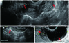
Sonographic examination showed a right ovary measuring 80×32 mm with multiple small follicles located over the uterus (Fig. 1A) and left ovary (74×42 mm) containing a 67×42 mm-sized anechoic cyst (Fig. 1B, C). Her pain became more aggravated as time went on without relief with parenteral analgesics. As the right ovary was suspected for torsion, we planned for an emergency laparoscopic operation.
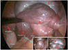
Laparoscopic findings revealed a dark blue-colored fallopian tube coiling around the utero-ovarian ligament two and half times in a counter-clockwise direction. The right ovary was enlarged, swollen, edematous, dark bluish discolored and seated over the uterus. A right paratubal cyst (>6 cm in diameter) which had been sonographically misinterpreted as a cystic component of the left ovary (Fig. 1B) was noted in the Douglas pouch, adjacent to the left ovary. The left ovary and tube were normal in appearance (Fig. 2).
The right ovary was uncoiled from the fallopian tube, and regained its color gradually. However, uncoiled tubal recovery was not as satisfactory as the right ovary. Furthermore, it was densely adherent to the cyst (Fig. 2B). We thought it would be nearly impossible to resect the cyst without damaging the tube. Due to the possibility of tubal damage which might increase the risk of a subsequent tubal pregnancy, we decided to perform a salpingectomy. The postoperative course was uneventful and the patient was discharged 4 days after the surgery.
Discussion
Torsion is the twisting of an object, such as a piece of metal or an organ of the body, into a spiral shape. Torsion of the uterine adnexa is one of the major causes of acute abdomen and requires immediate surgical intervention.
Causes of fallopian tube torsion are unknown, but there are noted factors influencing fallopian tube torsion, including intrinsic factors like congenital anomalies (excessive tube length), acquired pathology (hydrosalpinx, hematosalpinx, neoplasm), and autonomic dysfunction [4]. Extrinsic factors like trauma, pregnancy, adhesion and pelvic congestion also contribute [4]. The clinical symptoms are nonspecific (pain, nausea, vomiting, and sometimes voiding difficulties), so it is challenging for the clinician to recognize and differentiate from a number of other etiologies.
Distinct from the process of torsion, coiling causes objects to take on a different kind of form. For example, if you coil something, you wind it into a series of loops or into the shape of a ring. If something is coiled around another object, it can form a loop or a ring. In gynecologic structures, although rare, coiling can be induced by proximity between the ovary and the salpinx and by the salpinx acting like a rope, especially when containing a locking point like a paratubal cyst.
The striking aspect of our case is that the fallopian tube itself was not twisted, but coiled around the utero-ovarian ligament. The cause of fallopian tube coiling is unknown. It had been thought that a fallopian tube with a short or normal length should have been almost impossible to coil in this way [5]. The length of the fallopian tube in this patient was 11 cm, which is within the normal range. The paratubal cyst had worked as a locking point and the coiling fallopian tube tightened around the utero-ovarian ligament, affecting blood flow toward the affected ovary.
We searched the Cochrane Library, Embase, Google Scholar and PubMed using the keywords of fallopian tube, coiling, and paratubal cyst and found only one case report containing all three keywords.
Ultrasonographic signs of adnexal torsion include abnormal ovarian position, ovarian enlargement with edema, ovarian or adnexal mass, free fluid in the cul-de-sac, and decreased or absent Doppler flow in the ovary [6].
Unfortunately, we did not have precise ultrasound interpretations. Although we noted that the right ovary appeared enlarged, and was abnormally located above the uterus, we could not determine the pathologic implications in the preoperative period.
The definite diagnosis of tubal torsion or coiling is made retrospectively, usually after diagnostic laparoscopy. Currently, laparoscopy is the diagnostic tool of choice and laparoscopic detorsion/uncoiling is the treatment of choice [7].
Although coiling has a similar clinical presentation with torsion, surgical results can be significantly different. In the case of ovarian cyst torsion, detorsion and removal of the cyst is the main form of treatment, but in case of coiling, treatment is somewhat different. After uncoiling, a surgeon should decide whether to save the coiled tissue or not, and whether or not to perform salpingectomy, as the affected fallopian tube usually has a pathologic condition like a cyst and is stretched. There is very little information in the literature on coiling, and imaging studies on coiling are limited.
 XML Download