This article has been
cited by other articles in ScienceCentral.
Abstract
Bisphosphonate (BP) is a useful anti-resorptive agent which decreases the risk of osteoporotic fracture by about 50%. However, recent evidences have shown its strong correlation with the occurrence of atypical femoral fracture (AFF). The longer the patient takes BP, the higher the risk of AFF. Also, the higher the drug adherence, the higher the risk of AFF. It is necessary to ask the patients who are taking BP for more than 3 years about the prodromal symptoms such as dull thigh pain. Simple radiography, bone scan, and magnetic resonance imaging (MRI) are good tools for the diagnosis of AFF. The pre-fracture lesion depicted on the hip dual energy X-ray absorptiometry (DXA) images should not be missed. BP should be stopped immediately after AFF is diagnosed and calcium and vitamin D (1,000 to 2,000 IU) should be administered. The patient should be advised not to put full weight on the injured limb. Daily subcutaneous injection of recombinant human parathyroid hormone (PTH; 1-34) is recommended if the patient can afford it. Prophylactic femoral nailing is indicated when the dreaded black line is visible in the lateral femoral cortex, especially in the subtrochanteric area.
Keywords: Atypical femoral fracture, Bisphosphonate, Position statement
Epidemiology and definition of atypical femoral fracture (AFF)
Bisphosphonate (BP) is a potent anti-resorptive agent and was proven to be effective in prevention and treatment of osteoporosis through many randomized prospective studies. BP decreases the risk of osteoporotic vertebral and hip fractures by around 50%. However, excessive suppression of normal and pathologic bone remodeling has been a concern. Odvina et al.[
1] reported nonunion after low-energy injury among patients who took BP for several years for treatment of osteoporosis. Bone biopsy revealed lack of viable osteoblasts, osteocytes, and osteoclasts in the specimen. The authors thought that prolonged suppression of bone remodeling by BP caused frozen bone. Goh et al.[
2] reported atypical subtrochanteric fractures in their patients who took alendronate for several years. Fractures were caused by minor injuries and were associated with periosteal callus at the lateral femoral cortex. As the number of reports of this type of fracture increased,[
3456789] the American Society of Bone and Mineral Research (ASBMR) organized a task force team and reported the results on two occasions (
Fig. 1).
The criteria for the diagnosis of AFF are listed below (
Table 1).[
10]
AFF occurs even in those patients who are not exposed to BP therapy. However, in most of the cases, AFF occurs in the patients who are taking or who took BP for several years for treatment of osteoporosis or other metabolic bone diseases. Both oral and iv BP agents have been reported to cause AFF. It is difficult to estimate the incidence of AFF due to its low occurrence in BP users. The estimated incidence of AFF is 5 to 100 per hundred thousand patient-years. AFF occurs 4 to 5 times more frequently in Asian descendants compared to Caucasians. The longer the patient takes BP, the higher the risk of AFF. Also, the higher the adherence, the higher the risk of AFF. It is time to declare that BP is the causative agent of AFF although BP decreases the incidence of hip and vertebral fractures significantly.
Diagnosis
The incidence of AFF increases significantly when the patient takes BP for 4 years or more than 4 years.[
4] Prodromal symptoms such as dull pain and tenderness over the lateral aspect of the thigh are noted in 40% to 70% of the patients. It is recommended to check for prodromal symptoms if the patients are taking BP for more than 4 to 5 years. It is necessary to take a simple radiogram of the femur if AFF is suspected.
Simple radiogram often reveals a beak or flare like periosteal and/or endosteal callus in the lateral femoral cortex. Sometimes, the so-called 'dreaded black line' can also be detected in the lateral cortex. If the simple radiogram appears looks normal even though the patient complains of prodromal symptoms, then bone scan or magnetic resonance imaging (MRI) examination can identify tiny lesions in the lateral aspect of the femur. In Korea, dual energy X-ray absorptiometry (DXA) examination of axial bone is performed every year to obtain reimbursement from national insurance. We may occasionally detect a tiny periosteal callus in the lateral aspect of the femoral cortex (
Fig. 2,
3).[
11]
Medical treatment
BP should be stopped immediately after AFF is diagnosed and calcium and vitamin D (1,000 to 2,000 IU) should be administered. The patient should be advised not to put full weight on the injured limb. Daily subcutaneous injection of recombinant human parathyroid hormone (PTH; 1-34) is recommended if the patient can afford it. Prophylactic femoral nailing is indicated when the dreaded black line is visible in the lateral femoral cortex, especially in the subtrochanteric area.[
12]
Surgical treatment
Intramedullary nailing is the treatment of choice for AFF. However, excessive femoral bowing, which is often associated with AFF, is an obstacle during femoral nailing. It causes an iatrogenic fracture and straightening of the femur.(
Fig. 4) Delayed healing is reported in 28% of the cases, but the real incidence seems to be higher. In order to prevent delayed union or nonunion, restoration of correct anatomical alignment is recommended after intramedullary nailing. Minor malreduction, which is acceptable in cases of ordinary fracture, occasionally causes nonunion and metal failure in AFF. Daily subcutaneous injection of recombinant human PTH (1-34) is also recommended after surgical fixation. Careful examination of the contralateral femur is recommended because of the high incidence of bilateral lesions.[
12]
Figures and Tables
Fig. 1
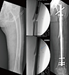
Periosteal callus (arrow and circle) on the lateral cortex of subtrochanter is noted in the pre- and post-operative radiograms. Fracture is not comminuted and caused by simple fall. The medial beak in the distal fragment is eminent.

Fig. 2
Simple radiograms shows bowing of the femoral shaft and dreaded black line in the apex of curvature. Fracture is associated with huge amount of endosteal callus. Bone scan shows hot spot at the lesion in the right femur. Hot spot in the left femur is due to femoral nailing after atypical femoral shaft fracture.

Fig. 3
Dual energy X-ray absorptiometry (DXA) image of the hip joint sometimes depicts periosteal callus (arrow) in the lateral femoral cortex.

Fig. 4
Atypical femoral fracture is frequently associated with excessive femoral bowing. Straightening and iatrogenic fracture frequently occurs during intramedullary nailing.

Table 1
American Society of Bone and Mineral Research (ASBMR) task force 2013 revised case definition of AFFs


 PDF
PDF ePub
ePub Citation
 Print
Print


 XML Download