

 PDF
PDF ePub
ePub Citation
 Print
Print


INTRODUCTION
Osteoporosis is an important public health problem. Osteoporosis is a deficiency of bone mineral density that increases fracture risk. Because the number of elderly people in the population is currently increasing, postmenopausal osteoporosis is an issue of growing importance.
The age-specific prevalence of osteoporosis is less than 2% in women younger than 50 years. Between the ages of 20 and 40 years, the prevalence of osteoporosis is only 1.2%.[1] Postpartum osteoporosis is a very rare event. Our case provides a perspective on the diagnosis of postpartum osteoporosis and complications such as compression fracture.
CASE
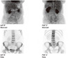
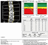
A 42-year-old multiparous woman arrived at the emergency room with back pain. One month earlier, she had a tertiary cesarean section and a left ovarian cystectomy. During the cesarean section, there were no other complications. She had breast-fed her baby after delivery. Her height was 159 cm and her weight was 56 kg. She had no other bone anomaly. She did not exercise on a regular basis. She had no past surgical or family histories, nor had trauma. On physical examination, she had diffuse back pain. The pain was so severe that she had difficulty in getting up for 7 days. The lumbar spine anteroposterior (AP) and lateral revealed recent compression fractures of T12, L1, and L3. A bone scan also revealed increased bone uptake, perfusion, and multiple recent compression fractures of T12-S1 (Fig. 1). Bone marrow density determined using dual energy X-ray absorptiometry (DXA) revealed that the T-scores for the lumbar vertebrae L1-L4, femoral neck, and femoral total were significantly low in the patient (Fig. 2). She was given calcium 500 mg daily and a chest brace for non-weight bearing.
DISCUSSION
Osteoporosis is a skeletal disease involving decreased density of the mineral portion of the bone. Osteoporosis increases the fracture risk because of the reduction of bone density. The causes of osteoporosis are most commonly age, menopause, hormone imbalance involving the thyroid or parathyroid, and alcohol intake.
Osteoporosis is diagnosed according to the 2007 International Society for Clinical Densitometry (ISCD) Official Positions or World Health Organization (WHO) criteria. We previously differentiated the two criteria and revealed that the WHO criteria are more tolerant than the ISCD Official Positions.[2]
Because osteoporosis is related to fractures, a fracture-risk assessment tool (FRAX) using clinical risk factors combined with femoral neck bone mineral density (BMD) can predict the 10-year fracture risk. We previously reviewed the Korean FRAX model with the general population and recommended that physicians use the Korean FRAX model in the general female population to predict fracture risk.[3] We already found that serum (25-[OH]D3) concentrations were significantly correlated with age (P=0.001), duration of menopause (P=0.005), and osteoporosis (P=0.044).[4]
Also, erythrocyte levels of n-3 polyunsaturated fatty acid (PUFA) and intake of fish were positively correlated with BMD of the femoral neck.[5] However, vitamin D and diet are not included as fracture risk factors. In our analysis of the BMD and previous obstetric history, the gravity was not statistically significantly associated with bone density.[6] Another study also revealed that postmenopausal women had a correlation between risk factors for osteopenia or osteoporosis and obstetric history including previous gravity.[7] A possible mechanism of postpartum osteoporosis involves ligament laxity due to endocrine changes such as parathyroid hormone (PTH)-like peptide, magnesium sulfate management, prolonged bed rest for preterm labor, and stress or fatigue fractures during delivery.[8] The fetus takes 30 g of calcium from the mother, and hypoestrogenemia during breast-feeding leads to bone mineral loss in women. As the gestational age proceeds, total calcium level decreases accordingly.[9]
Saudi women have significantly lower BMD levels than women in the USA. Because of their increased number of pregnancies, they have a longer duration of breast-feeding.[10] Therefore, correlations between the severity of BMD and breast-feeding duration with osteoporosis are unclear.
The most common symptom is severe back pain in the lower thoracic, lumbar region. Our patient could not carry her baby during daily activity.[10]
The most common site in pregnancy- and lactation-associated osteoporosis is the vertebrae, as in our case.[11]
A tendency toward systemic bone loss at the lumbar spine was found during pregnancy.[12] In particular, more than 1 standard deviation (SD) of bone loss was found. Eight vertebral fractures were found without any other risk factors.[10]
The recommended management of postpartum osteoporosis includes vitamin D and calcium supplementation and weaning. Bisphosphonate therapy is effective for increasing BMD.[11] Back pain is a common complaint for women during pregnancy and lactation. Postpartum osteoporosis, however, is a very rare event.
We should recognize the potential risk of postpartum osteoporosis and carefully differentiate back pain after delivery to prevent osteoporotic fractures.
 XML Download