Abstract
Objectives
Chitosan has been widely investigated and used. However, the literature does not refer to the shelf life of this solution. This study evaluated, through the colorimetric titration technique and an analysis of dentin micro-hardness, the shelf life of 0.2% chitosan solution.
Materials and Methods
Thirty human canines were sectioned, and specimens were obtained from the second and third slices, from cemento-enamel junction to the apex. A 0.2% chitosan solution was prepared and distributed in 3 identical glass bottles (v1, v2, and v3) and 3 plastic bottles (p1, p2, and p3). At 0, 7, 15, 30, 45, 60, 90, 120, 150, and 180 days, the specimens were immersed in each solution for 5 minutes (n = 3 each). The chelating effect of the solution was assessed by micro-hardness and colorimetric analysis of the dentin specimens. 17% EDTA and distilled water were used as controls. Data were analyzed statistically by two-way and Tukey-Kramer multiple comparison (α = 0.05).
Results
There was no statistically significant difference among the solutions with respect to the study time (p = 0.113) and micro-hardness/time interaction (p = 0.329). Chitosan solutions and EDTA reduced the micro-hardness in a similar manner and differed significantly from the control group (p < 0.001). Chitosan solutions chelated calcium ions throughout the entire experiment.
Chitosan is a natural hetero-polysaccharide obtained from the deacetylation reaction of chitin and is mainly extracted from crab and shrimp shells.1 Its properties of biocompatibility, biodegradability, bioadhesion, and non-toxicity to the human body23 have provided the basis for its application in different fields. In both the pharmaceutical and the medical fields, chitosan has been used as an anti-tumor substance and antibacterial agent.4 In agriculture, it is used as a preservative in seeds, facilitating the absorption of iron and calcium, and in the food and cosmetic industries, it is used as a source of fiber.56 Biologically, chitosan promotes weight reduction, contributes to cholesterol control, accelerates the healing of connective tissue, operates in the hemostasis process, and induces bone tissue formation.789
Being a natural substance with applications in different sectors of the health industry, chitosan was soon introduced in dentistry. Chitosan in the gel form showed promising results in periodontal regeneration in cases of intraosseous defects.1011 Because intra-canal medication promotes the prolonged release of calcium ions when combined with a calcium hydroxide paste, it prevents biofilm formation when incorporated into a root canal sealer.1213 However, its association with a composite resin confirmed its antibacterial property without any interference with the mechanical properties of the material.14 A recent study used chitosan particles to inhibit the growth of pathogens and in the modulation of the inflammatory response in human gingival fibroblasts.15 The incorporation of chitosan into a mouthwash interfered with the adhesion of microorganisms and prevented biofilm formation.16
Hence, the application of chitosan in dental environment has expanded rapidly, in spite of different specialties. In endodontics, the chelating effect of polysaccharide has drawn attention because the substance presents a high affinity with various metal ions under acidic conditions.17 Such information has stimulated the possibility of this substance being a very feasible alternative of ethylenediaminetetraacetic acid (EDTA), because this acid is considered a pollutant.18 Previous studies have shown that chitosan promotes the demineralization of dentin in a manner similar to EDTA and demonstrates a capacity for cleaning the dentin walls equivalent to that of citric acid and EDTA.1920 The chelating ability of chitosan on dentin tissue was observed, even when used in very low concentrations and for a short period of time.21
The efficiency of demineralizing agents is dependent on the application time, pH, concentration of the substance, and the amount of solution available.2223 However, irrespective of any factor that could possibly interfere with the properties of the substance, when it is employed beyond the expiration date, it usually does not achieve the expected result. Because chitosan is a biodegradable polysaccharide, it is worth clarifying the extent of its shelf life, in order to identify its validity, which would provide greater security and reliability of its use. The null hypothesis of this study is that the properties of this solution do not break down over time.
The chitosan solution was dispensed with an analytical reagent grade and water purified by a reverse osmosis system with ultraviolet light (Quimis Aparelhos Cientificos, Diadema, Brazil), with an electrical conductivity of less than 1 µS/mm2, as follows: in a 500 mL beaker, 0.6 g of chitosan (ACROS Organics, Geel, Belgium) was weighed, and then, 300 mL of 1% acetic acid was added. The final pH was 3.2, as measured by a digital pH meter.
The total volume of 300 mL of the solution obtained from 0.2% chitosan was divided into 6 equal volumes (50 mL) and added to 6 separate bottles, 3 made of amber glass and 3 made of milky white plastic. The glass (v1, v2, and v3) and plastic (p1, p2, and p3) bottles were identified by means of adhesive labels and stored in a wooden cabinet free from light at room temperature (25 ± 2℃).
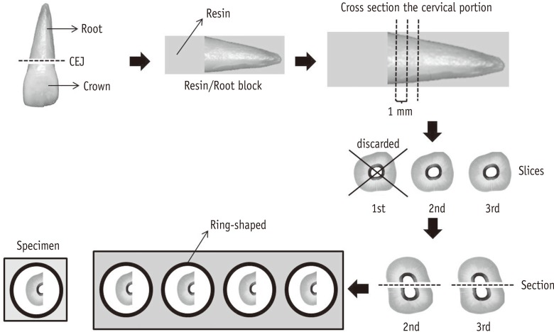
After the submission of this project and its approval by the local Ethics and Research Committee (Protocol No. 06846712.8.000.5419), 30 maxillary canine teeth were selected, which remained immersed in a 0.1% thymol solution and stored in the refrigerator at 9℃, until their time of use. Initially, the teeth were washed under running water for 24 hours and the crowns were sectioned, transversely, at the cementoenamel junction by using a carborundum disk (Labordental Ltda., São Paulo, Brazil). The remaining portion of the root was placed in a silicone mold, which was embedded into an auto-polymerizing acrylic resin, involving the entire root structure. Once the polymerization was completed, the resin/root formed a block and a hard-tissue cutting machine (Struers ApS, Ballerup, Denmark) was used for cross-sectioning the cervical portion. Three 1 mm thick slices were obtained in this region. The first slice was discarded, and the remaining slices were divided into two equal halves, by means of a scalpel blade. Each half had its cervical surface coated with liquid vaseline and embedded into the center of a ring-shaped silicon device fixed onto a wax plate. The inside of the silicone ‘ring’ was filled with the auto-polymerizing acrylic resin, thereby providing 4 specimens from a single tooth. The surface of each specimen was sanded and polished (Figure 1).
The experiment was performed in triplicate, and 3 samples from the same group were used for each test. For the analysis of dentin micro-hardness, 12 specimens were made in order to evaluate the chelating effect of solutions for each established time period.
The groups to be evaluated, through dentin micro-hardness testing, consisted of a 0.2% chitosan solution stored in 3 plastic bottles (p1, p2, and p3) and 3 glass bottles (v1, v2, and v3), in addition to 17% EDTA and distilled water (control). At the end, 30 specimens (n = 30) were assigned to each of the 4 groups of solutions.
An automatic pipette deposited 50 µL of the test solution on to a specimen for 5 minutes. Then, the specimens were washed under running water and dried with gauze. The test was carried out in triplicate as follows: with 4 specimens derived from a single tooth, we tested v1, p1, and EDTA solutions, and the control group. The other 4 specimens from the second tooth were used for evaluating v2, p2, and EDTA solutions, and the control group. Four more specimens derived from the third tooth were used for evaluating v3, p3, and EDTA solutions, and the control.
The dentin microhardness was measured with a Knoop indenter (Shimadzu HMV-2000, Shimadzu Corp., Kyoto, Japan), under a 10 g load for 15 seconds. For each specimen, three indentations were obtained, with a pre-programmed distance of 200 µm. The measurement started from the portion of dentin closest to the cementum following a straight line towards the root canal. The 3 values provided by the device were recorded and added, establishing an average for each specimen. The protocol described above was carried out, always in the same manner, for the proposed periods of 0, 7, 15, 30, 45, 60, 90, 120, 150, and 180 days after the dispensing of the 0.2% chitosan solution.
Initially, 300 µL of a 0.3% calcium carbonate standard solution (CaCO3, Omya, Três Lagoas, MG, Brazil) was deposited into a glass container (cell). The following was added to the solution: 600 µL of deionized water (ASFER, São Caetano do Sul, SP, Brazil), 50 µL of a buffer solution (Synth, Diadema, SP, Brazil), and 5 µL of a calcium ion indicator (Eriochrome Black T, Sigma-Aldrich Brasil Ltda., São Paulo, SP, Brazil). Each of these substances was added to the CaCO3 solution with specific objectives. The deionized water was added to fill the minimum liquid volume of the cell. The buffer solution maintained the CaCO3 ions available to the dye, facilitating the coloring process. Eriochrome T, on the other hand, promoted the identification of calcium ions, coloring them, resulting in a violet-colored complex. This initial stage was aimed at establishing the amount of the 0.2% chitosan solution (analyte) required to complex all the calcium ions present in the total volume of the cell. The amount of analyte was designated as the standardized volume of chitosan and obtained as follows: immediately after the preparation of the 0.2% chitosan solution and before being put into plastic and glass bottles, 10 µL volumes of the chelator were deposited in the cell by an automatic pipette of 100 µL. The micro-dripping was performed until the violet-colored CaCO3 solution turned blue, indicating the complete chelation of all the calcium ions. The change in color was detected by an ultraviolet-visible (UV/Vis) spectrophotometer (HP Agilent 8452a, Hewlett Packard, Palo Alto, CA, USA), which adapted itself to the cell, immediately after each micro-dripping, in order to measure the absorbance. The standardized volume of chitosan was determined to be 400 µL.
The preliminary colorimetric analysis, in which the standardized volume was identified, was considered the starting point for determining the shelf life of chitosan. Thus, for each time period set for the experiment (0, 7, 15, 30, 45, 60, 90, 120, 150, and 180 days after dispensing), 400 µL of chitosan was deposited inside the cell containing CaCO3, and the container was used in the spectrophotometry. After each analysis of solution coloring, the instrument emitted an absorbance value, expressed by means of a graph (wavelength/absorbance). After the colorimetric analysis of the three samples from each group (v1, v2, v3, p1, p2, and p3), an average absorbance value was calculated for each group and for each period studied.
A data file was assembled from the averages obtained from the original 3 microhardness measurements. Considering the test in triplicate, 3 averages were obtained for each solution, and for each period of time; totaling 120 data were analyzed. The data file was submitted to the two-way ANOVA test (α = 0.05), using the SPSS Statistic software, version 17.0 (SPSS Inc., Chicago, IL, USA). In addition, Tukey-Kramer test was performed post hoc to clarify which chelating solutions were different from the others.
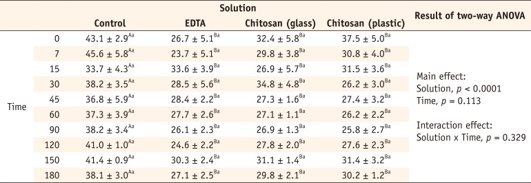
The data showed a similarity among the groups tested, with respect to the time of study (p = 0.113) and there was no interaction effect between the micro-hardness and time (p = 0.329, Table 1). This result showed that during the period of 180 days, the evaluated solutions (chitosan in glass, chitosan in plastic, EDTA, and water) individually maintained the same effect on the dentin micro-hardness: from the day of dispensing the solutions (time zero) to 180 days later, there was no change in the behavior of each solution alone. In the case of chitosan, in particular, it was found that both the chelator stored in glass and that in a plastic bottle maintained the chelation property over time. There was only a significant difference in the dentin micro-hardness (p < 0.0001). This result demonstrated that the chelating ability was different among the groups (Table 1). Tukey-Kramer post hoc test showed that the groups formed by chitosan in glass, chitosan in plastic, and EDTA reduced the dentin micro-hardness similarly to each other (p > 0.05) and different from the control group (p < 0.001, Table 1).
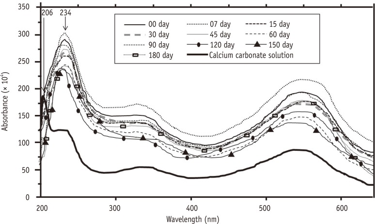
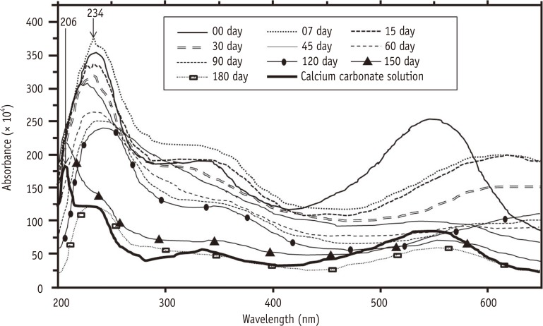
The colorimetric analysis resulted in two graphs (wavelength/absorbance), one for the chitosan stored in plastic bottles (Figure 2) and the other for chitosan stored in glass (Figure 3), as per the UV/Vis spectrophotometer. The average absorbance values provided by the instrument were displayed on the same wavelength (234 nm) for all of the times studied.
Chitin and chitosan have been widely used in studies because of their biodegradability, biocompatibility, antibacterial properties, and hydrophilicity, besides the fact that they come from abundant renewable natural resources.3510112425262728 The conditions for the alkaline deacetylation of chitin required to produce chitosan are responsible for the biopolymer's main characteristics, such as the average molecular weight and the degree of deacetylation.29 From such characteristics, different ways to use chitosan have been determined. In the present study, we used a commercial product with guaranteed purity and degree of deacetylation of more than 90%. The degree of purity, solubility, viscosity, degree of deacetylation, and molecular weight of commercial chitosan vary according to the dispensing process. The homogeneous deacetylation of chitin results in chitosan with a higher capacity for adsorption of metals.30 This property, as well as the ion exchange and chelation capacity, are the probable mechanisms responsible for the formation of complexes between chitosan and metal ions.2531
The pH of the 0.2% chitosan solution evaluated here was set at 3.2. Previous studies have shown that a chitosan solution having this concentration and pH is similar to 15% EDTA with respect to the promotion of the removal of the smear layer and the reduction of the dentin microhardness.192021 On the basis of this information, 17% EDTA was added to the experimental groups of this study, for the purpose of comparison.
A previous study on the validation of the stability of different analytical solutions revealed that 0.1 Mol (≈ 2.9%) EDTA remains stable for a period exceeding 6 months. When the concentration is reduced to 0.01 Mol (≈ 0.2%), the stability period decreases to less than 3 months. The authors argue that solutions containing the same analyte, but of different molarity, and of a lower concentration, are generally less stable.32 Most manufacturers of EDTA do not establish the product expiration date. Therefore, for greater quality assurance of the solution, the responsible chemist in the research laboratory of the university dispensed the EDTA used in this experiment.
In the present study, two different methods were used for verifying whether the chitosan solution maintained its chelating property during the research period. During a period of 6 months, the dentin micro-hardness was measured before and after the application of the chelating solutions. The comparison between the measurements showed that the solutions retained their chelation capacity. During the colorimetric analysis or spectrophotometry, a standard volume of the chelating solution was added to a solution containing calcium ions, and the equipment determined whether the tested solution chelated the calcium ions. Although the methods did not show a direct relationship, their results served to suggest a relationship.
The results of the analysis showed that the chelating effect of the 0.2% chitosan solution stored in the glass and plastic bottles was similar to that of the 17% EDTA. The similarity of the demineralization capacity between the chelating agents, during the 180 day analysis, allowed us to conclude that the chelating property of 0.2% chitosan maintained stability for at least 6 months. It is worth noting that the chitosan used was diluted in 1% acetic acid during its dispensation. The acid employed may be primarily responsible for the chelating effect of the solution; however, it is important to highlight that the concentration of 1% acetic acid had no effect on the dentin demineralization.20
Chitosan, under acidic conditions, presents an affinity to various metal ions including Ni2+, Zn2+, Co2+, Fe2+, Mg2+, and Cu2+.17 It would be no surprise if the chitosan stored in the glass bottles reacted with the silica, compromising its demineralization capacity. However, in the period studied, the chitosan packaged under these conditions showed no significant change with respect to this property. On the other hand, the care taken while adopting plastic packages for storing the EDTA and the recommendation to use plastic syringes for clinical applications could lead to a chelation reaction between the solution and the metallic components of the glass. Despite the preventive measures taken, no scientific evidence proves the existence of this reaction. A previous study demonstrated that EDTA can promote structural changes in glass, such as bubbles or cracks; however, whether these modifications can compromise the demineralization capacity of dentin tissue has not been proven.33
The absence of the chelating reaction of chitosan with glass may be related to the pH or the selective affinity to the substance,31 which is similar to what occurs with EDTA. This acid presents a higher affinity for ions Ca2+, Sr2+, Ba2+, and Mg2+ at alkaline pH between 8 and 10. For the ions Pb2+, Cu2+, Zn2+, Mn2+, Al2+, and Sn2+, the optimum pH is slightly acidic, between 4 and 6.34 The evaluated chitosan solution was prepared with a very acidic pH (3.2). At this pH, chitosan was probably not the most appropriate means to chelate the metal ions from glass, such as Mg2+ and Zn2+, thus maintaining its demineralization effect on dentin. As a supplement, the colorimetric analysis showed that both chitosan solutions (plastic and glass) chelated the calcium ions of the CaCO3 solution during the 6 months of the evaluation. The test showed that the chitosan retained its chelating property during the 180 day study, irrespective of the storage form. The colorimetric analysis is the means by which the amount of a certain substance is obtained on the basis of its light absorption capacity. In the present experiment, we used the CaCO3 standard solution, which was stained with Eriochrome T, assuming a violet color. The color change of this solution to a bluish color meant the absence of calcium ions. Thus, in the different scheduled times considered in the study, a standard amount of chitosan was added to the CaCO3 solution and the change in color was measured, in terms of the absorbance values, by using the UV/Vis spectrophotometer. The wavelength (λ) identified by the spectrophotometer for violet color is 206 nm; the CaCO3 solution was presented with all the calcium ions available for chelation. When the calcium solution was chelated, the color changed from violet to blue, and the wavelength demonstrated by the instrument was 234 nm. According to Figures 2 and 3, for all the proposed times, the absorbance value was visualized at a wavelength of 234 nm. If there is no chelation of the calcium ions in the case of one of the proposed times, the instrument would have emitted a band with a wavelength equal to or very close to that of the violet color (206 nm). It is worth pointing out that the analysis performed was qualitative, which allowed for the observation of whether there was any chelation of the calcium ions from the test solutions. Further, the results obtained using colorimetry were supported by the micro-hardness test.
Based on the methodology used and under the conditions of this experiment, we were able to confirm that 0.2% chitosan maintains its chelating property on dentin tissue for a period of at least 6 months. Such information, although it can be used to guarantee the shelf life of the product for future ex vivo studies, does not ensure its biological properties or authorize its use in clinical therapy. Considering the limitations of this study, we could not determine whether chitosan at lower or higher concentrations would react equally with respect to shelf life.
Acknowledgment
This study was supported by CAPES - National Council for the Improvement of Higher Education (Brazil).
References
1. Uragami T, Yamamoto S, Miyata T. Dehydration from alcohols by polyion complex cross-linked chitosan composite membranes during evapomeation. Biomacromolecules. 2003; 4:137–144. PMID: 12523858.
2. Senel S, Kas HS, Squier CA. Application of chitosan in dental drug delivery and therapy. In : Muzzarelli RAA, editor. Chitosan per os: from dietary supplement to drug carrier. Grottammare, Italy: Atec;2000. p. 241–256.
3. Akncbay H, Senel S, Ay ZY. Application of chitosan gel in the treatment of chronic periodontitis. J Biomed Mater Res B Appl Biomater. 2007; 80:290–296. PMID: 16767723.
4. Dash M, Chiellini F, Ottenbrite RM, Chiellini E. Chitosan - a versatile semi-synthetic polymer in biomedical applications. Prog Polym Sci. 2011; 36:981–1014.
5. Jeon YJ, Shahidi F, Kim SK. Preparation of chitin and chitosan oligomers and their applications in physiological functional foods. Food Rev Int. 2000; 16:159–176.
6. Zhang A, Mu H, Zhang W, Cui G, Zhu J, Duan J. Chitosan coupling makes microbial biofilms susceptible to antibiotics. Sci Rep. 2013; 3:3364. PMID: 24284335.
7. Muzzarelli RA, Mattioli-Belmonte M, Tietz C, Biagini R, Ferioli G, Brunelli MA, Fini M, Giardino R, Ilari P, Biagini G. Stimulatory effect on bone formation exerted by a modified chitosan. Biomaterials. 1994; 15:1075–1081. PMID: 7888578.
8. Park SS, Kim SG, Lim SC, Ong JL. Osteogenic activity of the mixture of chitosan and particulate dentin. J Biomed Mater Res A. 2008; 87:618–623. PMID: 18186071.
9. Walsh AM, Sweeney T, Bahar B, O'Dohert JV. Multi-functional roles of chitosan as a potential protective agent against obesity. PLoS One. 2013; 8:e53828. PMID: 23342013.
10. Park JS, Choi SH, Moon IS, Cho KS, Chai JK, Kim Ck. Eight-week histological analysis on the effect of chitosan on surgically created one-wall intrabony defects in beagle dogs. J Clin Periodontol. 2003; 30:443–453. PMID: 12716338.
11. Boynueğri D, Özcan G, Şenel S, Uç D, Uraz A, Ogüş E, Cakilci B, Karaduman B. Clinical and radiographic evaluations of chitosan gel in periodontal intraosseous defects: a pilot study. J Biomed Mater Res B Appl Biomater. 2009; 90:461–466. PMID: 19145627.
12. Ballal NV, Shavi GV, Kumar R, Kundabala M, Bhat KS. In vitro sustained release of calcium ions and pH maintenance from different vehicles containing calcium hydroxide. J Endod. 2010; 36:862–866. PMID: 20416434.
13. DaSilva L, Finer Y, Friedman S, Basrani B, Kishen A. Biofilm formation within the interface of bovine root dentin treated with conjugated chitosan and sealer containing chitosan nanoparticles. J Endod. 2013; 39:249–253. PMID: 23321239.
14. Kim JS, Shin DH. Inhibitory effect on streptococcus mutans and mechanical properties of the chitosan containing composite resin. Restor Dent Endod. 2013; 38:36–42. PMID: 23493656.
15. Arancibia R, Maturana C, Silva D, Tobar N, Tapia C, Salazar JC, Martínez J, Smith PC. Effects of chitosan particles in periodontal pathogens and gingival fibroblasts. J Dent Res. 2013; 92:740–745. PMID: 23788611.
16. Costa EM, Silva S, Madureira AR, Cardelle-Cobas A, Tavaria FK, Pintado MM. A comprehensive study into the impact of a chitosan mouthwash upon oral microorganism's biofilm formation in vitro. Carbohydr Polym. 2014; 101:1081–1086. PMID: 24299877.
17. Kurita K. Chemistry and application of chitin and chitosan. Polym Degrad Stabil. 1998; 59:117–120.
18. Spanó JC, Silva RG, Guedes DF, Sousa-Neto MD, Estrela C, Pécora JD. Atomic absorption spectrometry and scanning electron microscopy evaluation of concentration of calcium ions and smear layer removal with root canal chelators. J Endod. 2009; 35:727–730. PMID: 19410093.
19. Pimenta JA, Zaparolli D, Pécora JD, Cruz-Filho AM. Chitosan: effect of a new chelating agent on the microhardness of root dentin. Braz Dent J. 2012; 23:212–217. PMID: 22814688.
20. Silva PV, Guedes DF, Nakadi FV, Pécora JD, Cruz-Filho AM. Chitosan: a new solution for removal of smear layer after root canal instrumentation. Int Endod J. 2013; 46:332–338. PMID: 22970844.
21. Silva PV, Guedes DF, Pécora JD, da Cruz-Filho AM. Time-dependent effects of chitosan on dentin structures. Braz Dent J. 2012; 23:357–361. PMID: 23207849.
22. Çalt S, Serper A. Time-dependent effects of EDTA on dentin structures. J Endod. 2002; 28:17–19. PMID: 11806642.
23. Hülsmann M, Heckendorff M, Lennon A. Chelating agents in root canal treatment: mode of action and indications for their use. Int Endod J. 2003; 36:810–830. PMID: 14641420.
24. Guibal E, Milot C, Eterradossi O, Gauffier C, Domard A. Study of molybdate ion sorption on chitosan gel beabs by different spectometric analyses. Int J Biol Macromol. 1999; 24:49–59. PMID: 10077272.
25. Guibal E, Milot C, Roussy J. Influence of hydrolysis mechanisms on molybdate sortion isotherms using on chitosan. Sep Sci Technol. 2000; 35:1021–1038.
26. Carvalho TS, Lussi A. Combined effect of a fluoride-, stannous- and chitosan-containing toothpaste and stannous-containing rinse on the prevention of initial enamel erosion-abrasion. J Dent. 2014; 42:450–459. PMID: 24440712.
27. Wang J, Chen C. Chitosan-based biosorbents: modification and application for biosorption of heavy metals and radionuclides. Bioresour Technol. 2014; 160:129–141. PMID: 24461334.
28. del Carpio-Perochena A, Bramante CM, Duarte MA, de Mora MR, Aouada FA, Kishen A. Chelating and antibacterial properties of chitosan nanoparticles on dentin. Restor Dent Endod. 2015; 40:195–201. PMID: 26295022.
30. Li Q, Dunn ET, Grandmaison EW, Goosen MFA. Applications and properties of chitosan. J Bioact Compat Polym. 1992; 7:370–397.
31. Rhazi M, Desbrières J, Tolaimate A, Rinaudo M, Vottero P, Alagui A, Meray MEL. Influence of the nature of the metal ions on the complexation with chitosan. Application to the treatment of liquid waste. Eur Polym J. 2002; 38:1523–1530.
32. Nogueira JMF, Seródio P, Agudo R. Validação de estabilidade de soluções. Bol Soc Port Quim. 2001; 81:51–53.
33. Silva BCB, Irala LED, Grazziotin-Soares R, Salles AA. Verification of structural changes of glass when submitted to 17% EDTA. Rev Bras Odontol. 2011; 8:48–53.