

 PDF
PDF ePub
ePub Citation
 Print
Print


Introduction
The objectives of endodontic therapy are to eliminate microorganisms from the root canal system, and to establish an effective barrier to prevent further passage of microorganisms or their byproducts to periapical tissues.1234 Thus, complete obturation of the root canal system and creation of an ‘apical’ seal are important for successful endodontic treatment.5678
The standard root canal obturation materials are a combination of sealer with a central core material, which has, thus far, generally been gutta-percha (GP).9 Various chemical formulations have served as bases for sealers such as zinc oxide-eugenol, glass-ionomer, silicone, and epoxy or methacrylate resin.910 Among them, epoxy resin-based sealers are popular due to their low solubility, apical seal, and micro-retention to root dentin.21112 AH Plus (Dentsply DeTrey, Konstanz, Germany) is an epoxy-amine resin-based sealer that has become popular among dental clinicians due to its excellent properties, including adhesion to dentin and good sealing ability.1011
After root canal therapy, the placement of a post and core is often necessary when the coronal tooth structure is inadequate for retaining the crown.135713 The integrity of the remaining obturating material, which provides an apical seal, should not be disrupted during post space preparation.1415 However, it often deteriorates, causing endodontic treatment failure as a result of reinfection.1
When a rotary instrument is introduced into the canal to remove GP, the rotational forces may affect the integrity of the remaining obturating material depending on the rigidity of the sealer, thereby possibly breaking the root canal wall-GP interface.14 According to the manufacturer, AH Plus has a setting time of 8 hours. However, little research has been done on the extent of epoxy polymerization as a function of time, which may affect the bonding integrity to the GP and/or root canal wall when the post space is prepared.14
The aim of this in vitro study was to investigate the apical leakage when a post space was prepared at different times after root canal obturation with GP and AH Plus, in order to determine the optimal post space preparation timing in terms of the polymerization of the resin-based sealer. The maximum time period of one week after root canal obturation for post space preparation was used.1314 Prior to this dye leakage study, the epoxy polymerization of AH Plus as a function of time was evaluated using two different techniques: attenuated total reflectance-Fourier transform infrared (ATR-FTIR) spectroscopy and microhardness measurements. Differential scanning calorimetry (DSC) was also used for determining the glass transition temperature (Tg) of AH Plus with time. The null hypothesis tested was that the post space preparation timing does not affect the quality of the apical seal, irrespective of the extent of epoxy polymerization.
Materials and Methods
Fourier transform infrared (FTIR) spectroscopy
Cylindrical molds (diameter = 5 mm, thickness = 1 mm) were placed on a mylar strip over a glass slide. Freshly mixed AH Plus was filled into the mold and then, covered with another mylar strip and glass slide. The assemblies were stored in a dark incubator at 37℃ and 100% relative humidity (RH), and the extent of the epoxy polymerization was determined at four specified times (8, 24, and 72 hours, and 1 week) by using ATR-FTIR spectroscopy (ATR, MIRacle, Pike Technologies Inc., Madison, WI, USA; FTIR, IRPrestige-21, Shimadzu Corp., Kyoto, Japan). The absorbance spectrum was acquired by scanning the specimens 64 times over a 1,350 - 850 cm-1 range (resolution, 4 cm-1). The degree of epoxy conversion (DEC) was calculated by using the following formula1617: DEC (%) = [1 – (A916,t)(A1183,0)/(A916,0)(A1183,t)] × 100, where A1183,0 and A1183,t denote the areas of the reference peak at 1,183 cm-1 while A916,0 and A916,t refer to the areas of the epoxy peak at 916 cm-1, at time zero (0) and at the specified time (t), respectively (n = 10).
Vickers microhardness
For the microhardness test, 10 specimens per specified time (8, 24, and 72 hours, and 1 week) were prepared and stored in the same manner as in the case of the FTIR spectroscopy. Using a Vickers hardness tester (HMV-2, Shimadzu Corp.), three indentations were made on each resin specimen along the middle line, with each indentation separated by approximately 0.5 mm.17 Ten second dwell time and 10 g load were chosen to make the indentations. The Vickers hardness of each specimen was recorded as the average of three readings.17
Differential scanning calorimetry
Tg was measured by using DSC (DSC-60, Shimadzu Corp.) at four specified times after mixing (8, 24, and 72 hours and 1 week, n = 10) according to ASTM E1356.18 A small amount (approximately 60 mg) of each mixed material was transferred to a standard aluminum pan, which was then stored in a dark container at 37℃ with 100% RH until the corresponding specified time. After storage, each material was subjected to a DSC analysis after measuring the weight using a digital balance (AP210S, Ohaus Corp., Pine Brook, NJ, USA). The analysis was initiated at room temperature and then, the samples were heated to 200℃ at a heating rate of 10 ℃/min under nitrogen flow. For each specimen, the midpoint temperature of the temperature range, bounded by the tangents to the two flat regions of the heat flow curve, was used as the Tg.18
Apical dye penetration
A total of 50 human single-rooted premolars with a similar root structure, extracted for reasons such as advanced periodontitis or orthodontic treatment, were collected after obtaining the patients' informed consent under a protocol approved by the Ethics Committee (document number: BMRI 74005-452). For each tooth, a radiograph was taken to check for a single canal. The teeth were disinfected in 0.5% chloramine solution, stored in distilled water, and used within 2 months of extraction.19
The crowns were sectioned out with a low-speed diamond saw (TechCut 4, Allied High Tech Products Inc., Rancho Dominguez, CA, USA) under water cooling in order to create a root length of approximately 14 mm. The root canals were enlarged to an apical file size of 40/0.06 taper, using K3 nickel-titanium rotary instruments (SybronEndo, Orange, CA, USA), 0.5 mm from the anatomic apex.20 RC-Prep (Premier Dental Products, Plymouth, PA, USA) was used for lubricating the root canals during preparation. The canals were irrigated with 2.5% NaOCl at each change of file.2 After root canal preparation was completed, the canals were finally irrigated with 17% ethylenediaminetetraacetic acid for 1 minute, followed by distilled water, and dried with absorbent paper points (DiaDent, Cheongju, Korea).2
AH Plus (Dentsply DeTrey) that had been freshly mixed according to the manufacturer's instructions, was coated onto the canal walls by using a Lentulo spiral (Dentsply Maillefer, Ballaigues, Switzerland) and paper points. An ISO size 40 GP master cone (DiaDent) coated with the AH Plus sealer was placed into the canal at working length. After root canal obturation using the continuous wave compaction technique, the quality of obturation was radiographically checked.
The obturated specimens were randomly divided into five groups (n = 10/group) based on their post space preparation times. In GI, the post space was prepared using a Parapost drill, size 1, (Coltène/Whaledent Inc., Cuyahoga Falls, OH, USA) immediately after root canal obturation, leaving 5 mm of GP apically.20 The prepared post spaces were irrigated in the same way as for the canal enlargement. All the other specimens were stored in a dark incubator at 37℃ and 100% RH after the coronal orifices of the filled root canals had been immediately sealed with a temporary filling material (Cavit G, 3M ESPE AG, Seefeld, Germany). The stored specimens were taken out of the incubator at four specified times (8, 24, and 72 hours, and 1 week after root canal obturation), and the post space was prepared in the same manner as in GI (group codes: G8H, G24H, G72H, and G1W, respectively). Then, the coronal opening was sealed with light-cured glass-ionomer cement (GC Fuji II LC, GC Corp., Tokyo, Japan), which was light-activated for 40 seconds using a light-curing unit (Elipar TriLight, 3M ESPE, output intensity = 750 mW/cm2). The entire surface for each root specimen was covered with two coats of fingernail polish except for the last apical 1 mm.21 The specimens were immersed in a 2% methylene blue solution under reduced pressure conditions for 24 hours.21 The reduced pressure was obtained with a suction pump (Gast Manufacturing Inc., Benton Harbor, MI, USA). After immersion, the teeth were washed under running tap water to remove the excess dye material. After being grooved longitudinally in the buccolingual direction using a low-speed diamond saw without disturbing the root canal obturation material, each tooth specimen was split with light taps of a mallet and chisel. Specimens were washed under running tap water to remove the excess dye material.
For each tooth specimen, ×30 magnified photographs were taken using a stereomicroscope (SZ61, Olympus Corp., Tokyo, Japan) next to a millimeter ruler for reference.14 Apical dye leakage was measured as the linear penetration of the stain.1 The evaluation of each specimen was performed by two examiners simultaneously, with an agreed result.21
Statistical analysis
All statistical analyses were performed using SPSS 17.0 for Windows (SPSS Inc., Chicago, IL, USA) at α = 0.05. Each dataset, which was normally distributed as determined by Shapiro-Wilk test (p > 0.05), was statistically analyzed with a one-way analysis of variance (ANOVA) and Tukey's post hoc test.
Results
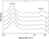
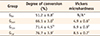
Figure 1 and Table 1 show the polymerization of AH Plus as a function of time after mixing, as assessed by FTIR spectroscopy and microhardness measurements. In both analyses, gradual increases in the values with time were observed. Significant increases in the microhardness were detected at three specified times (p < 0.05), although the values for the G8H specimens were not measurable due to their considerable surface softness.
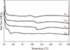
Representative DSC scans are presented in Figure 2. The Tg values for G8H, G24H, and G72H were 105.3 ± 6.0, 109.4 ± 5.6, and 114.7 ± 9.7℃, respectively, showing a gradual increase as a function of time after mixing. However, the G1W specimens did not show a clear Tg value on the DSC scans.
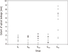
The results of the apical dye penetration test are shown in Figure 3. G1W had significantly higher values than the other groups (p < 0.05), which had no significant difference from one another (p > 0.05).
Discussion
The optimal timing of the post space preparation in root canals filled with GP and AH Plus still seems controversial.15 The present in vitro study was performed to determine an optimal timing for the post space preparation after root canal obturation by using AH Plus as a sealer, in terms of its polymerization and influence on apical leakage. Solano et al.14 reported that the post spaces made immediately after root canal obturation showed less leakage than those prepared one week after obturation. Corrêa Pesce et al. suggested that post space preparation did not affect the apical seal quality.22 In contrast, Nagas et al. reported significantly better apical sealing in delayed post space preparation than in immediate post preparation.13 However, most of the studies regarding the apical leakage of a root canal filled with sealers did not investigate the epoxy polymerization of AH Plus as a function of time.51114
In this study, the breakage of apical sealing due to post space preparation by using a rotary instrument was investigated using a dye leakage test. Dye penetration tests are fast and easy, and methylene blue is a small molecule that penetrates further than other dyes or radioisotopes.23 A comparison of the findings from the present dye leakage study with other analyses on sealing capacity is hampered by the absence of a standardized methodology.22 Moreover, although dye penetration tests are more sensitive than bacterial leakage tests, there may be no significant measure of agreement between dye and bacterial penetration along root fillings.6823
There is a general consensus that the length of the remaining root canal obturation material is inversely proportional to the probability of apical leakage.1 Many previous studies have stated that keeping 5 mm of the obturating material in the apical region constitutes a safe margin.13 Thus, the present study left 5.0 mm as the baseline remnant filling material.
In the present in vitro study, delayed post space preparations until 72 hours did not affect leakage values despite gradual progress in the epoxy polymerization of the sealer (Figure 3). However, when post space preparation was delayed until 1 week, at the time point when the sealer was very hard and brittle (Figures 1 and 2), apical leakage values were significantly higher. These findings suggest that the breakage of an apical seal is related to the extent of epoxy polymerization, although the flexibility of a partially polymerized sealer seems to maintain the integrity of the apical seal until 72 hours despite the rotational force generated during the removal of the GP transferred to the region. Thus, the null hypothesis that the post space preparation timing does not affect the quality of the apical seal was rejected.
When the epoxy prepolymer and hardener are mixed, the C–O bond in the original epoxide group is converted to a reactive –CH2 site and subsequently, crosslinks to the amine molecule, forming a highly ‘crosslinked’ 3 dimensional polymer network.4 The cure time of an epoxy resin system is dependent on the reactivity of the amine hydrogen atoms and thus, determined by the kinetics of the particular amine used in the hardener.24 AH Plus is composed of an epoxide paste containing an epoxy resin and an amine paste containing three different types of amine hardeners.10 A long setting time of 8 hours allows good flow and penetration into the orifices of the dentinal tubules of the root canal wall during obturation by clinicians.10 Thus, it forms good adhesion to the root dentin through mechanical interlocking although the material does not chemically bind to GP or the root dentin.1025 Although extensive crosslinking makes the epoxy resin hard and brittle, the DEC is usually less than 100% in actual epoxy reactions.24 The FTIR and microhardness analyses demonstrated that AH Plus was polymerized to some extent (DEC 51.2%) after 8 hours of mixing, but still remained too soft to allow microhardness measurements due to the presence of unreacted epoxy groups (Figure 1 and Table 1). The dye leakage test, which presented no significant difference between GI and G8H (Figure 3), suggests that when the rotary instrument is introduced into the obturated canal to remove GP, the sealer is still within its working time (GI) or still flexible (G8H), allowing the sealer to set without causing microfractures.14 The epoxy polymerization reaction continued after the setting time (8 hours) over the experimental period (up to 1 week), implying a post-setting conversion.26 Nonetheless, the leakage value did not significantly increase with time (until 72 hours) except under the 1 week condition (G1W).
The physical and mechanical properties of epoxy resin are influenced by DEC.1227 Tg, the temperature range where the transition of a polymer from a hard, glassy material to a soft, rubbery one,27 has been used as an indicator to estimate the extent of polymerization of thermoset resins such as an epoxy resin. High values of Tg indicate high rigidity of the polymer chains due to their restricted molecular motion, whereas low Tg values indicate flexible chains with a lower crosslink density and higher molecular mobility.282930 Although Tg is a range of temperatures rather than a specific temperature, a single midpoint temperature corresponding to 1/2 of the heat flow difference between the extrapolated onset and the extrapolated end was determined according to ASTM E1356.18 As seen in Figure 2, Tg was characterized by a drastic change in the baseline, indicating a change in the heat capacity of the resin, but with no accompanying change in the enthalpy associated with the transition. The FTIR and microhardness analyses presented gradual progress of the epoxy polymerization with time during the experimental period (even until 1 week). On the other hand, the DSC had a distinct Tg until 72 hours after mixing, suggesting that the material was still relatively soft and flexible. In contrast, the G1W specimens did not present a clear Tg value, indicating that the material 1 week after mixing formed a very hard and brittle polymer network. As a result, it seems that the rotary force applied to create a post space resulted in the formation of microcracks in the material and the breakage of the bond at the interface,14 leading to significantly higher leakage values than in the other cases.
Based on the results of this study, we recommend that the post space preparation of root canals filled with a GP/epoxy resin sealer be performed prior to extensive epoxy conversion in order to avoid potential microfractures of the sealer during GP removal using a rotary instrument. Solano et al. suggested immediate post space preparation following root canal obturation in clinical practice because the clinician is intimately familiar with the root canal anatomy.14 However, such post space preparation procedures involving drilling and rinsing with water may dilute the uncured epoxy resin and potentially damage the apical seal. Thus, to avoid such complications, some delayed post space preparation may produce a better apical seal than an immediate one as long as the epoxy sealer is flexible. However, further in vitro and in vivo studies are required to clarify the optimal clinical post space preparation timing of a root canal sealed with epoxy resin-based sealers.
Conclusions
Although the Tg values of AH Plus gradually increased with time, the preparation 1 week after mixing specimens presented no clear Tg value. When the post space was prepared 1 week after root canal obturation, the leakage was significantly higher than in the other groups, among which there was no significant difference in leakage.
 XML Download