

 PDF
PDF ePub
ePub Citation
 Print
Print


INTRODUCTION
In the clinical settings, it is common to find teeth that have lost part of their structure, affecting at least one of the proximal areas. The reconstruction of such cavities is a challenge for the operator, not only in the endodontic and restorative field, but also in the periodontal field [1]. The correct choice of treatment plan is crucial to success, but it is difficult because of the scarcity of clinical protocols and lack of consensus on the optimal method of restoration in such cases.
It is accepted that endodontically treated teeth are more prone to fracture [2]. Although this fragility was attributed to loss of water and collagen of the dentin at first, we now know that the most important changes in the biomechanics of the tooth are attributed to the loss of tissue [2]. Thus, it is essential to consider the amount of healthy tooth structure remaining in the decision of the type of treatment to execute. In 2008, Fichera et al. [3] set the following order of structures that contribute, from most to least, to resistance in the tooth: 1) interaxial dentin, localized from the occlusal surface to the roof of the pulp chamber, 2) marginal ridges, 3) pulp chamber roof, and 4) the enamel-dentin complex of each intact cusp. Taking this order into account, it can already outline a concept of the resistance of the tooth to be restored. For example, an endodontically treated tooth that has lost the roof of the pulp chamber but still has its marginal ridges should have the least significant structural weakening, versus a tooth with an intact chamber roof but loss of one or both marginal ridges (as in a class II cavity).
However, not only are the dimensions of the cavity or lack of tissue important, but the depth of the aggression that could reach the periodontal tissues is also a key point in the restoration. It must be kept in mind that class II restorations with deep cervical margins are particularly complex. Beyond the loss of tissue, the subgingival position hinders clinical management, and sealing can be challenged not only in the absence of enamel [4], but also by the complexity of the isolation, sometimes becoming almost unreachable. It is these extreme cases in which we perform a deep margin elevation (DME) [5] or periodontal treatments, such as crown lengthening, in an attempt to return a manageable cavity margin.
In this study, we present a decision tree for decision making for class II cavities on molars with increasingly large vertical dimensions. We aim to classify schematically from a multidisciplinary perspective, the best treatment option in each case, including both the choice of restoration (direct or indirect) and the management of the cavity margin (DME or surgical crown lengthening [SCL]) if necessary.
CASE REPORTS
Description of the different clinical scenarios
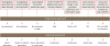
Schematically, we show a series of clinical conditions based on the depth of the lesion and the affected tissue (Chart 1).
Scenario 1: class II that does not reach the gingival margin and does not affect the pulp tissue
Such situations are relatively simple, because a sectional matrix with a ring to form a good point of contact with the adjacent tooth will be enough for success in our treatment [6] (Figure 1A).
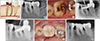
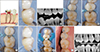 | Figure 1Scenario 1. (A) Diagram: supragingival caries does not reach the pulp. (B) Initial photography: caries cavity in molar tooth #16 does not invade the pulp tissue or periradicular tissues. (C) Preoperative bite-wing radiography. (D) Isolation using a rubber dam and wedged matrix to prevent damage to the adjacent tooth. (E) Exposure of caries. (F, G) Clean cavity: note that the cavity does not reach the periodontal tissues. (H) Obturation using the Palodent Plus system (DeTrey Dentsply, Konstanz, Germany). (I) Final photograph after checking for occlusion. (J) One-year control radiograph.
|
The case shown is a class II case in a 38-year-old male patient with discomfort when eating sweet foods. After a clinical and radiographic examination (Figure 1B and 1C), proximal caries on tooth #16 that did not affect the pulp space was observed. After isolation of the operative field (Figure 1D), we proceeded to remove the caries (Figure 1E-1G). Once removed, a proximal filling was performed using a sectional matrix, a wedge, and a Palodent Plus ring (DeTrey Dentsply, Konstanz, Germany) using a composite Ceram.X mono M2 (DeTrey Dentsply; Figure 1H). Once the procedure was completed, the occlusion was checked (Figure 1I). Radiological control: bitewing at 1 year to check that the treatment was working successfully (Figure 1J).
Scenario 2: class II that does not reach the gingival margin but did affect the pulp tissue
Such cases are also relatively easy to restore. First, the pulp issue is addressed by either a vital treatment or root canal therapy [7], and then the restorative treatment is performed. Depending on the proximal width, restoration may be made directly or indirectly [8], with or without coverage of the cusps [9]. Indirect restoration would be more appropriate when the loss of structure is greater, the thickness of the remaining walls is too weak, some cusps are lost, or parafunctional habits exist [48] (Figure 2A).
 | Figure 2Scenario 2. (A) Diagram: supragingival caries invading the pulp tissue. (B) Initial radiograph showing cavities in molar tooth #36. (C) Pulp chamber in which the entrance of 5 canals is noted. (D) Obturation of the root canal system. (E) Note that the distal cavity margin is above the gingival sulcus. (F) Use of the Palodent Plus system (DeTrey Dentsply, Konstanz, Germany) to reach a good contact point. (G) Completed distal wall. (H) Final photograph after anatomical characterization. (I) One-year X-ray control: correct root canal treatment and direct reconstruction.
|
The case shown is a male patient, aged 51 years, who presented with constant pain in tooth #36. In a radiographic examination, distal caries and the presence of an additional second distal root were observed (Figure 2B). Irreversible pulpitis was diagnosed, and a canal treatment was planned. After isolation of the operative field, the presence of 5 canals was observed (2 mesial and 3 distal; Figure 2C). Negotiation of the canals were performed using a size 10 K-file, and then rotary instrumentation was performed using a Proglider (Dentsply Maillefer, Ballaigues, Switzerland) and the ProTaper Next file system (Dentsply Maillefer), profusely irrigating sodium hypochlorite throughout the procedure. A final irrigation was made by activating the sodium hypochlorite and ethylenediaminetetraacetic acid (EDTA) manually using a master gutta-percha cone (manual dynamic irrigation). Then, the 5 canals were obturated using Thermafil filling system (Dentsply Maillefer; Figure 2D), and we proceeded with restoration. Because molars have thick walls (3 mm), a direct reconstruction with partial cuspal coverage was made in the vestibular interproximal area. This was achieved using a sectional matrix that allowed simultaneous attachment of a wedge and a ring (Palodent Plus, DeTrey Dentsply) to reach a good point of contact (Figure 2E and 2F). Stratification was performed to begin rebuilding the distal wall using composite Ceram.X duo (DeTrey Dentsply) (Figure 2G). Afterwards, a bulk composite, SDR (Smart Dentine Replacement, DeTrey Dentsply) was used in the area of the pulp chamber. A microhybrid composite for the posterior cusps was used, creating future grooves using a probe. Finally, a brown tint for characterization was used (Figure 2H). During the 1 year of radiological control, we observed proper functioning of the treatment (Figure 2I).
Scenario 3: class II arriving at the gingival margin and affecting the pulp tissue
In such cases, appropriate pulp treatment should be performed (in this case, endodontics), and then it must be rehabilitated, based on the remaining tooth structure. Because the lesion has reached the gingival area, it will be associated with larger cavities, in which the walls have become increasingly weakened. The functional stress to which these walls are subjected, together with polymerization shrinkage due to a direct composite in large cavities, imply that indirect restorations (polymerization shrinkage will occur outside) should have a greater role [4] (Figure 3A).
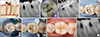
 | Figure 3Scenario 3. (A) Diagram: juxta-gingival decay that reaches the pulp. (B) Initial radiography: filtered restoration of tooth #26 and large cavities under the amalgam on tooth #27. (C) Root canal treatment of tooth #27. (D) Elimination of the filtering margin for tooth #26 and build-up on tooth #27. Note the margins at the juxta-gingival level. (E) Final photograph after composite endocrown cementation on teeth #26 and #27. (F) X-ray control of the correct operation at 1-year after treatment.
|
The case shown is a 60-year-old patient who presented to the office with constant acute pain in the second quadrant. On radiographic examination, mesial decay was observed under an amalgam restoration on tooth #27 (Figure 3B). Irreversible pulpitis was diagnosed, and a canal treatment was planned (Figure 3C), which was carried out largely as described above, using rotary instruments, copious irrigation, and thermoplastic filling.
We subsequently proceeded with the restoration. Due to the interproximal position of the caries, tooth #26 was also affected, and thus it was decided to remove both caries and both restorations to provide a completely aseptic situation (Figure 3D). No endodontic retreatment of tooth #26 was performed, because there was no clinical or radiographic abnormality, and composite infiltration was provided, without affecting the root canal. On tooth #27, a build-up was made using composite Ceram.X (DeTrey Dentsply) to provide a basis for a future endocrown. With proper absolute isolation, due to rubber dam invagination on the gingival sulcus, margin elevation was not necessary. Finally, we performed cementing of 2 composite endocrowns (Lava Ultimate CAD/CAM, 3M ESPE, St. Paul, MN, USA) (Figure 3E), with a good point of contact and precise fit, which would have been difficult to reproduce directly. The 1 year radiological control showed correct operation of the treatment (Figure 3F).
Scenario 4: class II located in the sulcus, affecting the pulp tissue
Such cases are generally difficult to restore. First, appropriate pulp treatment must be performed (endodontically in this case), and subsequently, rehabilitation is performed based on the remaining tooth structure. Generally, when lesions reach the gingival sulcus, they are already large lesions, making indirect restorations [10] with cuspal coverage [11] more important than direct restorations. In cases for which an indirect restoration is chosen, it should generally be a DME [5] (Figure 4A).
 | Figure 4Scenario 4. (A) Diagram: caries invades the gingival sulcus and the pulp chamber. (B) Initial radiograph: presence of apical focus on the distal root of tooth #36. (C) Initial photograph: note the clean margin within the gingival sulcus. (D, E) Retreatment of the distal canal. (F) Final photograph after cementation of the composite endocrown. (G) X-ray control at 1-year. Note the complete healing of the lesion and fit of the restoration.
|
The case shown is a 53-year-old female patient, who presented with pain upon chewing. The tooth was positive for percussion, and the existence of an apical lesion at distal root was observed by radiography (Figure 4B). Acute apical periodontitis in tooth #36 was diagnosed. Distal root retreatment was performed, removing the metal post and continuing with a canal retreatment of the root (Figure 4C).
It was observed initially (Figure 4D) that the cavity margin is located completely at the level of sulcus. However, in this case, due to the existence of a bone defect between teeth #36 and #37, there was still some distance to the bone crest. This would allow the use of a circumferential matrix, such as Tofflemire Matrix Bands (Waterpik Oral Health, Fort Collins, CO, USA) or AutoMatrix (DeTrey Dentsply), to raise the margin of the cavity with a composite under absolute isolation (DME), creating a basis for the future restoration. Because the loss of structure was large (note the opening class II at the vestibulo-lingual level, which has already implicated the disto-buccal and disto-lingual cusps) and the depth of the cavity was deep, overlay-endocrown was reconstructed using the protection of the cusps to contribute appropriate resistance to the fracture. After performing retreatment of the distal root (Figure 4E), a build-up of composite using the matrix AutoMatrix (DeTrey Dentsply) was performed. Finally, an endocrown (Lava Ultimate CAD/CAM, 3M ESPE) was cemented using a resin cement (Calibra, Dentsply Maillefer). In Figure 4F and 4G, the clinical and radiological controls are shown at 1 year.
Scenario 5: class II found in the junctional epithelium and affecting the pulp tissue
Such cases are also generally complex to restore. First, the appropriate pulp treatment must be made (endodontically in this case), and subsequently, it must be rehabilitated based on the remaining tooth structure. Usually, this type of tooth has lost significant tooth structure, such that an indirect restoration is usually the restorative treatment of choice. For adhesion, isolation is essential, and to achieve that, it is important to increase the margins [5]. As long as field can be isolated and a matrix placed, a DME can be performed [5]. If this type of treatment cannot be performed, then a crown lengthening can be used, provided there is a sufficient periodontal margin [12] (Figure 5A).
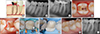 | Figure 5Scenario 5. (A) Diagram: caries with a margin in the junctional epithelium and reaching the pulp. (B) Initial radiograph: mesial and distal cavities in tooth #46. (C) Radiograph of the root canal treatment. (D, E) Mesial and distal margins free of caries. Note margins have already invaded the epithelial tissue. (F) DME with AutoMatrix (DeTrey Dentsply, Konstanz, Germany) and under total isolation. (G) Build-up of the future endocrown. Note buccal and lingual cusp protection. (H) Final photograph. (I) X-ray control at 1-year after treatment.
DME, deep margin elevation.
|
A male patient, 32 years of age, presented to the office with acute pain in the right mandibular quadrant. On initial radiographs (Figure 5B), the presence of deep mesial and distal caries approaching pulp cavities were observed on tooth #47. As a result of the diagnostic tests, increased pain with cold, palpation, percussion was observed with negative mobility. This was diagnosed as irreversible pulpitis, and we proceeded with a root canal treatment, performed largely as described above, using rotary instruments, copious irrigation, and a thermoplastic filling.
Once the root canal treatment was complete (Figure 5C), the restoration margins were found to be subgingival; they invaded the sulcus and epithelial tissue (Figure 5D and 5E). In such cases, the decision to perform crown lengthening is determined by the ability to place a matrix and isolate correctly. In the case presented, using a rubber dam and AutoMatrix (DeTrey Dentsply), it was possible to create an optimal operating field for adhesion that allowed accurate adjustment. Thus, stratification with composite Ceram.X (DeTrey Dentsply) at the margin was performed, obtaining a DME as the final result (Figure 5F). Thus, the margin became supragingival and much easier to handle.
Since the loss of tooth structure was critical, leaving only the buccal and lingual walls with insufficient thickness, protection of cusps (Figure 5G) was performed to increase the assurance of fracture resistance. It was rehabilitated using an endocrown-type overlay (Lava Ultimate CAD/CAM, 3M ESPE) that was cemented adhesively because of the possibility of absolute isolation. As seen in the final photograph (Figure 5H), the indirect restoration had a greater guarantee of good performance over the long term allowing easier marginal and occlusal adjustment, a point of contact, and a better anatomy compared with a direct restoration. In the radiological control at 1 year, the success of treatment over time can be seen (Figure 5I).
Scenario 6: class II found in the gingival connective tissue and affecting the pulp tissue
Usually, this type of tooth has lost quite a lot of structure; thus, an indirect restoration is typically the restorative treatment of choice. Such cases are also generally complex to restore. First, the appropriate pulp treatment must be made (in this case, endodontically), and subsequently it is rehabilitated based on the remaining tooth structure (large cavity or lost of one or more cusps, will be the criteria for choosing indirect restoration). Cement requires a perfectly isolated operative field. Due to the close proximity to bone crest, it is necessary to perform a crown lengthening [1] to isolate, and then an indirect restoration can be cemented with a degree of assurance. Other options would be an orthodontic or surgical extrusion [12] (Figure 6A).
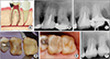
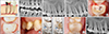 | Figure 6Scenario 6. (A) Diagram: caries invades the connective tissue and pulp. (B, C) Initial bitewing and periapical radiography, respectively; mesial caries in tooth #17 under an amalgam filling. (D) Periapical view after root canal treatment. (E) Remaining tooth structure after root canal treatment. Note how the margin is invading the connective tissue. (F, G) Tooth #17 after crown lengthening. Note how it gained at least a 2-mm supragingival margin. (H, I) Composite endocrown Lava Ultimate CAD/CAM (3M ESPE) on a laboratory model. (J) Final photograph. (K) X-ray control at 1-year after treatment.
|
In this case, a female patient, 33 years of age, who presented with acute pain in the right maxillary quadrant is shown. When observed by radiograph (Figure 6B and 6C), the presence of deep mesial caries under an old amalgam restoration on tooth #17, and caries in mesial of tooth #16 can be seen. As a result of diagnostic tests, irreversible pulpitis was diagnosed for tooth #17. We proceeded to clean the cavity and remove the amalgam restoration. This required the completion of a provisional pre-endodontic reconstruction of the mesial wall to allow proper isolation and irrigation during the root canal treatment. After negotiation of the canals, they were instrumented using the ProTaper Next system (Dentsply Maillefer) and filled with the Thermafil filling system (Dentsply Maillefer) (Figure 6D).
Once the root canal treatment was completed and the tooth cleaned completely, the margins of the healthy tissue of the cavity were found to be completely subgingival, invading the biological width almost in its entirety (Figure 6E). Using a periodontal probe, the distance of the tooth to the bone crest was less than 1 mm. In such cases, achieving a satisfactory margin elevation is practically impossible, because the space available for the matrix is minimum and, the chances of leakage are high. Thus, crown lengthening is indicated in such cases. By removing 2–3 mm of bone at this level, the margins of the cavity become supragingival (Figure 6F and 6G), greatly facilitating clinical management during the restorative phase. Again, the loss of tooth structure was critical, and therefore an overlay-endocrown indirect restoration to protect the cusps was planned. Thirty days after removal of the stitches, with complete healing observed, the build-up was performed using composite Ceram.X mono (DeTrey Dentsply), and impressions were made.
After Lava Ultimate CAD/CAM (3M ESPE) endocrown cementation (Figure 6H and 6I), crown lengthening of teeth #16 and #17 and the final restoration of tooth #17, with a right point of contact and marginal fit, can be seen in the radiograph at 1 year (Figure 6K) and in the final photograph (Figure 6J) of the completed root canal therapy. The mesial cavity obturation on tooth #16 was also noted (Figure 6K).
Scenario 7: class II reaching the bone crest and affecting the pulp tissue
Such cases are complex to restore, or, in many cases, require tooth extraction. First, the best treatment plan must be established based on the specific case. It must be determined whether root canal treatment or crown lengthening is possible, whether it is possible to make an orthodontic/surgical extrusion, or whether the best treatment plan is simply extraction. When crown lengthening is planned, it must be evaluated carefully to prevent excessive biological cost (the ostectomy) [12]. A possible alternative, not too aggressive in such cases, is crown lengthening combined with DME [5]. Thus, by creating a 2-mm osteotomy and subsequent DME only, we achieved cementing of indirect restoration (Figure 7A).
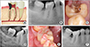 | Figure 7Scenario 7. (A) Diagram: decay that reached the bone level and invaded the pulp chamber. (B) Initial radiograph: deep filtered obturation in tooth #46. (C) Photograph after root canal treatment. Note that the distal margin of the cavity is completely at the bone level. (D) Radiograph after crown lengthening and after the DME. (E) Photograph after cementation of the endocrown and after occlusal check. (F) Periapical radiography control at 1-year. Correct operation of the treatment.
DME, deep margin elevation.
|
The case presented here was a 55-year-old woman who presented with acute pain in the right mandibular quadrant. Shown in a periapical view, the presence of a class II extensive restoration of tooth #46 distally filtered and secondary caries reaching the bone crest and the pulp chamber can be seen (Figure 7B). Irreversible pulpitis was diagnosed, and planning was carried out. It was essential at this time to assess the viability of a future restoration. It is not logical to perform a root canal on a tooth that cannot subsequently be restored with a certain guarantee. We thus assessed the possibility of performing a crown lengthening, evaluating the position of the furcation, crown-root relationship, the vicinity of the roots, the attached gingiva, and the muco-gingival line. This was an extreme case, and a multidisciplinary endodontic, periodontal, and restorative treatment was planned.
Endodontic treatment was performed using the ProTaper Next system (Dentsply Maillefer) and the Thermafil filling system (Dentsply Maillefer). After ensuring appropriate cleaning of the distal margin (infraosseous) (Figure 7C), crown lengthening was performed. Based on the remaining tooth structure and considering that the mesial wall was still intact, an indirect reconstruction was chosen. Thirty days after the removal of the stitches, with complete healing observed, the build-up and a DME (Figure 7D) were performed using composite Ceram.X mono (Ceram.X, DeTrey Dentsply) under absolute isolation with the aid of an AutoMatrix (DeTrey Dentsply). Finally, the Lava Ultimate CAD/CAM (3M ESPE) endocrown was cemented. The radiological and clinical control reflected the success of treatment at 1 year (Figure 7E and 7F).
DISCUSSION
The preservation of tooth structure is the basic principle in current restorative dentistry, because it maintains a balance among biological, mechanical, functional, and esthetic parameters [13]. Thus, adhesive restorations with complete cuspal coverage, like endocrowns or overlays, have been proposed as an alternative to more traditional methods, such as full crowns [14].
The choice of options will be determined primarily by the amount of healthy tooth structure remaining [815], while taking into account of other characteristics potentially present in the patient, such as parafunctional habits, occlusal forces, and esthetic demands [16]. Thus, in a molar class II that retains most of its structure, direct reconstruction would still be indicated [415], provided that the thickness of the remaining walls is appropriate and functional cusps are not affected. Cases A, B, and C belong to this group.
However, when the loss of structure is greater, the thickness of the remaining walls is too weak, some cusps are lost, or parafunctional habits exist, the most appropriate restoration is indirect restoration (an endocrown in devitalized teeth, as in cases D, E, F, and G) [4816]. The cavity of endocrowns sould be designed with divergent walls, with interproximal margin below contact point and providing sufficient thickness to the restoration but respecting as much tooth as possible. Both clinical and in vitro studies have shown a high success rate using this type of restoration [1718] although more long-term studies are needed.
The process of cementation can be made with a resin cement or with preheated resin composite while the most important point is the isolation of the field. The choice of material in these indirect restorations is also important, which will be defined primarily by the occlusal environment [19]. While, traditionally, porcelain has predominated in durability and biocompatibility [14], composites have gained ground due to their significant evolution in recent years. A recent alternative that is related to manufacturing techniques is CAD/CAM technology restoration, made in milled ceramic or composite blocks with superior mechanical properties [19]. In the cases described, composite blocks were chosen because no wearing contralaterally and it can be repaired. Moreover, composite has low elastic modulus, which results in lower rates of catastrophic fracture, and it can absorb functional stress by deformation [14].
In extreme cases of loss of structure, i.e., more than two-thirds of the tooth, a crown is indicated for treatment. Evidence shows at least 1.5–2 mm crown ferrule is necessary for predictable long-term success [20], so the case planning plays a major role. However, there are many cases with loss of subgingival structure, in which simple extraction is the treatment of choice. This article focuses on non-extreme tooth structure loss, as in most clinical practice cases, and for which an endocrown would be indicated.
In the reconstruction of an endodontic tooth, in addition to the assessment of the remaining tooth structure, it is essential to determine the distance of the carious lesion to bone. The bone crest must be detected, and special attention should be paid to determine the distance thereof to the cavity margin after removing the carious tissue. Thus, it requires performing proper periodontal probing. Thus, it is also important to supplement with radiological techniques. A radiograph using a correct parallel technique, especially bitewing radiography, can reproduce the biological dimensions accurately [21]. This information will be important when planning the treatment, because biological width invasion can lead to failure of the restoration.
Traditionally, a minimum vertical distance of 3 mm between the restoration margins and the alveolar bone crest has been established as a requirement [12]. Respecting the biological width is considered essential in maintaining gingival health. Invasion is associated with problems because the tooth-restoration interface does not allow establishment of the tissues that make up the biological width (biological constant) as well as causes an accumulation of plaque that will react accordingly with the tissues [22]. However, it should be noted that this marginal space at the interface is unavoidable solely in non‑adhesive restorations. Conversely, subgingival composite obturation with a perfectly adapted marginal seal is generally well tolerated by the surrounding tissues (if not reaching to invade the connective tissue) [23]. Martins et al. [23] showed the binding capacity of the fibers of epithelial tissue to the surface of resin restorations, provided that the adaptation, polishing, and hygiene conditions are optimal. However, if developed in depth, and the connective tissue is violated with a restoration, the literature shows the appearance of an intense inflammatory infiltrate, followed by bone resorption [24]. In 2012, Magne and Spreafico [5] proposed DME as a non-invasive alternative to crown lengthening. This technique can elevate the deep cervical margin with composite resins, usually leaving 1–1.5 mm supragingival [19], which will facilitate taking impressions, preparing an emergency profile, correct rubber dam isolation, and proper disposal of excesses at the time of cementation of an indirect restoration. Note that, three criteria must be met to make a DME: 1) ability to completely isolate the operative field, 2) ability to place a matrix that is capable of properly isolating margins and ensuring a perfect seal, and 3) no invasion of the connective tissue space by the matrix. Otherwise, crown lengthening would be indicated, which will be true whenever the alveolar crest is close [125].
Crown lengthening is a periodontal surgical technique to gain supracrestal tooth length. The periodontal health, crown-root ratio, the position of the furcation, the closeness between the roots of adjacent teeth, and the shape of the root (molars tend to adopt S ways at the junction of roots) should be evaluated [1]. Once this is performed, a minimum of 4 weeks of post-surgical healing is recommended before performing restoration in non-esthetic posterior areas [12].
Orthodontic extrusion is another option to achieve supragingival margins. It has certain advantages over crown lengthening, such as a lower insertion loss and better final crown-root ratio, and non-involvement of the neighboring tooth [1]. These benefits are useful mainly in the anterior sector, where esthetic value and harmony of margins are required. An other alternative is surgical extrusion, which involves creating an atraumatic extraction followed by reimplantation of the tooth in a new position [26]. Its indications are restricted to extreme cases, usually anterior, for which the other 2 treatment options are not viable.
CONCLUSIONS
Controlled clinical trials should be conducted to achieve a high level of evidence in this field. However, within the limits of this study, the type of restoration in molars of class II will depend on the amount of healthy tooth structure remaining. Whenever we can use a matrix that allows raising the margin predictably, we can perform a DME. In cases with invasion of the connective tissue or bone crest or those with deeper margins, crown lengthening surgery is indicated (whenever periodontally possible), as an attempt to achieve the highest possible long-term success of the restoration.
 XML Download