Journal List > Restor Dent Endod > v.42(3) > 1090222

Singh, Nagpal, and Singh: Effect of dentin biomodifiers on the immediate and long-term bond strengths of a simplified etch and rinse adhesive to dentin

Abstract

Objectives

This in vitro study evaluated the effect of dentin biomodifiers on the immediate and long-term bond strengths of a simplified etch and rinse adhesive to dentin.

Materials and Methods

Flat coronal dentin surfaces were prepared in 120 extracted human molars. Teeth were randomly divided into 5 groups (n = 24) according to 5 different surface pre-treatments: No pre-treatment (control); 1M carbodiimide (EDC); 0.1% epigallocatechin-3-gallate (EGCG); 2% minocycline (MI); 10% sodium ascorbate (SA). After surface pre-treatment, adhesive (Adper Single Bond 2 [SB], 3M ESPE) was applied. Composite was applied into transparent plastic tubes (2.5 mm in diameter), which was placed over the bonded dentin surface. From each group, 10 samples were subjected to shear bond strength (SBS) evaluation at 24 hours (immediate) and remaining 10 samples were tested after 6 months (delayed). Additionally, 4 samples per group were subjected to scanning electron microscopic analysis for observation of resin-dentin interface. The data were statistically analysed with Shaperio‑Wilk W test, 2-way analysis of variance (ANOVA), and post hoc Tukey's test.

Results

At 24 hours, SBS of all surface pre-treatment groups were comparable with the control group, with significant differences found between EDC and SA groups only (p = 0.009). After 6 months storage, EDC, EGCG, and MI pre-treatments preserved the resin-dentin bond strength with no significant fall.

Conclusions

Dentin pre-treatment with all the dentin biomodifiers except SA resulted in significant preservation of resin-dentin bond over 6 months storage period, without negatively affecting the immediate bond strength of the etch and rinse adhesive tested.

INTRODUCTION

It is well accepted that resin-dentin bonds created by contemporary dentin adhesive systems deteriorate over time [12]. Hence, thorough understanding of all the mechanical, physical, and biochemical factors that affect the stability of hybrid layers is important. Dentin adhesion is like in situ tissue engineering, in which collagen fibrils exposed by acid etching (either etch and rinse or self-etch) act as scaffold for micro-mechanical interlocking of monomers leading to formation of hybrid layer. To achieve a stable hybrid layer, this resin infiltration into the filigree of exposed collagen fibers should be as complete as possible. However, sub-optimal infiltration of the denuded collagen matrix is quite common, especially with etch-and-rinse adhesives [3]. Moreover, the moisture of demineralized dentin also impairs the infiltration of hydrophobic monomers [4]. This discrepancy between the depth of demineralised collagen layer and resin infiltration leads to denuded exposed collagen fibrils at the bottom of hybrid layer, lacking the protection of polymerized resin.
The lack of resin protection and presence of water makes the exposed collagen fibrils vulnerable to hydrolytic degradation by host-derived proteases (matrix metalloproteinases [MMPs] and cysteine cathepsins) at the bottom of the hybrid layer [3]. MMPs are secreted as proenzymes (zymogens), these are inactive forms which later on get activated by the acidic agents during adhesive bonding procedures. These activated MMPs can slowly hydrolyze the collagen fibrils in the hybrid layer that anchors resin composites to the underlying mineralized dentin, thereby decreasing the longevity of bonded restorations.
Recently, the concept of dentin biomodification has been employed to achieve a more stabilized and durable adhesive interface [5]. It involves the use of several natural and synthetic agents, acting as MMP inhibitor and collagen cross-linker to bio-modify and enhance the mechanical properties of the dentin substrate [6]. MMP inhibitors are either endogenous (tissue inhibitors of metalloproteinases [TIMPs]) or exogenous. Various exogenous MMP inhibitors and collagen cross-linkers have been used as dentin bio-modifiers. They can be either used to pre-treat the demineralized dentin surface or have been incorporated into the bonding components [789]. Green tea is a natural MMP inhibitor obtained from the Camellia sinensis plant. Epigallocatechin-3-gallate (EGCG) is the major polyphenol present in green tea. It inhibits MMP-2 and MMP-9 and improves the mechanical properties of collagen matrix to resist proteolytic degradation [10]. Tetracyclines are antibiotics with cationic chelating properties. Chemically modified tetracyclines (minocycline [MI] and doxycycline) lack antibacterial activity, but have some MMP-inhibitory property [11]. Carbodiimide (EDC) is a synthetic, less cytotoxic cross-linking agent. It inhibits endogenous proteases by inactivating the active sites by reducing their molecular mobility and also improves the resistance of cross-linked collagen matrices to degradation by inducing exogenous cross-links and thereby increasing their stiffness [712]. Ascorbic acid or sodium ascorbate (SA) suppresses the denaturing effect of etching on dentin collagen and found to be a potent inhibitor of MMPs, offering protection against the degradation of composite-dentin bond [13].
As MMP inhibition and collagen cross-linking capabilities vary amongst different dentin biomodifiers, they may also differ in the extent of dentin stabilization achieved for improving resin-dentin bond. Apart from this, results may also vary with the specific adhesive system used, application time, and concentration of the dentin biomodifier [68101314]. The conflicting results reported in the literature require more new studies to be conducted in this field. Hence, the aim of this study was to investigate the effect of pre-treatment with EDC, EGCG, MI, and SA on the immediate and long-term bonding efficacy of an etch and rinse adhesive to dentin. The null hypothesis tested was that there is no effect of different dentin biomodifiers on the immediate (24 hours) and long-term (6 months) resin-dentin bond strengths compared to control group obtained with a simplified etch and rinse adhesive.

MATERIALS AND METHODS

Experimental design

The present study has a factorial design and 2 factors are evaluated 1) dentin pre-treatment in 5 levels: no pre-treatment (control group), 1M EDC solution, 0.1% green tea solution (EGCG), 2% MI hydrochloride solution, and 10% SA solution; 2) measurement time of shear bond strength (SBS) in 2 levels: after storage in distilled water for 24 hours and in artificial saliva (Wet Mouth, ICPA Heath Products Ltd., Mumbai, MH, India) for 6 months. The quantitative outcome variable was the SBS value, in MPa. The average bond strength of half the samples (n = 10) became the value for the particular experimental subgroup at the particular time (immediate at 24 hours or delayed at 6 months). The failure patterns were described in terms of percentages.

Specimen preparation

The study was conducted after taking ethical approval from Institutional Ethical and Review Board (IERB) with reference No. KDCRC/IERB/ENDO/2014/16. One hundred and twenty freshly extracted non-carious, human molars were used in this study. The teeth were examined under stereomicroscope (SZX10, Olympus, Tokyo, Japan) and teeth free of caries, cracks, or any developmental defects were included. Teeth were cleaned and stored in 0.5% chloramine T trihydrate (Sigma Aldrich, Bangalore, KA, India) for no more than 3 months. Tooth crowns were flattened occlusally using a low-speed diamond saw (Isomet, Buehler Ltd., Lake Bluff, IL, USA) under water irrigation to expose superficial dentin. A standardized smear layer was created with 600 grit silicon-carbide (SiC) paper. The samples were embedded in an autopolymerizing resin at the level of cementoenamel junction with long axis perpendicular to the acrylic resin surface.

Experimental solutions

All chemicals (Sigma-Aldrich) were of analytical grade and were used without further purification. All the required concentrations were prepared by diluting the extract in distilled water (pH 7.4): 1M EDC-hydrochloric acid (HCl) solution was prepared by dissolving 958 mg EDC into 5 mL distilled water (pH 6.0); 0.1% green tea solution (EGCG) was prepared by dissolving 5 mg EGCG into 5 mL distilled water (pH 6.91); 2% MI solution was prepared by dissolving 100 mg MI into 5 mL distilled water (pH 5.0); 10% SA solution was prepared by dissolving 500 mg SA into 5 mL distilled water (pH 7.76). The prepared diluted solutions were filtered with paper filter No. 6 (Whatman, London, UK).

Adhesive procedures

An adhesive tape with a 2.5 mm central hole was positioned on the flat dentin surface to demarcate the pre-treatment and bonding area. All the samples were subjected to acid etching procedure with Scotchbond universal etchant (3M ESPE Dental Products, St. Paul, MN, USA) for 15 seconds followed by rinsing with water for 10 seconds and lastly blot dried. Then the following treatments were done on acid etched dentin surface with corresponding pre-treatment solutions (groups 2–5) with a microbrush for 60 seconds except for the control group. Excess liquid on dentin surface was gently blotted with filter papers to leave a visibly moist dentin surface. Following this, 2 coats of Adper Single Bond 2 (SB, 3M ESPE Dental Products) adhesive were applied on pretreated dentin surface, gently air thinned for 5 seconds, and light-cured for 10 seconds.
  • Group 1 (control): SB adhesive was applied following manufacturer's instructions.

  • Group 2: 1M EDC (E6383, Sigma Aldrich).

  • Group 3: 0.1% EGCG (E4143, Sigma Aldrich).

  • Group 4: 2% MI (M9511, Sigma Aldrich).

  • Group 5: 10% SA (A7631, Sigma Aldrich).

Transparent plastic tubes (TYGON laboratory tubing, Saint Gobain, Akron, OH, USA) with 2.5 mm in diameter and 2 mm in height were placed perpendicular to the previously etched, pre-treated and bonded dentin surface similar to previous studies [1516]. A nanohybrid resin composite (Filtek Z350 XT, Body Shade A1; 3M ESPE Dental Products) was filled into the precut tubes. Each bonded specimen was light-cured for 20 seconds using quartz-tungsten-halogen (QTH) light curing unit (Spectrum 800, Dentsply Caulk, Milford, DE, USA) at a light intensity of 600 mW/cm2. The plastic tubes were gently cut and carefully removed with a number 11 surgical blade after polymerization.

Storage of samples before testing

Half of the specimens (immediate testing group) were then stored in distilled water at 37°C for 24 hours for completion of polymerization before immediate testing and scanning electron microscopy (SEM) analysis. The remaining half samples from each group (delayed testing group) were stored in artificial saliva (Wet Mouth, ICPA Heath Products Ltd.) at 37°C in an incubator for 6 months before SBS and SEM evaluation [17]. Artificial saliva (pH 7.11) was composed of 0.5% w/v sodium carboxymethyl cellulose, 30% w/v glycerine, and flavoured base. Point 2 percent sodium azide (pH 7.31) was further added to prevent bacterial growth [18]. This was confirmed through the maintenance of the clear color of artificial saliva during the storage period. Moreover, storage solution was changed every 2 weeks, and its pH was monitored at every change.

Determination of SBS

SBS was determined using a universal testing machine (Instron, ADMET, Enkay Enterprises, New Delhi, DL, India) using the corresponding computer software. The specimens were placed and stabilized by the jig, while a straight knife-edge rod (2.0 mm) was applied at the tooth-restoration interface at a crosshead speed of 1 mm/min until fracture occurred. SBS in MPa was calculated by dividing the peak force (N) by the cross-sectional area of the failed interface (mm2), measured by a digital caliper.

Fractographical analysis

The mode of failure of all 20 samples from each group was determined by observation under a stereomicroscope (SZX10, Olympus) at × 10 magnification and classified into adhesive (A), mixed (M), and cohesive (C) failures in either dentin or resin.

SEM evaluation

Four samples per group (2 for immediate testing subgroup and 2 for delayed testing subgroup) were used for SEM (EVO18 Special Edition, Carl Zeiss, Jena, Germany) evaluation. The restored samples were sectioned mesiodistally and polished with a series of increasingly finer SiC abrasive papers up to 1,200 grit and highly polished with a diamond paste. Acid-base treatment (6N HCl for 30 seconds followed by 4% NaOCl for 10 minutes) was done, and the samples were dehydrated in ascending ethanol concentrations (50%, 75%, and 95% for 20 minutes each and 100% for 1 hour) and then transferred to a critical point dryer for 30 minutes. The specimens were then gold sputter coated and the resin-dentin interface was examined under SEM.

Statistical analysis

Data showed normal distribution as tested using the Shaperio‑Wilk W test (p > 0.05). Therefore, an analysis was performed using the parametric tests i.e., independent t-test, 2-way analysis of variance (ANOVA) test, and post hoc Tukey's test, with SPSS version 21.0 (Statistical Package, SPSS Inc., Chicago, IL, USA) at a significance level of p < 0.05. Prior to the ANOVA test, Levene's test for equality of variances was also performed. Levene's test uses the level of significance set a priori for the ANOVA (i.e., α = 0.05) to test the assumption of homogeneity of variance.

RESULTS

SBS

Levene's test of equality of error variances was 0.501. It states that the error variance of the bond strength (dependent variable) is equal across groups. The results for the 2-way ANOVA indicated a significant effect with factors: various ‘pre-treatments’ (p < 0.05, F = 21.2) and ‘duration’ (p < 0.05, F = 45.5; Table 1). Additionally, the interaction effect between various pre-treatments and duration was also found to be significant (p < 0.05, F = 1,500.4).
Table 1

Shear bond strength (SBS) of Adper Single Bond 2 adhesive after different surface pre-treatments

rde-42-188-i001
Dentin pre-treatment SBS p value Result of 2-way ANOVA
Immediate (24 hr) Delayed (6 mon storage)
1. No additional surface treatment (control) 32.4 ± 5.5Aa 16.9 ± 5.0Ab < 0.001 Main effect
2. 1M EDC 38.6 ± 6.1Aa 35.1 ± 6.2Ba 0.119 Treatment groups, p < 0.001
3. 0.1% green tea (EGCG) 32.8 ± 5.8Aa 29.9 ± 5.3Ba 0.125 Duration, p < 0.001
4. 2% MI 34.5 ± 6.8Aa 31.4 ± 6.8Ba 0.158 Interaction effect
5. 10% SA 29.4 ± 5.1Ba 15.8 ± 4.2Ab < 0.05 Groups × duration, p < 0.001
Different uppercase superscript letters mean that there is a significant difference between the mean values within the row (p < 0.05). Different lowercase superscript letters mean that there is a significant difference between the mean values within the column (p < 0.05).
ANOVA, analysis of variance; EDC, carbodiimide; EGCG, epigallocatechin-3-gallate; MI, minocycline; SA, sodium ascorbate.
Mean SBS value and standard deviation of all the groups are presented in Table 1. At 24 hours, SBS of all surface pre-treatment groups (EDC, MI, EGCG, and SA) were comparable to the control group, with EDC showing maximum immediate bond strength as compared to other 4 groups (Table 1). However, a significant difference was found between only EDC and SA groups.
At 6 months storage period, significant reduction in bond strength was observed for the control group (p < 0.05) and SA treated group (p < 0.05). However, EDC, EGCG, and MI pre-treatment groups preserved the resin dentin bond strength with no significant fall over a period of 6 months (p = 0.119, p = 0.125, and p = 0.158, respectively).

Fractographical analysis

The effect of different dentin pre-treatment and different time intervals on the distribution of failure pattern were compared using the χ2 test (Figure 1). Most of the failures encountered were mixed in all of the pre-treatment groups tested immediately, but it failed to reach the level of significance (p = 0.818). At 6 months, an increase in the number of adhesive failures was observed in all the groups but the difference was not significant (p = 0.707). However, EDC, EGCG, and MI treated groups showed predominantly mixed failures.
Figure 1
Failure patterns of experimental groups according to the dentin pre-treatments and to the time intervals (represented in percentage).
EDC, carbodiimide; EGCG, epigallocatechin-3-gallate; MI, minocycline; SA, sodium ascorbate.
rde-42-188-g001

SEM

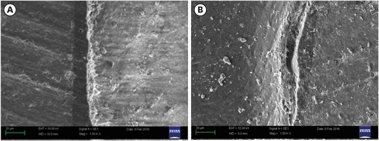
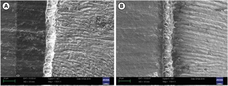
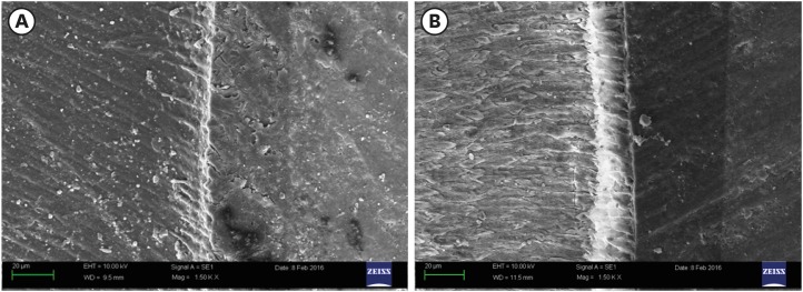
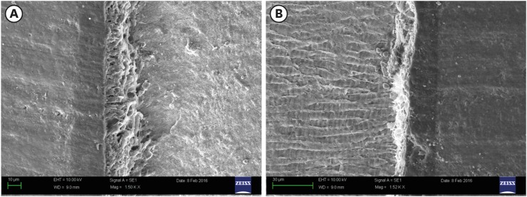
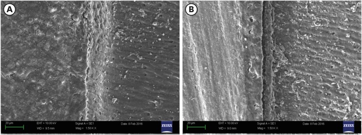
The representative SEM photomicrographs of each pre-treatment group are shown in Figures 2, 3, 4, 5, 6. Figures 2A, 3A, 4A, 5A, 6A show the resin-dentin interface of different groups observed at 24 hours after bonding. Perfect interfacial adaptation with absence of gap was observed in all the groups. Figures 2B, 3B, 4B, 5B, 6B show resin-dentin interface of different groups observed after 6 months of storage in artificial saliva. The interfacial gap was negligible in the samples pretreated with all dentin biomodifiers except SA and control groups which showed the presence of interfacial gap suggesting bond deterioration.
Figure 2
Scanning electron micrograph of a representative specimen from the control group. (A) Good interfacial adaptation with no gaps can be seen at 24 hours. (B) Poor interfacial adaptation with a large gap can be seen after 6 months.
rde-42-188-g002
Figure 3
Scanning electron micrograph of a representative specimen from the carbodiimide (EDC) treated group. (A) Good interfacial adaptation can be seen at 24 hours. (B) Absence of gaps indicating a good adaptation after 6 months.
rde-42-188-g003
Figure 4
Scanning electron micrograph of a representative specimen from the epigallocatechin-3-gallate (EGCG) treated group. (A) Good interfacial adaptation can be seen at 24 hours. (B) Absence of gaps indicating a good adaptation after 6 months.
rde-42-188-g004
Figure 5
Scanning electron micrograph of a representative specimen from the treated minocycline (MI) treated group. (A) Good interfacial adaptation can be seen at 24 hours. (B) Absence of gaps indicating a good adaptation after 6 months.
rde-42-188-g005
Figure 6
Scanning electron micrograph of a representative specimen from the sodium ascorbate (SA) treated group. (A) Good interfacial adaptation can be seen at 24 hours. (B) Absence of gaps indicating a good adaptation after 6 months.
rde-42-188-g006

DISCUSSION

Different strategies have aimed to improve the bond durability by applying various dentin biomodifiers (MMP inhibitors and collagen cross-linkers) as a pre-treatment before resin infiltration or by admixing enzyme inhibitors to primers. The biomodification of collagen can be done with various agents like EDC, EGCG, MI, and SA having both cross-linking and MMP inhibitory potential.
In this study, artificial saliva has been used for 6 months storage similar to previous studies [1718] and water was used for 24 hours storage so that a valid comparison could be made with immediate bond strength values of adhesives reported in previous studies. A meta-analysis reported that water as storage media presented the higher heterogeneity, while artificial saliva and other aging methods presented lower heterogeneity [19]. Moreover, Tezvergil-Mutluay et al. [20] reported that using water as a storage medium underestimates the hydrolytic activity of endogenous dentin MMPs, because it promoted the loss of calcium and zinc ions from the dentin matrices, rather than restoring them. Therefore, artificial saliva was used for long-term storage to better simulate the clinical conditions.
In this study, 1M EDC concentration was used as previous studies have found better results with 1M EDC/2M EDC as compared to 0.5M EDC [2122]. In this study, distilled water (pH 7.4) was chosen as solvent for preparing EDC solution as Seseogullari-Dirihan et al. [23] found that EDC water solution caused the least amount of collagen degradation at around neutral pH. In this study, EDC when used as dentin biomodifier preserved the bond strength even after 6 months storage in artificial saliva. Our results are supported by various studies which reported improvement in resin-dentin bond durability with EDC pre-treatment [172425]. EDC causes stiffening of collagen making it more difficult for MMPs to unwind the collagen triple-helix structure. Apart from causing cross-linking of dentin collagen, EDC cross-links matrix-bound MMPs as well and cross-linking occurs more rapidly in MMPs than in collagen [2627]. It inactivates the catalytic site of proteases and reduces the mobility of enzymes by creating a new peptide bond across adjacent peptides. This could be explained by the better accessibility of carboxyl and amino groups in MMPs than in collagen [28]. It shows that EDC is a potent MMP inhibitor and that its MMP inhibitory effect is much faster than its collagen cross-linking effect. Therefore, it can also be anticipated that MMP inactivation by cross-linking agents like EDC should last much longer than the inhibition by MMP inhibitor alone.
EGCG is a potent inhibitor of MMPs and cysteine cathepsins, and their inhibitory effect is concentration-dependent. In this study, 0.1% EGCG was chosen, as linear polymer chains are formed at a low concentration during polymerization of dental adhesive and with higher concentrations of EGCG, formation of these linear polymer chains might get disturbed leading to inadequate polymerization of the adhesive. In our study, EGCG pre-treatment was able to prevent the decrease in bond strength after 6 months of in vitro aging without affecting the immediate bond strength. Our results are in accordance with other authors who also demonstrated that EGCG prevents decrease in bond strength of etch-and-rinse adhesives over long-term [293031]. This beneficial effect could be attributed to 2 MMP inhibitory mechanisms. First, EGCG binds to zinc ion, which plays an important role in protecting collagen against degradation by creating a new conformation, protecting the cleavage sites of collagen from metalloproteinases [31]. Second, it causes irreversible degradation of the MMP-2 molecule by exhibiting hydrogen bonding and hydrophobic interactions with it, or by masking the catalytic region of MMP-2 [32]. In accordance with da Fonseca et al. [29], we also found that 0.1% EGCG pre-treatment on acid etched dentin showed higher immediate bond strength as compared to 10% SA although it was not statistically significant. However, contrary to our study Monteiro et al. [6] found that green tea solution adversely affected the immediate bond strength. It may be due to the higher concentration (2%) of green tea used in their study and the use of green tea extract prepared from its herb, which might contain many unwanted impurities.
Chemically modified tetracyclines (doxycycline and MI) are considered broad-spectrum MMP inhibitors. Similar to Stanislawczuk et al. [33] and Li et al. [34], we also found that the application of 2% MI, as dentin pre-treatment did not affect the immediate bond strength and also prevented the decrease in bond strength of the etch-and-rinse adhesives even after storage for 6 months in artificial saliva. This beneficial effect could be attributed to their ability to chelate calcium and zinc ion, which play an important role in maintaining MMP structure and functional active sites. By binding to the zinc ion present on catalytic domain of the enzyme, modified tetracyclines can inhibit the MMPs by altering their conformation at the molecular level and thus blocking their catalytic activity in the extracellular matrix.
We chose SA as one of the dentin biomodifying agent in our study as it has been reported to stabilize collagen and promote collagen biosynthesis besides having an antioxidant effect [35]. Moreover, ascorbic acid has an inhibitory effect on dentin MMP-2 activity [13], which participates in resin-dentin hybrid layer degradation. However, in our study we could not observe any beneficial effect of SA on the durability of resin-dentin bond, although it did not affect the immediate bond strength. This may be attributed to its lower MMP inhibitory potential and lack of cross-linking ability as compared to other dentin biomodifiers used in the study.
Contrary to our study, 2 previous studies reported that SA maintained the bond strength over the long-term but decreased the immediate bond strength [814]. They attributed this decrease to its hydrophilic character, which causes phase separation and inhibition of polymerisation when incorporated into adhesives [814]. Moreover, SA causes decrease in degree of conversion and slows down the polymerization process, resulting in continued polymerization which might be the reason for increased SA doped-adhesives resistance against degradation over long-term.
Despite promising results obtained in the present study, there are some limitations such as SBS testing was done instead of microtensile bond strength testing which might alter the bond strength, as stress distribution along a large cross sectional area is not as uniform as for small cross-sectional area. Bond strength testing and storage of samples can be done considering the intrapulpal pressure to simulate the conditions clinically. Caries-affected dentin has not been used in the present study which usually produces lower bond strength and poor quality of the hybrid layer, as it is different in morphological, chemical and physical characteristics from normal dentin. The pH of dentin biomodifier/distilled water solutions was varying and no buffer system or solution was used in the study to neutralize the solution. Therefore, results might be affected by these factors.
All the dentin biomodifiers (EGCG, EDC, and MI) except SA significantly preserved the resin-dentin bond strength after 6 months storage as compared to the control group, thus null hypothesis was rejected. This provides the justification for further studies that are required to develop bio-active adhesives with integrated MMP inhibition and collagen cross-linking potential to improve the durability of the resin-dentin bond. Moreover, optimum concentration, time, pH and form of application of these dentin-biomodifying agents still needs to be determined in which they show maximum efficacy without interfering with the polymerization process.

CONCLUSIONS

All dentin biomodifiers (EDC, EGCG, and MI) except SA significantly preserved the resin-dentin bond strength even after 6 months of storage. None of the dentin biomodifiers significantly affected the immediate (24 hours) bond strength of SB adhesive.

Acknowledgement

We would also like to sincerely thank Dr. Nabila Sharif for helping us throughout this study in editing process and final preparation of the manuscript.

Notes

Conflict of Interest: No potential conflict of interest relevant to this article was reported.

Author Contributions:

  • Conceptualization: Singh P, Nagpal R, Singh UP.

  • Data curation: Singh P, Nagpal R.

  • Formal analysis: Singh P, Singh UP.

  • Funding acquisition: Singh P.

  • Investigation: Singh P, Nagpal R.

  • Methodology: Singh P, Nagpal R.

  • Project administration: Singh P, Singh UP.

  • Resources: Singh P.

  • Software: Singh P, Singh UP.

  • Supervision: Nagpal R.

  • Validation: Singh P, Singh UP.

  • Visualization: Singh P.

  • Writing - original draft: Singh P, Nagpal R.

  • Writing - review & editing: Singh P, Nagpal R, Singh UP.

References

1. Hashimoto M, Ohno H, Kaga M, Endo K, Sano H, Oguchi H. In vivo degradation of resin-dentin bonds in humans over 1 to 3 years. J Dent Res. 2000; 79:1385–1391. PMID: 10890717.
2. Yang B, Adelung R, Ludwig K, Bössmann K, Pashley DH, Kern M. Effect of structural change of collagen fibrils on the durability of dentin bonding. Biomaterials. 2005; 26:5021–5031. PMID: 15769538.
crossref
3. Pashley DH, Tay FR, Yiu C, Hashimoto M, Breschi L, Carvalho RM, Ito S. Collagen degradation by host-derived enzymes during aging. J Dent Res. 2004; 83:216–221. PMID: 14981122.
crossref
4. Pashley DH, Tay FR, Breschi L, Tjäderhane L, Carvalho RM, Carrilho M, Tezvergil-Mutluay A. State of the art etch-and-rinse adhesives. Dent Mater. 2011; 27:1–16. PMID: 21112620.
crossref
5. Bedran-Russo AK, Pauli GF, Chen SN, McAlpine J, Castellan CS, Phansalkar RS, Aguiar TR, Vidal CM, Napotilano JG, Nam JW, Leme AA. Dentin biomodification: strategies, renewable resources and clinical applications. Dent Mater. 2014; 30:62–76. PMID: 24309436.
crossref
6. Monteiro TM, Basting RT, Turssi CP, França FM, Amaral FL. Influence of natural and synthetic metalloproteinase inhibitors on bonding durability of an etch-and-rinse adhesive to dentin. Int J Adhes Adhes. 2013; 47:83–88.
crossref
7. Castellan CS, Bedran-Russo AK, Antunes A, Pereira PN. Effect of dentin biomodification using naturally derived collagen cross-linkers: one-year bond strength study. Int J Dent. 2013; 2013:918010. PMID: 24069032.
crossref
8. Erhardt MC, Osorio R, Viseras C, Toledano M. Adjunctive use of an anti-oxidant agent to improve resistance of hybrid layers to degradation. J Dent. 2011; 39:80–87. PMID: 21035517.
crossref
9. Khamverdi Z, Rezaei-Soufi L, Rostamzadeh T. The effect of epigallocatechin gallate on the dentin bond durability of two self-etch adhesives. J Dent (Shiraz). 2015; 16:68–74.
10. Hiraishi N, Sono R, Sofiqul I, Yiu C, Nakamura H, Otsuki M, Takatsuka T, Tagami J. In vitro evaluation of plant-derived agents to preserve dentin collagen. Dent Mater. 2013; 29:1048–1054. PMID: 23942145.
11. Sorsa T, Tjäderhane L, Konttinen YT, Lauhio A, Salo T, Lee HM, Golub LM, Brown DL, Mäntylä P. Matrix metalloproteinases: contribution to pathogenesis, diagnosis and treatment of periodontal inflammation. Ann Med. 2006; 38:306–321. PMID: 16938801.
crossref
12. Hass V, de Paula AM, Parreiras S, Gutiérrez MF, Luque-Martinez I, de Paris Matos T, Bandeca MC, Loguercio AD, Yao X, Wang Y, Reis A. Degradation of dentin-bonded interfaces treated with collagen cross-linking agents in a cariogenic oral environment: an in situ study. J Dent. 2016; 49:60–67. PMID: 27106766.
13. Roomi MW, Ivanov V, Kalinovsky T, Niedzwiecki A, Rath M. Inhibition of matrix metalloproteinase-2 secretion and invasion by human ovarian cancer cell line SK-OV-3 with lysine, proline, arginine, ascorbic acid and green tea extract. J Obstet Gynaecol Res. 2006; 32:148–154. PMID: 16594917.
crossref
14. Gotti VB, Feitosa VP, Sauro S, Correr-Sobrinho L, Leal FB, Stansbury JW, Correr AB. Effect of antioxidants on the dentin interface bond stability of adhesives exposed to hydrolytic degradation. J Adhes Dent. 2015; 17:35–44. PMID: 25625137.
15. Khamverdi Z, Rezaei-Soufi L, Kasraei S, Ronasi N, Rostami S. Effect of Epigallocatechin Gallate on shear bond strength of composite resin to bleached enamel: an in vitro study. Restor Dent Endod. 2013; 38:241–247. PMID: 24303360.
16. Meharry MR, Moazzami SM, Li Y. Comparison of enamel and dentin shear bond strengths of current dental bonding adhesives from three bond generations. Oper Dent. 2013; 38:E237–E245. PMID: 23802638.
crossref
17. Scheffel DL, Delgado CC, Soares DG, Basso FG, de Souza Costa CA, Pashley DH, Hebling J. Increased durability of resin-dentin bonds following cross-linking treatment. Oper Dent. 2015; 40:533–539. PMID: 25764044.
crossref
18. Mobarak EH. Effect of chlorhexidine pretreatment on bond strength durability of caries-affected dentin over 2-year aging in artificial saliva and under simulated intrapulpal pressure. Oper Dent. 2011; 36:649–660. PMID: 21864128.
crossref
19. Montagner AF, Sarkis-Onofre R, Pereira-Cenci T, Cenci MS. MMP inhibitors on dentin stability: a systematic review and meta-analysis. J Dent Res. 2014; 93:733–743. PMID: 24935066.
20. Tezvergil-Mutluay A, Agee KA, Hoshika T, Carrilho M, Breschi L, Tjäderhane L, Nishitani Y, Carvalho RM, Looney S, Tay FR, Pashley DH. The requirement of zinc and calcium ions for functional MMP activity in demineralized dentin matrices. Dent Mater. 2010; 26:1059–1067. PMID: 20688380.
crossref
21. Scheffel DL, Hebling J, Agee KA, De Souza Costa CA, Pashley DH. Collagen degradation and MMP activity in dentin biomodified by cross-linkers. Dent Mater. 2014; 30(Supplement 1):e158.
crossref
22. Scheffel DL, Hebling J, Scheffel RH, Agee KA, Cadenaro M, Turco G, Breschi L, Mazzoni A, Costa CA, Pashley DH. Stabilization of dentin matrix after cross-linking treatments, in vitro. Dent Mater. 2014; 30:227–233. PMID: 24332989.
23. Seseogullari-Dirihan R, Tjaderhane L, Breschi L, Vallittu P, Pashley DH, Tezvergil-Mutluay A. Effect of pretreatment pH on dentin protease inactivation by carbodiimide. Dent Mater. 2012; 28(Supplement 1):e64.
crossref
24. Angeloni V, Mazzoni A, Frassetto A, Cadenaro M, Falconi M, Manzoli L, Pashley DH, Breschi L. EDC stabilize the adhesive interface over time. Dent Mater. 2013; 29(Supplement 1):e65–e66.
crossref
25. Cadenaro M, Fontanive L, Navarra CO, Gobbi P, Mazzoni A, Di Lenarda R, Tay FR, Pashley DH, Breschi L. Effect of carboidiimide on thermal denaturation temperature of dentin collagen. Dent Mater. 2016; 32:492–498. PMID: 26764172.
crossref
26. Tezvergil-Mutluay A, Mutluay MM, Agee KA, Seseogullari-Dirihan R, Hoshika T, Cadenaro M, Breschi L, Vallittu P, Tay FR, Pashley DH. Carbodiimide cross-linking inactivates soluble and matrix-bound MMPs, in vitro. J Dent Res. 2012; 91:192–196. PMID: 22058118.
27. Scheffel DL, Hebling J, Scheffel RH, Agee K, Turco G, de Souza Costa CA, Pashley D. Inactivation of matrix-bound matrix metalloproteinases by cross-linking agents in acid-etched dentin. Oper Dent. 2014; 39:152–158. PMID: 23786610.
crossref
28. Perumal S, Antipova O, Orgel JP. Collagen fibril architecture, domain organization, and triple-helical conformation govern its proteolysis. Proc Natl Acad Sci USA. 2008; 105:2824–2829. PMID: 18287018.
crossref
29. da Fonseca BM, Pleffken PR, Balducci I, Pucci CR, Tay FR, de Araujo MA. New trends in dentin bonding: treatment with Chlorhexidine, Hyaluronic acid, vitamin C and green tea. Braz Dent Sci. 2013; 16:56–62.
30. Santiago SL, Osorio R, Neri JR, Carvalho RM, Toledano M. Effect of the flavonoid epigallocatechin-3-gallate on resin-dentin bond strength. J Adhes Dent. 2013; 15:535–540. PMID: 23560257.
31. Zheng P, Zaruba M, Attin T, Wiegand A. Effect of different matrix metalloproteinase inhibitors on microtensile bond strength of an etch-and-rinse and a self-etching adhesive to dentin. Oper Dent. 2015; 40:80–86. PMID: 24815915.
crossref
32. Carvalho C, Fernandes FP, Freitas VP, França FM, Basting RT, Turssi CP, Amaral FL. Effect of green tea extract on bonding durability of an etch-and-rinse adhesive system to caries-affected dentin. J Appl Oral Sci. 2016; 24:211–217. PMID: 27383701.
crossref
33. Stanislawczuk R, Costa JA, Polli LG, Reis A, Loguercio AD. Effect of tetracycline on the bond performance of etch-and-rinse adhesives to dentin. Braz Oral Res. 2011; 25:459–465. PMID: 22031061.
crossref
34. Li H, Li T, Li X, Zhang Z, Li P, Li Z. Morphological effects of MMPs inhibitors on the dentin bonding. Int J Clin Exp Med. 2015; 8:10793–10803. PMID: 26379873.
35. Pinnel SR, Murad S, Darr D. Induction of collagen synthesis by ascorbic acid. A possible mechanism. Arch Dermatol. 1987; 123:1684–1686. PMID: 2825607.
crossref
TOOLS
Similar articles