

 PDF
PDF ePub
ePub Citation
 Print
Print


INTRODUCTION
Advances in endodontic bio-materials are at the forefront of endodontic research. Gray mineral trioxide aggregate (GMTA) was introduced to the field of endodontics at Loma Linda University as a root-end filling material [1]. More recently, a white version (white mineral trioxide aggregate [WMTA]) was developed to address esthetic concerns. Many studies have demonstrated the favorable biological properties, good sealing ability, and hard tissue formation and induction of WMTA [2345]. However, prolonged setting time continues to be the main shortcoming [67].
The long setting time of mineral trioxide aggregate (MTA) can make handling of the material rather challenging [8]. To overcome this drawback, a number of setting accelerators applied at different concentrations have been examined [69101112]. CaCl2 is the most common setting accelerator examined as a potential additive to MTA [691314151617]. Investigations have proven the ability of the MTA/CaCl2 combination to achieve good sealing ability and high pH values, especially at the concentration of 10% [1819]. However, contradictions in the biological profiles of this combination have been reported. Bortoluzzi et al. [20] and Jafarnia et al. [21] reported favorable biological responses to the WMTA/CaCl2 combination when applied in an animal model and to L929 mouse cells, respectively. A recent study has reported favorable cytotoxic properties of this combination on dental pulp stem cells [17]. Conversely, another report demonstrated undesirable cellular responses when this combination is applied to challenge MG-63 cells [22].
According to the discussion above, available reports on the biological properties of WMTA/CaCl2 combination are mostly based on non-dental cell lines and animal models, which may respond differently compared to human dental cells [7]. Therefore, the present study aimed to examine the chemical composition of the WMTA/CaCl2 combination (fast-set WMTA [FS WMTA]), and assess the biological properties of this FS formulation to the human periodontal ligament fibroblasts (HPLFs). The research hypotheses were that: 1) the addition of calcium chloride dihydrate (CaCl2·2H2O) to WMTA affects the surface morphology and chemical composition; 2) WMTA and FS WMTA show comparable cell viability values and cell attachment properties on HPLFs.
MATERIALS AND METHODS
Preparation of normal and FS formulations
1. Normal set formulation
One gram of WMTA (ProRoot MTA, Dentsply Tulsa Dental Specialties, Tulsa, OK, USA) was mixed with 250 μL of the liquid provided by the manufacturer. The mix was then introduced into acrylic molds (5 mm in diameter and 2 mm in height) [23].
Scanning electron microscope/Energy dispersive X-ray microanalysis (SEM/EDX)
SEM/EDX microanalysis was performed after one day of curing [25]. The study samples were fitted onto aluminium stubs, coated with gold using a sputter coating machine (Leica EM SCD005, Leica Microsystems, Vienna, Austria), and then viewed under SEM (Quanta 450 FEG, FEI Netherlands, Eindhoven, The Netherlands) at 2 magnification steps (× 600 and × 20,000). For elemental analysis using EDX, 3 stubs were made for each group, and each stub was examined at 3 different areas [26]. The mean weight percentages for every element were then calculated.
X-ray diffraction (XRD)
The phase analysis of samples was examined using XRD (PANalytical X'Pert Pro MRD PW3040, PANanalytical B. V., Almelo, The Netherlands). The generator of the machine was operated at 30 mA and 40 kV, and the detector was rotated between 10 and 80 (°2Th.), at 0.05 (°2Th.) per 2 seconds. Phase identification was accomplished by the use of search-match software utilizing International Centre for Diffraction Data (ICDD) database.
Cytotoxicity evaluation using extraction dilution method (methyl-thiazol-diphenyltetrazolium [MTT] assay)
1. Culture of HPLFs cell line
The complete growth medium — 500 mL of stromal cell growth medium (Lonza, Walkersville, MD, USA), fetal bovine serum (Lonza), human fibroblast growth factor-B (Lonza), human recombinant insulin (Lonza), and gentamycin sulphate (Lonza) — was prepared for the culture of HPLFs cell line (Lonza). HPLFs were then thawed and cultured.
2. Preparation of the materials
The study samples were prepared as described in the chemical analysis section, and then sterilized using ultraviolet light in a class II biological safety cabinet (Labconco, Kansas, MO, USA) for 30 minutes [27]. Subsequently, the samples were introduced into sterile 15 mL centrifugation tubes, and the prepared media were then added into each tube before incubation for 7 days at 37℃.
3. Cells preparation
Prepared media which is 5 × 10³ cells in 100 µL were added into each well of sterile 96-well plate (Nunc 96-well plate, Thermo Fisher Scientific, Roskilde, Denmark). Five replicates were prepared for each concentration (each group has 5 concentrations) then incubated at 37℃ and 5% CO2 for 24 and 72 hours. Each experiment was performed 3 times on 3 successive passages (P10–12).
4. Application of the extracts
On day 7, the extracts were passed through a sterile 0.2 µm filter (Pall Corp., Ann Arbor, MI, USA). The extracts were then prepared at 5 serial dilutions (50, 25, 12.5, 6.25, 3.125 mg/mL). The maximum concentration (50 mg/mL) was selected based on a pilot study. The media in the seeded 96-well plate were then replaced by the materials extracts, and the last row served as the control. The plates were then incubated at 37℃ and 5% CO2 for 24 and 72 hours.
5. Application of MTT assay
MTT solution which is 30 µL was added into each well at each interval of time. After 3–4 hours, the content of each well was replaced by 100 µL of dimethyl sulfoxide (DMSO). The optical density (OD) was measured using an enzyme linked immunosorbent assay (ELISA) reader (Sunrise Remote/Touch Screen, Tecan, Grödig, Austria) at a test wavelength of 570 nm, and a reference wavelength of 600 nm. The cell viability values were then calculated:
where A is the OD of test group, B is the OD of blank wells, and C is the OD of control group.
The cytotoxicity profile was classified according to Zhang et al. [28] (> 90%, non-cytotoxic; 60%–90%, slight cytotoxicity; 30%–60%, moderate cytotoxicity; 0%–30%, severe cytotoxicity). Mann-Whitney U test was performed for statistical analysis (p = 0.05).
Cell attachment properties
The cell attachment properties were evaluated as described in previous studies [2329]. Sterile acrylic molds filled with the study samples were prepared, and 250 µL of prepared medium having 105 cells was added onto the top of every sample and left for 30 minutes. Subsequently, 5 mL of prepared medium was added, and the plate was incubated at 37℃ and 5% CO2 for 24 and 72 hours. After each time interval, the samples were washed, 2.5% gluteraldehyde (Merck) was added and the samples were then dehydrated in ethanol. The samples were then fitted onto aluminium stubs, coated with gold, and then viewed under the SEM. Empty acrylic molds and acrylic molds filled with a dental amalgam filling material served as control groups.
RESULTS
Surface morphology
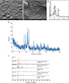
The surface morphology of WMTA was characterized by the presence of amorphous, globular, and crystalline particles in a general ground mass of finer amorphous material. At high magnification, WMTA exhibited areas of coarse amorphous crystalline structures. The addition of CaCl2·2H2O resulted in a more homogenous and less porous surface morphology compared to the normal set formulation (Figures 1A, 1B, 2A, and 2B).
Figure 1
(A, B) Surface morphology of WMTA (A: × 600, B: × 20,000); (C) EDX microanalysis. Mean of weight percentages in WMTA: Ca = 32.26%, O = 40.76%, C = 7.82%, Si = 5.47%, Al = 0.32%, Mg = 0.06%, and Bi = 13.31%; (D, E) XRD analysis of WMTA.
WMTA, white mineral trioxide aggregate; EDX, energy dispersive X-ray microanalysis; XRD, X-ray diffraction.
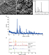
Figure 2
(A, B) Surface morphology of FS WMTA (A: × 600, B: ×20,000); (C) EDX microanalysis. Mean of weight percentages in FS WMTA: Ca = 30.85%, O = 34.19%, C = 3.28%, Si = 4.90%, Al = 0.47%, Mg = 0%, Bi = 19.30%, and Cl = 7.01%; (D, E) XRD analysis of FS WMTA.
FS WMTA, fast-set white mineral trioxide aggregate; EDX, energy dispersive X-ray microanalysis; XRD, X-ray diffraction.
Chemical analysis
EDX results showed that WMTA comprised Ca, O, C, Si, Bi, Al, and Mg. Cl was only identified in FS WMTA because of the addition of CaCl2·2H2O. The peaks and mean weight percentage for every element are listed in Figures 1C and 2C. XRD analysis showed that WMTA and FS WMTA composed of calcium silicate (Ca3SiO5/Ca3[SiO4]O), calcite (calcium carbonate [CaCO3]), and bismuth oxide (Bi2O3). Small peaks of calcium aluminum silicate (CaAl2Si2O8/Ca3Al6Si2O16) were also detected in both formulations. FS WMTA differed from WMTA by the presence of calcium silicate chloride (Ca2SiO3Cl2) (Figures 1D, 1E, 2D, and 2E).
Cytotoxicity evaluation
Results showed that although FS WMTA exhibited favorable cytotoxicity profile, the addition of CaCl2·2H2O to WMTA reduced the cell viability values, and the difference was significant in 7 out of 10 concentrations at 2 time intervals (p < 0.05; Figure 3). In terms of cytotoxicity classification, both groups were severely cytotoxic at 50 mg/mL after 24 and 72 hours. At concentrations ≤ 25 mg/mL, both WMTA and FS WMTA exhibited a non-cytotoxic profile at both time intervals except for one concentration of WMTA (3.125 mg/mL at 24 hours), which demonstrated a slight cytotoxic activity (Figure 3).
Figure 3
Intergroup comparisons between the cell viability values according to the concentrations of WMTA and FS WMTA extracts (A) after 24 hours; (B) after 72 hours.
WMTA, white mineral trioxide aggregate; FS WMTA, fast-set white mineral trioxide aggregate.
*There is a statistically significant difference between WMTA and FS WMTA at the concentration.

Cell attachment properties
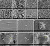
After 24 hours of incubation, HPLFs adhered over the surface and at the interface of WMTA. The cells showed prominent cytoplasmic processes, especially at the interface (Figure 4A and 4B). After 72 hours of incubation, the HPLFs increased in number with prominent cytoplasmic processes interacting with the surface of the study samples and the neighbouring cells (Figure 4C and 4D). FS WMTA showed similar findings, but the cells were not as confluent as with WMTA (Figure 4E-4H). Figure 4I-4N show the cell attachment properties of the control groups (favorable and unfavorable profiles with acrylic mold and amalgam, respectively).
Figure 4
Cell attachment properties of WMTA and FS WMTA (white arrows). (A) WMTA at the interface (24 hours); (B) WMTA on the top (24 hours); (C) WMTA at the interface (72 hours); (D) WMTA on the top (72 hours); (E) FS WMTA at the interface (24 hours); (F) FS WMTA on the top (24 hours); (G) FS WMTA at the interface (72 hours); (H) FS WMTA on the top (72 hours); (I) Acrylic mold (control, 24 hours); (J) Acrylic mold (control, 72 hours); (K) Amalgam at the interface (control, 24 hours); (L) Amalgam on the top (control, 24 hours); (M) Amalgam at the interface (control, 72 hours); (N) Amalgam on the top (control, 72 hours). Yellow arrows indicate surface perforations.
WMTA, white mineral trioxide aggregate; FS WMTA, fast-set white mineral trioxide aggregate.
DISCUSSION
The chemical composition, physical properties, and biological profile of WMTA have been examined in previous studies. However, information on the properties of WMTA when combined with CaCl2 in the dihydrate formula is scarce. The present study showed that addition of CaCl2·2H2O to WMTA results in a more homogenous and less porous surface morphology compared to WMTA. The porosity in the set calcium silicate-based formulations has been identified as a potential drawback for the material [718]. It can potentially allow the penetration of bacteria or their by-products; however, studies have shown that after the placement of MTA, a layer of hydroxyapatite forms over the material that fills the voids and surface defects [7]. The influence of CaCl2 on the surface morphology of calcium silicate cements has been reported, and this effect appears to mainly depend on the concentration of CaCl2 and hydration time [3031].
EDX is a chemical microanalysis technique used to study the compositions of dental materials, including the cements [32]. Numerous studies have shown several detectable variations in the elemental composition of WMTA via EDX. Although Ca, O, Si, and Bi were identified in all related investigations [263334353637], consistent with the current study, the presence of Al [2633343637], S [33343637], C [263336], and to a lesser extent, Mg and K [33] has also been reported. The present study demonstrated the presence of small/moderate peaks of C as well as small peaks of Al and Mg. S was not detected in any WMTA or FS WMTA samples.
The heterogeneity of elemental compositions of WMTA among different studies could be mainly attributed to 2 reasons. First, the formula of commercial WMTA products vary chemically, probably because of distinct manufacturing practices in different countries [38], and/or the continuous attempts of manufacturers to enhance the quality of the material. Similar observations have been reported with GMTA [3538]. Second, the sample size used for EDX examination is critical for determining minor/trace elements. Two studies did not mention the sample size for EDX analysis [3436]. Other studies prepared 2 stubs for each sample, and each sample was examined twice [3539]. In the present study, 3 samples were analyzed 3 times, and each sample was analyzed at 3 different areas.
XRD is an analytical technique used to study cements [40]; it enables identification of the major crystalline products in a cement sample. XRD analysis showed that WMTA primarily comprises tricalcium silicate, CaCO3, and Bi2O3, similar to findings in previous studies [353641]. Addition of CaCl2·2H2O to WMTA resulted in formation of a number of components, including Ca2SiO3Cl2. To the best of our knowledge, the available information on Ca2SiO3Cl2 is limited [42], and the formation of a chloride complex during the hydration reaction of WMTA requires further investigation. Based on the results and discussion above, the research hypotheses with regards to surface morphology and chemical composition can be accepted.
In vitro cytotoxicity tests are essential stages of the biocompatibility screening process [43]. The decision to use a given protocol for cytotoxicity evaluations must be based on its consonance with the chemical nature of the material being tested. Because MTA is a hydrophilic substance likely to release ionic components, it is apt to interfere with intracellular enzyme activities [44]. Therefore, MTT assay was selected for this study. The extraction dilution method was selected in this study to examine the cytotoxic effects of leachable elements from study samples on cells that are both distant to and in close contact with the cement [44]. This method can also simulate the clinical scenario where toxic elements of the materials leach into the surrounding fluids and bony crypt.
During extraction, the color of the samples notably changed from white to black after incubation in the culture medium of HPLFs. This finding has previously been reported in the literature [454647], and may be attributed to the presence of Bi in WMTA. HPLFs showed higher cell viability values after exposure to WMTA than FS WMTA, although the cytotoxic profiles of both formulations appear favorable. This observation is consistent with 2 studies [1721].
Cell adhesion must occur before cells can proliferate, differentiate, and produce an extracellular mineralized matrix on a substrate [48]. The quality and quantity of cell attachment onto retrograde filling materials is generally agreed to be a valid criterion for evaluations of the biocompatibility of dental bio-materials [4950]. Despite differences in methodologies, the favorable attachment properties of WMTA to HPLFs observed in this work are consistent with others [435152].
Few studies have examined the cell attachment properties of WMTA mixed with CaCl2. One investigation compared 3 combinations of FS WMTA including CaCl2 [22]. SEM observations of MG-63 osteosarcoma cells demonstrated unfavorable attachment to the surfaces of WMTA mixed with CaCl2. Despite this unfavorable biological response, stating that this combination is not suitable for clinical application may be inappropriate because the cell types that would come into contact with this combination in the clinical setting may react differently [4], which has been confirmed in the current study. A recent study showed favorable cell attachment properties of this FS formulation on dental pulp stem cells isolated from teeth extracted from young adult patients [17].
Based on the results, the research hypotheses with regards to biological properties (cytotoxicity and cell attachment properties) can be partially accepted. The favorable sensitivity of HPLFs towards FS WMTA may have an impact on its clinical application for repair of perforation defects. Future in vivo studies are warranted to validate the findings demonstrated in this experimental investigation.
 XML Download