

 PDF
PDF ePub
ePub Citation
 Print
Print


Introduction
Casting gold alloys have been most widely used as an indirect restorative material. Recently, owing to the increasing demand for esthetic restoration, tooth-colored indirect restorative materials such as composite resin inlays, ceramic inlays, and zirconia inlays have been used as alternatives to casting gold alloys. Among these tooth-colored indirect restorative materials, zirconia has attracted the maximum attention because it has the highest flexural strength and fracture toughness of all the existing ceramics.1 A spontaneous phase transformation occurs internally in zirconia upon the application of an external stress, which tightens the crack tip and prevents crack propagation, leading to an increase in the fracture toughness and flexural strength.234
The most important aspect of tooth-colored indirect restorative materials is that they have to be firmly bonded to the underlying tooth substrate. In the bonding process, the use of luting cements is essential. The earliest luting cements (also described as non-adhesive luting cements), such as polycarboxylate and zinc phosphate, provided only a mechanical bond to the tooth.5 Subsequent studies have focused on increasing the bond strength, and decreasing microleakage and technique sensitivity.6 A new generation of luting cements, including glass ionomer, resin-modified glass ionomer, and resin cements, has been developed.7 Among these luting cements, adhesive resin cements have been widely used clinically for the bonding in indirect restoration89 because of their stronger bonding ability, lower microleakage, and more accurate fit than those of non-adhesive luting cements.1011
Several studies revealed that when indirect restorations that use a ceramic or a composite resin were bonded to the tooth substrate with adhesive luting cements, the fracture resistance was improved as compared to that with the use of non-adhesive luting cements.121314 Further, as zirconia, it does not contain silica (SiO2), the well-established micromechanical interlocking of silica-silane bonds cannot be achieved.151617 Many studies have been conducted to improve the bonding strength of zirconia and the tooth substrate.18192021222324 To prove the improvement of the bonding strength of yttria-stabilized tetragonal zirconia polycrystal (Y-TZP), most of the above-mentioned studies measured the bonding strength applying the shear force. Furukawa et al.25 reported that the fracture resistance of a restoration is dependent upon the bond strength between a restoration and the tooth substrate. However, few studies have been conducted to investigate the effect of adhesive luting cements on the fracture resistance of zirconia as compared to that of non-adhesive luting cements.
The purpose of this study was to evaluate the effect of adhesive luting on the fracture resistance of zirconia as compared to that of a composite resin and a lithium disilicate glass ceramic by measuring the fracture resistance after non-luting, non-adhesive luting, and adhesive luting.
Materials and Methods
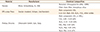
The materials listed in Table 1 were used in this study. Instead of pure zirconia, Y-TZP in which Y2O3 is added for controlling the volume expansion and phase stabilization2627 was used.
Specimen preparation
A bovine tooth and restorative materials were prepared for the flexural test. The bovine tooth measuring 3 mm × 17 mm (thickness and length without width) was placed in the empty space between the supporting parts of the jig. Both the top and the bottom of the bovine tooth were prepared to be parallel to the base by using a superfine grit diamond bur (SF 102R, Shofu Inc., Kyoto, Japan) and 1,000, 1,500, and 2,000 grit silicon carbide papers. However, if it was difficult to make it parallel to the base because of its anatomical structure, a vinyl polysiloxane material (Aquasil soft putty/regular set, Dentzply, Konstanz, Germany) was used for making it parallel.
On the other hand, the composite resin and the lithium disilicate glass ceramic (dimensions: 2 mm × 2 mm × 25 mm) were prepared using a customized Teflon mold and a low-speed diamond saw (Isomet, Buchler Ltd., Lake Bluff, IL, USA). The Y-TZP, specimens were prepared to be 20% larger in size considering the sintering shrinkage and then sintered in the sintering furnace. Each restorative material was polished using 1,000, 1,500, and 2,000 grit silicon carbide papers. Only Y-TZP specimens were sandblasted with Al2O3 (110 µm, four bars at a distance of 1 cm) after the silicon carbide paper polishing.
Luting procedure
A one-step adhesive system (Single Bond Universal, 3M ESPE, St. Paul, MN, USA) as the primer, a zinc phosphate cement (HY-Bond, Shofu Inc.) as the non-adhesive luting agent and a self-adhesive resin cement (G-cem LinkAce, GC Corp., Tokyo, Japan) as the adhesive luting agent were used in this study.
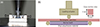
For the non-adhesive luting, only the zinc phosphate cement was used as the non-adhesive luting cements without any etchant or primer. According to the manufacturer's instructions, the powder was mixed with the liquid in a fixed ratio of one level scoop to four drops of the liquid. A cool and dry glass slab was used for slowing the exothermic reaction. The mixing procedure was performed by a single clinician. The hand-mixed zinc phosphate cement was placed on the prepared specimen of each restorative material and non-adhesive luting was performed under a constant load of 1 kg in a special clamp at room temperature (Figure 1).
For the adhesive luting of the composite resin and Y-TZP, both the one-step adhesive system and the self-adhesive resin cement were used. On the surface of the prepared specimens of the composite resin and Y-TZP, the one-step adhesive system as the primer was applied first and the self-adhesive resin cement was placed next (Figure 1). Under a constant load of 1 kg, light curing was performed using a visible light curing unit for 1 second and the excess cement was removed with hand instruments. While maintaining the pressure, all surfaces were light cured for 20 seconds each.
For the adhesive luting of the lithium disilicate glass ceramic, hydrofluoric acid and the silane coupling agent, the one-step adhesive system and the self-adhesive resin cement were used. The specimens of the lithium disilicate glass ceramic were etched with 4.9% hydrofluoric acid for 3 minutes and washed thoroughly for 30 seconds. The etched specimens were air-dried and coated with the silane coupling agent. Silane was allowed to remain in contact for 60 seconds, and then, the surface was air-dried. The one-step adhesive system and the self-adhesive resin cement were applied using the same method as that used for the adhesive luting of the composite resin and Y-TZP. The specimens that underwent the non-adhesive and adhesive luting procedures were stored in water at 37℃ for 24 hours before the flexural test.
Flexural test
The universal testing machine (Model 4201, Instron Corp., Canton, MA, USA), which was equipped with a load cell of 5 kN, was used for this test, and the machine's crosshead speed was set to 0.5 mm/min. For the non-luting group, the specimen of each restorative material was placed on the bovine tooth without any primer or luting agents. The fracture resistance of the non-luting group was measured by recording the force at which the specimen was fractured. Ten specimens of each restorative material were measured. In the non-luting group, the specimen was fixed on the bovine tooth by using sticky wax and the loading actuator such that it does not move.
For the non-adhesive luting and adhesive luting groups, the specimen of each restorative material was placed on the jig after achieving non-adhesive or adhesive luting between bovine tooth and each restorative material. The fracture resistance of the non-adhesive luting and the adhesive luting groups was measured using the same method as that used for the non-luting group. Ten specimens of each restorative material were measured.
Stereomicroscope and scanning electron microscope (SEM) analysis
For the adhesive luting group, failure patterns were categorized by observing the fractured specimens under a stereomicroscope. The failure patterns were broadly categorized into three stages, namely stage 1, stage 2, and stage 3. Stage 1 is the failure pattern of representing the partial fracture of the specimen and the adhesive luting layer. In the stage 1 failure pattern, one part of the fractured specimen still remained, but the other part of the fractured specimen fell out of the bovine tooth, thereby cracking the adhesive luting. Stage 2 is the failure pattern of representing a complete fracture of the specimen and a partial fracture of the adhesive luting layer. In other words, all parts of the fractured specimen fell out of the bovine tooth with only a part of the adhesive luting layer remaining. Stage 3 is the failure pattern of including the fracture of the bovine tooth as well as the fracture of the specimen and the adhesive luting layer.
In addition, by observing the fractured sections of the specimens using FE-SEM (S-5500, Hitachi, Tokyo, Japan) and AFM (Multimode-8, Bruker, Santa Barbara, CA, USA), we tried to clarify the relationship of the fractured-surface roughness of each restorative material with the fracture resistance.
Statistical analysis
Data obtained through experiments were processed statistically using SPSS (SPSS 18 for Windows, SPSS Inc., Chicago, IL, USA). The data were compared with a two-way analysis of variance (ANOVA) and the all pairwise multiple comparison procedure (Tukey's test) was used for determining the significant differences among the groups. All statistical analyses were performed at α = 0.05.
Results
Changes in fracture resistance upon adhesive luting
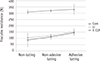
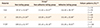
For the composite resin, the fracture resistance values of the non-luting, non-adhesive luting, and the adhesive luting groups were 87.67 ± 16.62 N, 111.63 ± 10.07 N, and 144.62 ± 38.70 N, respectively. For the lithium disilicate glass ceramic, the fracture resistance values of the non-luting, non-adhesive luting, and the adhesive luting groups were 105.59 ± 24.04 N, 122.05 ± 10.87 N, and 153.49 ± 23.86 N, respectively. For Y-TZP, the fracture resistance values of the non-luting, non-adhesive luting, and the adhesive luting groups were 309.52 ± 52.75 N, 321.45 ± 18.16 N, and 331.05 ± 19.81 N, respectively (Table 2). Y-TZP showed the highest fracture resistance among all the tested groups, followed by the lithium disilicate glass ceramic, and the composite resin had the lowest fracture resistance.
In comparison with the non-luting group, the fracture resistance of the composite resin was increased by about 27 and 65% in each non-adhesive luting and adhesive luting group, respectively. In the case of the lithium disilicate glass ceramic, the fracture resistance was increased by about 15 and 45% in each non-adhesive luting and adhesive luting group as compared to the non-luting group. For Y-TZP, the fracture resistance was increased by about 3 and 6% in each non-adhesive luting and adhesive luting group as compared to the non-luting group (Figure 2). The fracture resistance after adhesive luting was increased by approximately 29% in the composite resin, 26% in the lithium disilicate glass ceramic and 2% in Y-TZP, as compared to non-adhesive luting.
Stereomicroscope and SEM analysis
Stage 3 failure was not observed in any of the adhesive luting groups. Only stage 2 failure was observed in the case of Y-TZP in the adhesive luting group. In the case of the lithium disilicate glass ceramic and the composite resin of the adhesive luting group, both stage 1 and stage 2 failures were observed. Of the two failure patterns, stage 1 failure occurred more often in the lithium disilicate glass ceramic and stage 2 failure occurred more often in the composite resin (Table 2).
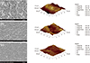
The roughness of the fractured-surface was measured using with AFM, and we observed that the fractured-surface roughness of Y-TZP was the smallest, followed by that of the lithium disilicate glass ceramic; the composite resin had the largest fractured-surface roughness (Figure 3). This result showed that the roughness of the fractured-surface reversed the order of fracture resistance.
Discussion
Since zirconia is not silica-based, the well-established micromechanical interlocking of silica-silane bonds cannot be achieved. Therefore, many studies have been conducted to improve the bonding strength of zirconia and the tooth substrate, and several methods have been proposed for the same.18192021222324 The first method was to achieve strong bonding by silanization using a silane coupling agent on a silica-coated surface by Rocatec or Cojet (3M ESPE, Seefeld, Germany).2021222324 According to the results from the studies using this method, the tribochemical silica coating seems to slightly improve the bonding strength of Y-TZP.222324 The other method was to achieve strong bonding by the use of a primer containing a phosphate monomer.28293031 The phosphoric acid monomer that has attracted the most attention with respect to adhesion is 10-methacryloxydecyl dihydrogen phosphate (10-MDP); it is known that improved bonding to Y-TZP is achieved by using two hydrogen groups derived from the phosphate group that reacts with oxygen in Y-TZP to form a stable covalent bond of Zr-O-P while releasing a water molecule.3233 Many related studies have reported that the primer containing the phosphate monomer significantly enhanced the bonding strength of Y-TZP.282930
In this study, the primer containing both 10-MDP phosphate monomer and silane was applied to the surface of Y-TZP and the self-adhesive resin cement was chosen as the adhesive luting agent, because a dual-cured resin cement is generally preferred to a light-cured resin cement, owing to its excellent self-curing capability, given that visible light cannot penetrate Y-TZP.3435 In other words, we tried to achieve maximum bonding to Y-TZP by using all the abovementioned methods.
A comparison of the changes in fracture resistance revealed that the fracture resistance of adhesive luting was increased by just 2% in the case of Y-TZP as compared to that in the case of non-adhesive luting. This implies that several methods that have thus far been applied to improve bonding to Y-TZP were ineffective under a compressive force. Therefore, we predict that because the fracture strength of the self-adhesive resin cement is less than that of Y-TZP, the force that causes a fracture of the Y-TZP specimens will cause a subsequent fracture throughout the entire self-adhesive resin cement. Consequently, we can conclude that the self-adhesive resin cement contributed only a little towards increasing the fracture resistance of Y-TZP. However, in the case of the composite resin, the fracture resistance of the adhesive luting group was increased by approximately 29%. Because the fracture strength of the self-adhesive resin cement is similar to that of the composite resin, the cracks from the fracture of the self-adhesive resin cement are likely to propagate gradually along the bonding interface rather than occurring at once. In the case of the lithium disilicate glass ceramic, we expect that because the fracture strength of the lithium disilicate glass ceramic is higher than that of the self-adhesive resin cement, the fracture resistance of lithium disilicate glass ceramic in the adhesive luting will increase as much as in the case of Y-TZP. However, in contrast to this expectation, the fracture resistance was increased by 26% in the adhesive luting group. This result can be explained by the failure pattern. Y-TZP showed the stage 2 failure representing the complete fracture of the specimen and a partial fracture of the resin cement in all the specimens. Thus, several methods used for maximizing the bonding to Y-TZP in this study did not increase the bonding strength between Y-TZP and the self-adhesive resin cement. In contrast, the lithium disilicate glass ceramic showed the stage 2 failure in just 1 specimen and the stage 1 failure pattern in the other 9 specimens. Stage 1 failure means that a part of the fractured-specimen still containing the adhesive luting with the bovine tooth remained after the specimen was fractured. This can be explained by the fact that the traditional ceramic bonding methods (hydrofluoric acid etching and silanization) are responsible for the strong bonding between the ceramic and the self-adhesive resin cement, which consequently enhances the fracture resistance.
The surface roughness of the material is closely related to its microstructure. Surface roughness is measured to be low if the microstructure is homogeneous and the singularity is high. In this study, the SEM and AFM images of the fractured surface illustrated in Figure 3 showed the lowest fractured-surface roughness of Y-TZP with the most homogeneous microstructure and the highest singularity, and the highest fractured-surface roughness of the composite resin with a heterogeneous microstructure and the poorest singularity. In addition, the microstructure of a material and its mechanical properties are closely related. The mechanical properties of a material increase with an increase in the homogeneity and density of the microstructure. Therefore, we believe that the fractured-surface roughness related to the microstructure is also related to the fracture resistance. These results that showed an inverse correlation between the fractured-surface roughness and the fracture resistance support this idea.
The limitations of this study are that it is an in vitro investigation and a single-direction loading test, which does not fully replicate oral conditions. Further studies should consider other self-adhesive resin cements, thermocycling and water storage aging conditions to challenge the adhesive interface.
Conclusions
Choosing Y-TZP as an indirect esthetic restorative material enables us to preserve as much of the tooth structure possible by minimizing the amount of tooth loss during cavity preparation. Although many studies have been conducted to improve the bonding strength of Y-TZP, in this study, the fracture resistance of Y-TZP did not increased significantly after adhesive luting as compared to that of the composite resin and the lithium disilicate glass ceramic.
 XML Download