

 PDF
PDF ePub
ePub Citation
 Print
Print


Introduction
Root canal treatment is a highly successful procedure.1,2 However, the treatment of teeth with immature roots and pulp necrosis presents significant difficulties for the dental clinician in regard to disinfection and filling of the wide root canal space and open apex. For the practitioner, the revascularization of immature necrotic teeth has become an alternative to apexification techniques.
Current techniques involve disinfecting irrigation, placement of calcium hydroxide or an antibiotic paste as a temporary medication, and the instigation of a blood clot within the root canal space to establish the revascularization. Unfortunately, a blood clot is difficult to produce, unstable and therefore susceptible to breakdown. Moreover, it is a desirable substrate for microbes left behind in the root canal or reintroduced through reinfection from the oral cavity. While or immediately after setting, it is also difficult to seal with a coronal restoration.
To overcome some of the difficulties associated with an intracanal blood clot, the use of scaffold materials was suggested. Examples of three-dimensional organic scaffolds include collagen,345 glycosaminoglycan, fibrin, cell-sponge constructs or combinations thereof. Synthetic scaffolds are PCL (polycaprolactone),6 PEG (polyethyleneglycol), PLA (polylactic acid),5 PGA (poly-glycolic acid), PLGA (poly-lacticco-glycolic acid),7 or hydrogels. For endodontic purposes, scaffolds must provide an adequate porosity and pore size to allow pulp stem or progenitor cells to migrate and/or organize within a three-dimensional structure, be capable to support cell organization, neuro-, angioor vasculogenesis and to provide necessary nutritional support.89
Collagen is a natural constituent of the dental pulp. Collagen scaffolds were shown to provide a suitable environment for pulp progenitor and adult stem cells with odontoblastic properties in vitro,10 and adequate new tissue development with rare histologic inflammation in vivo.11 Cell seeded collagen scaffolds have been used in animals for in vivo pulp regeneration after pulpotomies,12 and after pulpectomies,13 demonstrating regeneration of pulp tissue with angio- and neurogenesis.1213 Collagen and PLGA scaffolds also showed the regenerative capacity of swine dental pulp stem cells after reimplantation into minipig teeth.14 Development of dentin/pulp-like tissues was observed after subcutaneous implantation of rabbit dental pulp stem cells in PLGA scaffolds.15 PLGA was also used for growth factor or drug delivery, e.g. for pulp capping purposes containing TGF-β1 to initiate tertiary dentin formation,11 or as nanoparticles loaded with methylene blue to initiate photo-disinfecting action against Enterococcus faecalis.16
Antibiotic containing scaffold materials were suggested for pulpal regeneration.8 In periodontics, they were found to be suitable for guided tissue regeneration.17 Metronidazole or ciprofloxacin releasing polydioxanone polymer nanofiber scaffolds were recently introduced for pulp regeneration.181920 This study aimed to investigate the biocompatibility of two experimental antibiotic releasing scaffolds, based on either type-I collagen or on PLGA, with human dental pulp stem cells (hDPSCs) for the potential clinical use in revascularization or regeneration procedures.
Materials and Methods
Scaffolds
One resilient lyophilized collagen (COLL) scaffold, releasing metronidazole and clindamycin, was compared to an experimental injectable PLGA scaffold, releasing clindamycin. COLL scaffold was prepared at the University of Pennsylvania. Using cooled pipettes, 3.0 mg/mL type I bovine collagen (Ultrapure bovine collagen solution, Sigma-Aldrich, St. Louis, MO, USA) was mixed on ice with 9 mMol glucose (D-(+)-Glucose, Sigma-Aldrich), sterile ×10 phosphate buffered saline (PBS, Sigma-Aldrich), 1 N sodium hydroxide (NaOH, Sigma-Aldrich), distilled water, 10 mg/mL Metronidazole (Metronidazole, Sigma-Aldrich) and 25 mg/mL Clindamycin (Clindamycin, Sigma-Aldrich) to a final concentration of collagen 2.6 mg/mL. Two hundred microliter (µL) of the mixed solution were delivered each into one well of micro-well strips (Immuno Module Plate With PolySorp Surface, Thermo Fisher Scientific Inc., Waltham, MA, USA). The micro-well strips were then placed into an incubator for 30 minutes at 37℃ to allow gelation. Then, the collagen gels were cross-linked in a ultra-violet cross-linker (Stratalinker UV crosslinker 2400, Stratagene, La Jolla, CA, USA) for another 30 minutes. After the crosslinking process, the collagen gel was frozen at -80℃ for 24 hours and finally lyophilized in a freeze-dryer (Labconco Freezone lyophilizer, Labconco, Kansas City, MO, USA) for 12 hours until a solid and porous scaffold was observed.
The PLGA scaffold was provided by Skywalk Pharmacy (Milwaukee, WI, USA). While the exact manufacture is not being disclosed, this experimental scaffold contained 7.2 wt% Clindamycin, the remainder split between a liquid PLGA solution and N-Methyl-2-pyrrolidone (NMP), a solvent used in the pharmaceutical industry for the formulation of oral and transdermal drugs. The scaffold solidified on contact with moisture with a gel-like consistency.
Cell culture
hDPSCs were kindly provided by Dr. Sunday Akintoye (University of Pennsylvania School of Dental Medicine, Department of Oral Medicine). Cells were cultured in growth culture media consisting of α-modified minimum essential medium (α-MEM, GIBCO, Invitrogen, Carlsbad, CA, USA), supplemented with 20% fetal bovine serum (FBS, Equitech Bio, Kerville, TX, USA), 100 U/mL penicillin, 100 mg/mL streptomycin sulfate (GIBCO/BRL, Grand Island, NY, USA) and 2 mMol glutamine (GIBCO/BRL), and incubated in a humidified 5% CO2 atmosphere at 37℃. The growth media in the wells was changed every other day. Cells grown in medium only served as control group for all experiments.
Light microscopy
hDPSCs were seeded at a density of 5.0 × 10⁴ cells/well in 12-well plates (Corning Inc., Corning, NY, USA). Three hours after plating, one piece of COLL scaffold from a micro-strip well or one drop of PLGA scaffold of similar size as the COLL scaffold was completely immersed in the cell media in co-incubation with the cells. When the cells reached 90 - 95% confluence, light microscopy pictures were taken at ×100 and ×200 magnifications using a light microscope (TMS-F microscope, Nikon Instruments, Melville, NY, USA).
MTT assay
Actively proliferating hDPSCs were plated in 96-well plates at densities of 1.0 × 10⁴, 2.5 × 10⁴, and 5.0 × 10⁴ per well. Three hours after plating, one piece of COLL scaffold or one drop of PLGA scaffold of the same dimensions as mentioned above was immersed in form of co-incubation. After 48 hours of incubation, cell proliferation was assessed using the Colorimetric MTT Cell Survival and Proliferation Assay Kit (Millipore, Chemicon International Inc., Temecula, CA, USA) according to the manufacturer's protocol. Briefly, MTT reagents were added and incubated for 4 hours. Then 100 µL isopropanol with 0.04 N HCl was added to each well. After thorough mixing using a plate shaker for 10 minutes at room temperature, the scaffolds were carefully removed from the wells and a colorimetric analysis performed with a multiplate reader (Synergy HT, Biotek, Winooski, VT, USA) at 570 nm. A solution of growth media with isopropanol and HCl served as negative control. The optical density values were obtained from triplicate samples and the experiment was repeated twice.
IL-8 ELISA test
hDPSCs were seeded at a 5.0 × 10⁴ cells per well density in 12-well plates. Similarly, 3 hours after plating, one piece of COLL scaffold or one drop of PLGA scaffold were immersed with the cells for co-incubation. Supernatants were collected after 24 hours (Day 1 samples), 72 hours (Day 3 samples), and 6 days (Day 6 samples), and subsequently stored at - 80℃ until use. The amount of IL-8 was measured by an enzyme-linked immunosorbent assay (ELISA) using the Human IL-8 ELISA Kit II (BD OptEIA, BD Biosciences, Bedford, MA, USA) according to the manufacturer's instructions. Growth media served as a negative control. The absorbance was read at 450 nm in the multiplate reader for triplicate samples. The experiment was repeated twice.
Statistical analysis
Prism 6.0 software (GraphPad Software Inc., La Jolla, CA, USA) was used for statistical analysis. The results of the absorbance measured using multiplate reader were analyzed using two-way analysis of variance (ANOVA) and post hoc Bonferroni test for the type of scaffold, and cell number or time point. Statistical significance was accepted at p < 0.05.
Results
Light microscopy
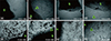
The exposure of hDPSCs to the two different scaffold materials after 7 days in co-culture revealed major morphological differences. Figure 1 shows the comparison of representative cell appearances under the light microscope at ×100 and ×200. In general, hDPSCs in the COLL group appeared healthier, well stretched and attached to the cell culture plate. Cells grew in direct proximity to the COLL scaffold with identical healthy appearance as in other areas of the culture wells (Figures 1a - 1d). Cells in co-incubation with the PLGA scaffold frequently demonstrated signs of degeneration and apoptosis, in particular in proximity to the actual material. An abundance of cell debris and rounded cell morphologies suggested non-attachment. (Figures 1e - 1h).
MTT assay
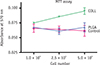
Table 1 and Figure 2 show the absorbance readings measured using the multiplate reader. Of the hDPSCs seeded at 3 different densities (1.0 × 10⁴, 2.5 × 10⁴, and 5.0 × 10⁴), with media only, COLL or PLGA scaffolds, at 2.5 × 10⁴ and 5.0 × 10⁴ cell densities, COLL demonstrated significantly higher cell proliferation rates than cells in media only (control) (p < 0.05 and p < 0.01, respectively) or in co-incubation with PLGA (p < 0.05 and p < 0.01, respectively). The effect of the different cell numbers was not statistically significant.
IL-8 ELISA test
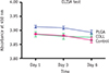
No significant differences were observed between cells with media only and COLL at 1, 3, and 6 days. Cells incubated with PLGA expressed significantly higher IL-8 than the control at all time points (p < 0.01) and compared to COLL after 1 and 3 days (p < 0.01, Table 2 and Figure 3). The effect of the different time points was found to be statistically significant (p < 0.001).
Discussion
To date, no final approach to clinical revascularization or a common strategy to achieve pulp regeneration has been agreed upon. While it was recognized that clinical revascularization does not provide pulp or pulp-like tissues, the clinical benefits of an ingrowth of vital tissue into a disinfected tooth with a history of pulp necrosis are greatly beneficial to the patient. Successful revascularization not only provides vitality, but also generates the formation of a closed apex and strengthens the tooth structure by thickening of the root walls, thus adding prevention from fracture. While a consensus seems to exist that complete regeneration of the pulp would involve exogenously introduced cells, as cell-free regeneration after complete loss of the dental pulp appears unlikely,21 it is still unclear, which types of cells, scaffold materials and bioactive factors would provide the greatest chances of successful regeneration. Thus, clinical revascularization remains the best strategy for a practitioner to treat immature teeth with pulp necrosis. However, creating a suitable blood clot remains a challenge in clinical practice and scaffold materials may prove an alternative for clinical practice.
This study investigated two different potential scaffolds containing antibiotics. In endodontics, local antibiotics were initially used as intracanal medication to aid in the disinfection of infected, pulpless teeth.22 However, this treatment fell into disfavor and was only reestablished with new techniques for revascularization to disinfect immature, necrotic teeth. A triple antibiotic paste consisting of metronidazole, ciprofloxacin and minocycline was introduced,23 and its efficacy to penetrate dentin demonstrated in vitro.24 Several case reports on revascularization using triple antibiotic paste were published.25,26 The effectiveness of in vivo disinfection was shown by Windley et al.27 Several clinical complications were described for revascularization procedures, including discolorations by minocycline. Cefaclor was suggested as replacement for minocycline.28,29 Another problem was the potential collapse of MTA into the fresh blood clot, which could be overcome by the additional placement of a solid resorbable collagen matrix to pack MTA against.28
The placement of scaffolds for current revascularization and, possibly, future regeneration procedures could serve several purposes. For revascularization, it can provide stability placing a suitable material against the blood clot. For regeneration, a matrix can supply stem or progenitor cells to the root canal and provide support for the ingrowth of vascularization and re-innervation, as well as aid stability for early stages of tissue regeneration. For both procedures, a scaffold can provide bioactive substances.30
To use bioactive materials for the delivery of growth factors or drugs such as antibiotics in endodontic regenerative procedures was suggested earlier on.3031 Nevertheless, only few endodontic studies introduced growth factor,32,33 or antibiotic-containing scaffolds.181920 Bottino et al.18 used metronidazole or ciprofloxacin releasing nanofiber-scaffolds and tested them against Porphyromonas gingivalis and Enterococcus faecalis. In accordance with our study the metronidazole containing scaffolds demonstrated good cytocompatibility. Metronidazole inhibited only growth of Porphyromonas gingivalis, while ciprofloxacin was effective against both strains. This supports the idea of combining antibiotics to achieve a wider antibacterial spectrum, similar to triple antibiotic paste or the COLL scaffold in this investigation, combining metronidazole with clindamycin, a lincosamide, used to treat dental infections with anaerobic bacteria.34 A commercially available triple antibiotic paste (PulpVAZ, Blaze Pharmaceuticals LLC, Ankeny, IA, USA) contains clindamycin as alternative to minocycline. Clindamycin demonstrated successful antibacterial activity as an intracanal dressing in vivo.35 In vitro, it showed significantly more antibacterial activity than tetracycline.36 Clindamycin releasing fibers from ethylene vinyl acetate significantly reduced bacteria in extracted human teeth and provided sustained antibiotic release of over 2 weeks.34 This surpassed the activity of metronidazole and ciprofloxacin for 48 hours.18 Even if not directly comparable, this substantial time difference suggests that clindamycin, if biologically acceptable, may be a good alternative for pulp regeneration.
The present study evaluated biocompatibility and cell proliferation of hDPSCs with the experimental scaffolds. Although both scaffolds incorporated clindamycin, and both collagen and PLGA are known to be suitable scaffold materials for endodontic procedures, significant differences between the materials existed. Light microscopy permitted to observe cell morphology after co-incubation with the scaffolds. Cells in contact with the PLGA scaffolds frequently showed signs of degeneration and apoptosis. Whereas no significant difference in IL-8 concentration were found between hDPSCs cultured in media only and COLL, cells incubated with PLGA expressed significantly higher IL-8 than the control at all time points and compared to COLL after 1 and 3 days. This implied the induction of inflammation by the experimental PLGA scaffolds, suggesting less biocompatibility than the COLL scaffolds.
Several factors may be responsible for the lower biocompatibility of the experimental PLGA scaffold. PLGA itself is biocompatible; however the experimental scaffold contained a very high percentage of NMP. This stabilizer, although widely used pharmaceutically may have adverse effects on hDPSCs. The concentration of the antibiotic component itself (7.2 wt%) may have influenced the biocompatibility. High concentrations of antibiotics in triple antibiotic paste of 1, 10, and 100 mg/mL resulted in significantly lower cell viability of stem cells from the apical papilla than concentrations of 0.1 and 0.01 mg/mL or any concentration of calcium hydroxide.37 This may appear to be contradictory to the good biocompatibility of the experimental COLL scaffold containing 10 mg/mL metronidazole and 25 mg/mL clindamycin. However, Chuensombat et al. demonstrated that the adverse cell viability effect of triple antibiotic paste, when tested against human dental pulp cells and apical papilla cells was related to minocycline and ciprofloxacin, whereas no significant differences between metronidazole and the control were found up to 25 mg/mL.38 Also, antibioticcontaining scaffolds with 10 wt% metronidazole and 25 wt% ciprofloxacin were successfully tested.18,19
Conclusions
Within the limitations of this study, experimental resilient lyophilized metronidazole and clindamycin releasing collagen scaffolds could be of potential clinical use for revascularization or pulp regeneration procedures. Whereas further investigations including detailed material characterization, antimicrobial activity, and antibiotic substantivity are needed to validate the use of these scaffold materials, this pilot study demonstrated the biocompatibility and the effects on cell proliferation of antibiotic-releasing scaffolds in regenerative endodontics and gives the practitioner the insight of the potential application of scaffold materials useful for revascularization, which are cost-effective, easy to manipulate and would provide predictable positive outcomes for clinicians.


 XML Download