

 PDF
PDF ePub
ePub Citation
 Print
Print


Introduction
The demand for esthetic restorations, even in posterior teeth, has increased in recent years.1 Composite resin restorations have certain advantages compared to amalgam restorations, including better esthetic results and favorable adhesive properties, resulting in decreased cavity size and strengthening of the remaining tooth structure.2 Composite resins are believed to be suitable materials for direct restorations, including class V cavities, in particular due to their good esthetic results.34 However, posterior composite resin restorations exhibit higher failure rates resulting from secondary caries.2 Additionally, a major problem with class V composite resin restorations is microleakage along the cervical wall in these restorations,4 which is referred to as marginal leakage and considered a major problem in restorative dentistry.5
Microleakage is defined as the penetration of bacteria, fluids, molecules, or ions into the spaces between the cavity walls and the restorative materials, resulting in sensitivity, recurrent caries, discoloration of the restoration margins, irritation of the pulp, and restoration failure. Therefore, measures should be adopted to prevent microleakage when an adhesive system is being developed for dental applications.6 Long-term adhesion of tooth-colored dental materials to tooth structures is important for their clinical success, and microleakage should therefore be evaluated when the success of restorative materials is assessed.78 A number of factors affect microleakage at the tooth-restoration interface, including the bond strength between the adhesive and the tooth structure, residual stresses due to polymerization shrinkage of the composite resin, discrepancies between the thermal expansion coefficients of enamel and dentin on one hand and that of the restorative material on the other hand, and occlusal forces.9 Therefore, it is important to fully adapt the restorative material to the cavity margins because it is one of the most important factors contributing to the success of a restoration.17
Currently, dental adhesive agents provide a favorable marginal seal and decrease marginal microleakage, especially at the cervical margins of the cavity. Adhesive agents are important factors that prevent microleakage in composite resin restorations.10 The early 1990s witnessed the introduction of three-step total-etch adhesive systems, ushering in a new era in adhesive dentistry.11 Adhesive agents are applied with composite resins to bring about a proper and durable bond between the tooth structure and the restorative material. Gap-free composite resin restorations with the use of adhesive systems depend on micromechanical bonds with tooth structures.1213
In recent decades, two different strategies, etch-and-rinse and self-etch adhesive techniques, have been used in dental bonding procedures.141516 Some studies have shown that self-etching primers displayed a similar degree of efficacy to conventional etch-rinse adhesive systems in preventing microleakage.1718 However, other studies have shown that separate etching, priming, and bonding steps are more effective in decreasing or preventing microleakage.192021 Therefore, it is necessary to separately evaluate the microleakage of each adhesive system with its specific formulation or procedural steps.6
Recent studies have confirmed that none of the adhesives presently available can completely prevent marginal microleakage in composite resin restorations.2223 Marginal microleakage is detected using a variety of techniques.722 The most commonly used technique is dye penetration,4724 which is inexpensive.22 In the conventional method, also referred to as the microscopic method, the restored tooth is immersed in a dye solution, followed by cutting through the center of the restoration to evaluate the leakage visually on the coronal and cervical margins under a stereomicroscope.7 Recently a three dimensional (3D) version of this technique has been introduced, in which the tooth is softened in an acid, followed by removing the entire restorative material to evaluate microleakage.525 The aim of this in vitro study was to compare the conventional two dimensional (2D) and the new 3D microleakage evaluation techniques in the same class V composite resin restorations with the use of two different adhesive systems. In addition, the effect of immediate and delayed microleakage was evaluated using the above-described techniques in order to compare their efficacy in measuring marginal microleakage. The null hypotheses of this study were that no significant differences exist in the marginal microleakage displayed by the two bonding systems, that thermocycling does not affect marginal microleakage at the tooth-restoration interface, and that no significant difference exists between the two different techniques of measuring microleakage.
Materials and Methods
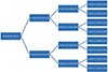
In this in vitro study, 48 sound human molars, with no carious lesions, restorations, abrasions, or cracks were evaluated in accordance with ethical standards (approval number, 292178). The teeth were cleaned with a brush after all the periodontal fibers and bone remnants were removed and then stored in 0.2% thymol solution at 4℃. Before the experiment was initiated, all the samples were stored in distilled water at 37℃ for 24 hours. In each tooth two class V cavities were prepared on the buccal and lingual surfaces with a diamond bur (No. 811 031 4, 2ML, Teez Kavan, Tehran, Iran) in a high-speed handpiece, with a new bur for every six cavities. The cavities were conical in shape, with divergent walls and a flat bottom, and the cavities measured 3 × 2 × 1.5 mm. Clearfil SE Bond (CSEB, Kuraray Co., Ltd., Osaka, Japan) two-step self-etch adhesive was randomly applied to half of the prepared cavities (without enamel etching) and Single Bond (SB, 3M ESPE, St. Paul, MN, USA) two-step etch-and-rinse adhesive was applied to the other half of the cavities, according to the manufacturer's instructions (Figure 1). The A3 shade of APX composite resin (APX, Kuraray, Tokyo, Japan) was used to fill the cavities with an oblique incremental technique in three layers, with the first oblique layer placed in the occlusal part of the cavity. Each layer was polymerized with a light-curing unit (Coltolux 50, Mod. C7950, Coltene/Whaledent Inc., Cuyahoga Falls, OH, USA), using a light intensity of 480 mW/cm2 for 20 seconds. After the restorative procedures, the specimens were placed in an incubator (Behdad, Tehran, Iran) for 24 hours in distilled water at 37℃ to decrease the stress resulting from the polymerization process. In the next stage, the restorations were polished with a flame-shaped polishing bur (No. 368/018, Teez Kavan), a polishing disk (3M ESPE) and a cup-shaped polishing rubber (SGG 1HB, Stoddard, Letchworth Garden City, England) from coarse to fine, respectively. In a random manner, half of the specimens in each group (n = 24) underwent a thermocycling procedure consisting of 10,000 cycles (Mp Based, KARA 1000, Tehran, Iran) at 5/55℃, with a dwell time of 30 seconds and a transfer time of 10 seconds. The root apices were sealed with sticky wax and all tooth surfaces were covered with three layers of nail varnish, except for a 1 mm periphery of the cavities for microleakage evaluation. All the specimens were then immersed in a contrast liquid (Acid-resistant dye, Rotring Ink, Stanford GmbH, Hamburg, Germany) for 24 hours,5 followed by rinsing of half of them under tap water. In a random manner, half of the specimens in each group were prepared for the 2D method and the other half were prepared for the 3D method of measuring microleakage.
Preparation of specimens for the 2D technique
In order to facilitate the cutting procedures, the specimens were mounted in self-cured acrylic resin (Acropars, Marlic Medical Co., Tehran, Iran). A diamond disk (No. 340.104.220, JOTA AG., Rüthi, Switzerland) was then used to section the specimens buccolingually parallel to the long axis of the tooth, yielding two sections for each specimen. Each specimen was scored for the penetration of dye under a stereomicroscope (MBC-10, Lomo, St. Petersburg, Russia) at a magnification of ×16 in each group.
Preparation of specimens for the 3D technique
The specimens were immersed in 5% nitric acid.5 After 72 hours, the restorations were easily removed from the cavities in the softened teeth with the use of a sharp excavator. Each removed restoration was observed to determine the leakage patterns under a dissecting microscope connected to a digital camera (Moticam 480 Digital Camera, Mod. SP. 10.0224, Motic Inc., Ltd., Richmond, BC, Canada). The inner surfaces (facing the tooth) of the specimens were photographed in three different positions (obtained through 120° rotations) to cover the entire (360°) restoration surfaces.5 A computer software program was used to observe the leakage pattern.
In both the 2D and 3D techniques, the maximum depth of dye penetration was determined, and leakage was scored from 0 to 3 as follows:526
Score of 0: no leakage
Score of 1: leakage depth up to one third of the internal surface
Score of 2: leakage depth up to two thirds of the internal surface
Score of 3: leakage through the entire lateral surface to the bottom of the filling
The data were analyzed with nonparametric tests, including the Kruskal-Wallis test and the Mann-Whitney U test as a post-hoc test at a significance level of 0.05.
Results
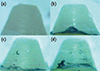
None of the groups were leakage-free at restoration-adhesive-tooth interface. The Kruskal-Wallis test showed significant differences in dye penetration between the groups (p = 0.001 for occlusal margin microleakage and p = 0.009 for cervical margin microleakage, Figures 2 and 3).
Microleakage in the 2D technique
For the 2D technique, the Mann-Whitney test showed a statistically significant difference between the two bonding agents (SB vs. CSEB) in occlusal margin microleakage (p = 0.021). However, the differences were not significant for cervical margin microleakage (p = 0.469, Table 1). The difference in the microleakage before and after thermocycling was not significant (p = 0.510 for occlusal margin microleakage and p = 0.469 for cervical margin microleakage, Table 2). The mean scores of microleakage at the cervical margins were greater than those at the occlusal margins (mean = 0.75 for the cervical margins vs. mean = 0.40 for the occlusal margins, p = 0.001).
Microleakage in the 3D technique
For the 3D technique, the Mann-Whitney test showed a statistically significant difference between the two bonding agents (SB vs. CSEB) in cervical margin microleakage (p = 0.001). However, the difference was not significant for occlusal margin microleakage (p = 0.183, Table 1). The difference between the groups with immediate and post-aging microleakage was not significant (p = 0.074 for occlusal margin microleakage and p = 0.128 for cervical margin microleakage, Table 2). The difference between the microleakage scores at cervical and occlusal margins was not significant (p = 0.747).
Microleakage in the Single Bond groups
In the cavities restored using SB, the Mann-Whitney test showed that the difference between the 2D and 3D techniques of measuring microleakage was significant for occlusal margin microleakage (p = 0.002). However, the difference was not significant for cervical margin microleakage (p = 0.330, Table 1). The difference between immediate and post-aging microleakage was not significant at the occlusal and cervical margins (p = 0.518 and p = 0.653, respectively, Table 3).
Microleakage in the Clearfil SE Bond groups
In the cavities restored with CSEB, the Mann-Whitney test showed that the difference between the 2D and 3D microleakage evaluation techniques was significant at both the occlusal and cervical margins (p = 0.017 and p = 0.002, respectively, Table 1). The difference between immediate and delayed microleakage was not significant at the occlusal and cervical margins (p = 0.056 and p = 0.075, respectively, Table 3).
Immediate microleakage
In the cavities that underwent immediate measurement, the Mann-Whitney test showed that the difference between the 2D and 3D microleakage evaluation techniques was significant at the occlusal margins (p = 0.003). However, the difference was not significant at the cervical margins (p = 0.534, Table 2). The differences in microleakage between SB and CSEB were not significant at the occlusal and cervical margins (p = 0.256 and p = 0.534, respectively, Table 3).
Post-aging microleakage
In the cavities that underwent delayed measurement, the Mann-Whitney test showed that the difference between the 2D and 3D microleakage evaluation techniques was significant at the occlusal margins (p = 0.001), but the difference was not significant at the cervical margins (p = 0.113, Table 2). The differences in microleakage between the SB and CSEB adhesive agents was significant at the occlusal and cervical margins (p = 0.020 and p = 0.040, respectively, Table 3).
Discussion
The first null hypothesis of this study that no significant difference would be found between the marginal microleakage of the two bonding systems was partially rejected. The present study compared a self-etch adhesive with an etch-and-rinse adhesive in relation to marginal microleakage. Based on our results, the frequency of occlusal microleakage was higher with the application of CSEB compared to SB when the 2D technique was used, consistent with the results of previous studies, in which these mild and ultra-mild self-etch adhesive systems exhibited poor adhesive capacity to enamel.101127 However, with the use of the 3D technique, cervical microleakage was found to be more severe when CSEB was applied than when SB was applied, contrary to previous reports, in which the mild-etching CSEB achieved early dentin bond strength values via a very effective three-step etch-and-rinse adhesive system.28 The 2D technique revealed more frequent and severe microleakage at the cervical wall than at the coronal wall, consistent with a previous report.7
The second null hypothesis of this study that thermocycling does not affect the marginal integrity of the tooth-restoration interface was partially rejected. In this study, the effect of thermocycling on both 2D and 3D techniques was compared by subjecting half of the specimens to a 10,000 round thermocycling procedure at 5/55℃, which represented 'aging' of the restoration, similarly to what occurs under clinical conditions. This was expected to exhibit an influence on the microleakage evaluation4 because it has been reported that the majority of bonding systems have proper primary bond strengths and marginal seal, but that their strength decreases due to a variety of causes. The results of 2D and 3D assessments in this study did not show significant differences between the CSEB and SB groups or between immediate and delayed microleakage. De Munck et al. reported that thermocycling and water storage had a minimal effect on microleakage compared with their effect on bond strength tests.29 The effect of thermocycling on microleakage to simulate clinical aging has been a source of controversy.3031 Specimens not undergoing thermocycling exhibited no significant differences in microleakage between SB and CSEB at both the occlusal and cervical margins. However, after application of thermocycling, the mean microleakage values with CSEB were higher than those with SB at both the occlusal and cervical margins, contrary to a previous report describing the cervical margin. Koshiro et al. reported that the dentin-bonding agent interface, using a mild self-etch adhesive, exhibited more stability over time than the dentin-bonding agent interface of a total-etch adhesive.3233
The third null hypothesis of this study that no significant difference would be observed between the two different techniques of measuring microleakage was partially rejected. A range of techniques can be used to detect marginal microleakage, including air pressure, scanning electron microscopy, chemical or radioactive tracers and dyes, bacteria, and fluid permeability.28 The conventional dye penetration technique is extensively used to evaluate the marginal seal of the restoration and is considered a standard technique by the International Organization for Standardization.7
In the present study, two different dye penetration techniques were used to evaluate the marginal microleakage of class V restorations. In the 2D technique, two sections through the restoration were used. The use of a single section through the center of the restoration underestimates in vitro microleakage. In a 3D technique, introduced as an alternative method for evaluation of microleakage, the entire restoration is removed to show the entire extent of dye penetration.11
The present study was undertaken to compare the efficacy of 3D and conventional 2D techniques in revealing leakage depth along the cervical and coronal walls around restorations. An important advantage of the 3D method is its non-destructive nature; no cutting is carried out, which allows the shape of the specimens to be preserved after the evaluations. Its other advantage is its ability to reveal microleakage around the entirety of the restoration.7 However, irrespective of the accuracy of the 2D technique, the results of these techniques might not be objective because they do not exactly reflect the microleakage pattern.5 For a more accurate evaluation of microleakage, scanning and transmission electron microscopy should be used in future studies.
In the present study, specimens using different types of adhesives as well as with and without aging processes showed more extensive microleakage using the 3D technique at coronal walls of the restorations than was identified using the 2D technique, indicating that the 3D technique might be more reliable in detecting marginal microleakage in enamel.
Previous studies have demonstrated less microleakage at the occlusal margins than at the cervical margins. The 3D technique shows the entire restoration and all microleakage patterns, meaning that this technique can reveal occlusal microleakage better and more accurately than the 2D technique. Microleakage seems to be less likely to occur at the center of the occlusal walls of restorations, which is where sections are made in the conventional technique. The 3D technique has the advantage of evaluating the entire enamel wall. It seems that more microleakage occurs at the restoration margins, especially at the junction between the occlusal and lateral walls (Figure 2) due to the higher chance of adhesive pulling or bubbling, and these areas are readily visible in the 3D technique.
The results of dye penetration studies might be affected by the type of the dye used. In addition, a concern about dye penetration technique is that these studies can overestimate the amount of microleakage that will occur in the clinic because of the smaller size of the tracer molecules.24 In the present study, Rotring Ink was used for both methods as a contrast liquid because of its acid resistance properties.5 However, in studies involving the 2D technique, no comparisons have been made between this dye and others, such as fuchsin and silver nitrate. Therefore, such comparisons are recommended in the future.
Some recent studies have used multiple parallel sections for microleakage assessment employing 2D methods, and doing so appears to provide better assessments. However, the sectioning processes in such procedures are destructive, which might influence the results. In contrast, in the 3D method, the dye substance should not be soluble in acidic solutions. In this method, the specimens should be immersed in a strong acidic solution, such as 5% nitric acid, for at least 72 hours in order to become soft enough for the removal of restorations from the cavities.5 This process should be carried out with great care to assess restoration margins meticulously. In addition, a consistent contrast liquid must be used. It seems that this method is more reliable than other methods. However, future studies comparing this method with other conventional techniques might further characterize the advantages and disadvantages of this technique.


 XML Download