

 PDF
PDF ePub
ePub Citation
 Print
Print


Introduction
Since the first methacrylate-based composites were developed, the volumetric contraction has been considered the major limitation of these materials.1,2,3 Silorane-based composites, which are obtained from the reaction of oxirane and siloxane molecules, present low polymerization shrinkage and greater hydrophobicity, making them practically insoluble in the oral environment, while methacrylate-based composites present higher rate of polymerization contraction stress, promoting restoration debonding on cavity walls.4,5,6,7,8
When the tooth is restored with the silorane-based composite, hybridization of dentin surface occurs with the use of a two-step self-etching adhesive system developed exclusively for this purpose.9 However, several studies have demonstrated that self-etching adhesive systems have lower bond strength than the conventional adhesives because of the low degree of adhesive infiltration into etched dentin, as the smear layer is not removed, but incorporated into the hybrid layer.10,11,12,13,14,15,16,17,18,19 Although the primer of the adhesive system of the silorane-based composite promotes intense dentin decalcification and exposes its collagen fibrils, the majority of the dentinal tubules remain obliterated by smear plugs, leading to the formation of a 2.0 µm-thick hybrid layer and few resin tags.12 Nevertheless, to increase bond strength, the manufacturer of silorane-based composite specific adhesive recommends pre-etching of dentin surface with phosphoric acid before application of the primer.
Therefore, the aim of this study was to evaluate the effect of pre-etching on bond strength of silorane-based composite specific adhesive system to dentin. The null hypothesis tested was that there would be no difference between bond strength for silorane adhesive system, irrespective of the dentin pre-etching.
Materials and Methods
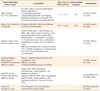
The materials used in this study were presented in Table 1.
Samples selection
Thirty human molars from the tooth bank of FORP/USP were selected for this study, with prior approval from the Research Ethics Committee of the University of São Paulo (Process No. 2011.1.468.58.7, CAAE No. 0028.0.138.000-11). Teeth with white spots, signs of demineralization, fractures, abrasions and carious lesions were excluded. After selection, the teeth were cleaned and stored in 0.5% chloramine solution at 5℃ for 48 hours. When the storage period elapsed, the teeth were horizontally embedded with self-polymerizing acrylic resin (Jet Clássico, São Paulo, SP, Brazil) into a silicone matrix in order to obtain acrylic resin blocks. After acrylic resin polymerization, the blocks were coupled to a metallographic cutter (SYJ - 150 Digital Diamond Low Speed Saw 4, MTI Crystal, Richmond, CA, USA), and the occlusal surfaces of the teeth were sectioned in the transverse direction to their long axis with the aid of a double-faced diamond disc No. 11-4243 (Buehler, Lake Bluff, IL, USA) under constant water cooling. Afterwards, the occlusal surfaces were flattened on a manual polisher (Struers, Ballerup, Denmark) using a 600-grit abrasive paper (Norton, São Paulo, SP, Brazil) to obtain a flat dentin surface.
Teeth restoration
The teeth were randomly separated into 5 groups (n = 6) according to the composites/adhesive systems used for restoration as described in Table 2.
For the Pre-etching/Silorane group, pre-etching of the dentin surface was performed with 37% phosphoric acid (Dental Gel, Dentsply, Petrópolis, RJ, Brazil). The acid was applied on the entire dentin surface for 15 seconds. Next, the surface was thoroughly washed with water, and then dried with absorbent paper to remove the excess of water. The specific self-etching primer (Filtek Silorane, 3M ESPE, Sumaré, SP, Brazil) was then applied and rubbed over the dentin surface for 15 seconds. A light jet of air was applied in order to spread the primer and form a uniform film over the dentin surface. Next, the primer was light activated with a LED type appliance (FlashLite 1401, Discus Dental, Culver City, CA, USA, light intensity, 1,100 mW/cm2 and wavelength in the band between 460 and 480 nm) for 10 seconds. The adhesive (Filtek Silorane) was applied, which was spread over the entire dentin surface and light activated for 10 seconds in accordance with the manufacturer's recommendations.
For the Silorane group, application of the adhesive system occurred in the same way as in the Pre-etching/Silorane group, with the exception of pre-etching.
For the Pre-etching/Methacrylate group, etching of the dentin surface was initially performed with 37% phosphoric acid as performed in the Pre-etching/Silorane group. Next, the dentin surface was completely dried with a jet of air for application of the self-etching primer (Clearfil SE Bond, Kuraray Medical Inc., Kurashiki, Japan). The self-etching primer was applied and spread over the entire dentin surface for 20 seconds. A light jet of air was applied to spread the primer uniformly, and the adhesive (Clearfil SE Bond) was applied and light activated with a LED type appliance (Flash Lite 1401) for 10 seconds in accordance with the manufacturer's instructions.
In the Methacrylate group, adhesive system was applied in the same way as in the Pre-etching/Methacrylate group, with the exception of pre-etching.
For the Three-step/Methacrylate group, the dentin surface was etched with 37% phosphoric acid (Dental Gel) the same as in the Pre-etching/Silorane and Pre-etching/Methacrylate groups. Afterwards, primer (Adper Scotchbond Multi-Purpose, 3M ESPE) was applied on the entire dentin surface and a light jet of air was applied for 5 seconds. The adhesive (Adper Scotchbond Multi-Purpose) was applied on the dentin treated with primer, and light activated for 10 seconds with a LED (Flash Lite 1401) type appliance, in accordance with the manufacturer's instructions.
After application of the adhesive systems, the flat dentin surfaces had a bulk of silorane-based composite (Filtek Silorane) built up to form a block for the Pre-etching/Silorane and Silorane groups, while methacrylate-based composite (Filtek Z250, 3M ESPE) was used for the Preetching/Methacrylate, Methacrylate, and Three-step/Methacrylate groups. Four horizontal layers of composite were applied on the dentin surface, and each layer was light activated with the LED (Flash Lite 1401) type appliance for 20 seconds.10
Next, the teeth were stored in a plastic receptacle containing distilled water at 5℃ for 24 hours. On conclusion of this period, the teeth were sectioned perpendicular to the adhesive interface using a double-faced diamond disk No. 11-4243 (Buehler) mounted in a metallographic cutter (SYJ - 150 Digital Diamond Low Speed Saw 4) into stick-shaped test specimens (1.0 × 1.0 mm), according to the 'non-trimming' technique.10 Afterwards, the specimens were fixed in the devices of the universal testing machine (Emic Model 1L - 2000, São José dos Pinhais, PR, Brazil) using cyanoacrylate adhesive (SuperBonder Gel, Henckel Locktite, Rocky Hill, CT, USA), and submitted to the microtensile test at a tension speed of 0.5 mm/min until fracture occurred. The bond strength was calculated using the following formula,
Ten stick-shaped test specimens per tooth were submitted to microtensile test. The average of the specimens from the same tooth was used as a single unit. The normal distribution of data was tested by the Kolmogorov-Smirnov test and the values obtained in the microtensile test were statistically compared (1-way ANOVA, Tukey test, p < 0.05) with the aid of Graphpad Prism 4.0 Software (GraphPad Software, La Jolla, CA, USA).
Results
The results for microtensile bond strength are presented in Table 3. The Pre-etching/Methacrylate group presented the highest bond strength values, with significant difference from the Silorane and Three-step/Methacrylate groups (p < 0.05). However, there was no statistically significant difference from the Pre-etching/Silorane and Methacrylate groups. Pre-etching promoted a significant increase in bond strength of the group restored with silorane-based composite (p < 0.05). Pre-etching did not increase the bond strength of the Methacrylate group. The Three-step/Methacrylate group presented the lowest bond strength (p < 0.05).
Discussion
In the present study, the influence of pre-etching on bond strength of silorane-based composite specific adhesive system to dentin was evaluated. Based on the results obtained, it could be stated that the tested null hypothesis was rejected since pre-etching promoted higher bond strength values for silorane specific adhesive system. According to the manufacturer (3M ESPE), this specific self-etching adhesive system is able to promote bonding between dental tissues of hydrophilic nature, and the silorane-based composite with hydrophobic property. This adhesive system is composed of a methacrylate-based self-etching primer, which has hydrophilic monomers, in addition to water and ethanol that promote ionization, and form the base for substrate/composite bonding.9 Also, according to the manufacturer, the specific adhesive is methacrylate-based, and contains hydrophobic monomers, whose function is to complement bonding between primer and the hydrophobic monomer of the composite. Thus, the bond strength between substrate and silorane-based composite is assured by the chemical reaction of the methacrylate-based and silorane molecules contained in the specific adhesive system.9
Several studies have reported that there is no significant difference in the bond strength values between composite and dentin promoted by self-etching and conventional adhesive systems.16,18 However, Sheets et al. demonstrated that pre-etching of the dentin surface with phosphoric acid increases the bond strength significantly in comparison with self-etching systems.19 For Van Landuyt et al., the fact that self-etching adhesive systems simplify the clinical procedure does not mean that these systems improve the performance of restorations made with composites.20 According to these same authors, self-etching adhesive systems, particularly the one-step type, tend to present lower bond strength than the conventional three-step systems, and they are not always simpler to apply.20 Moreover, interface analysis by TEM demonstrated a great variation among the one-step adhesives, depending on their composition and acidity.20 In this case, at least two different types of hybrid layers could be observed ultramorphologically: a) completely demineralized hybrid layer in which all hydroxyapatite was removed; and b) a partially demineralized hybrid layer with hydroxyapatite crystals.20 Also, light-microscopic evaluation of adhesive solution homogeneity demonstrated that one-step self-etching adhesives were prone to form droplets, which makes their application difficult, and their technique more sensitive.20 Nevertheless, two-step self-etching systems, such as the one used in this study, present advantages in comparison with conventional three-step systems because the critical phases during acid etching and its removal are avoided. Hence, their application becomes not only faster but easier as well.20
Moreover, acid etching does not always play a fundamental role in bond strength increase.20 The infiltration of resin monomers at the interface of the hybrid layer and dentin surface may be reduced due to the reduction in the gradient of diffusion of resin monomers in association with the blocking effect of the copolymer of polyalkenoic acid present in three-step adhesive system, such as the one used in this study.21,22 These findings are in agreement with the results of the present study, since pre-etching did not increase the bond strength for methacrylate-based composite submitted to two-step self-etching adhesive system application. Furthermore, the Three-step/Methacrylate group, which was hybridized with conventional three-step system, presented lower mean bond strength values than the Pre-etching/Methacrylate and Methacrylate groups. According to Van Landuyt et al., these values may be explained by the technique sensitivity which requires more steps, increasing the probability of errors by the operator.20 Short application procedures, such as the self-etching adhesive systems, is interesting for the professional since the number of application steps could determine the technique sensitivity. The more steps, the higher the risk of application failures.20
However, the same could not be said regarding the specific adhesive system used for silorane-based composite since the best results were obtained by means of pre-etching. The lower values of bond strength obtained with the specific adhesive system combined with the silorane-based composite could be explained by the poor chemical interaction between the components of the adhesive and the composite.4 The silorane specific adhesive system is presented in two bottles, with two different application steps.4 However, it is considered a one-step adhesive system, with an acid adhesive.4 Such fact may explain the results, since the specific adhesive system do not strongly bond to the silorane-based composite due to its hydrophilic nature, and hydrophobic of the composite.4,9
Moreover, the specific adhesive system of silorane-based composite requires separate light activation steps for the primer and the adhesive, different from the majority of two-step self-etching systems, in which the application of primer and adhesive is finalized by only one light irradiation.23 The hydrophilic primer of the specific adhesive system is composed of phosphated methacrylate, acid copolymers, methacrylate-based monomers of Bisphenol A-Glycidyl dimethacrylate (Bis-GMA) and Hydroxyethyl-methacrylate (HEMA), a system of solvents that contain water and ethanol to humidify and penetrate into the dentin surface, a photoinitiator system based on Camphorquinone and silane treated silica particles (3M ESPE). Due to the relatively high HEMA content, the previous application of primer would be very vulnerable to the absorption of water if the hydrophobic layer of the silorane adhesive were not applied.16 Therefore, according to Mine et al., in addition to providing a bond compatible with the resin matrix of silorane, the application of individual rounds of light activation on the components of the adhesive system is beneficial, which provides greater stability of the composite-tooth substrate bond and long term durability of the hybrid layer.16
Mine et al. characterized the ultra-structure of the hybrid layer formed between the enamel/dentin and the adhesive system of a silorane-based composite.16 An interesting fact observed was the infiltration of silver nitrate along the adhesive interface of the silorane system, between the primer and adhesive, and within the primer itself, indicating the presence of porosities in the hybrid layer. Silver nitrate is frequently used for the observation of spaces at the bond interface, which may be responsible for the nanoleakage of fluids, compromising the integrity of the hybrid layer.24 The phenomena observed by Mine et al. are probably related to the hydrophilicity of the HEMA and water containing primer, which is more difficult to remove with jets of air, resulting in greater failures at the primer/adhesive interface than at the adhesive/dentin interface.16,25 This water present in the primer of the adhesive system reacts with the phosphorylated methacrylate present in the adhesive systems, generating the hydrogen ions necessary for demineralizing the dentin.12 However, in areas where polymerization of the adhesive system does not occur completely, water molecules may persist within the collagen network or adhesive layer, degrading bonding over the course of time.12
The adhesive system of the silorane-based composite is considered a 'mild' self-etching system when compared with other self-etching systems due to its pH which is around 2.7. Nevertheless, it produces intense intertubular demineralization.12 According to Duarte et al., the silorane-based composite specific adhesive system promotes intense intertubular decalcification exposing the collagen fibrils.12 However, the dentinal tubules still remain blocked by smear plugs. Therefore, few resin tags are observed due to the presence of remaining smear plug within the dentin tubules, which makes adhesive infiltration difficult.
When applied on smear layer-free dentin, the adhesive system forms a hybrid layer with larger resin tags due to the greater penetration of the primer into the dentinal tubules. This is different from when it is applied on thick smear layers, which interrupt the resin tags formation, and affect the efficacy of the adhesive bond, decreasing the bond strength values, as observed in the present study.12 However, according to Lohbauer et al., the length of resin tags does not influence the bond strength of self-etching adhesive systems since the resin monomers present in the adhesive penetrate into the exposed collagen fibrils network, forming an interlock, which micromechanically guarantees satisfactory bond strength.26
Pre-etching with phosphoric acid prior to silorane-based composite specific adhesive system application demonstrated a significant increase in the bond strength values in comparison with the group in which the adhesive system was applied in accordance with the manufacturer's instructions. However, the same cannot be said for methacrylate-based composite. The different characteristics of silorane and conventional dimethacrylate-based composites could explain such conflicting results, since the two composites used self-etching adhesive systems. The silorane specific adhesive system involves the application of an initial hydrophilic solution - the primer - to form the base for bonding with the dental tissues; and a second solution - the adhesive agent - which has a hydrophobic nature in order to complement bonding to the primer and unite with the hydrophobic monomer of the composite.16 However, according to Fernandes et al., such components have some degree of incompatibility.4 The efficiency of dentin etching depends more on the chemical composition of the adhesive than the effect of the phosphoric acid itself.27,28,29 Thereby, the results of the present study suggest a greater chemical interaction between the components of methacrylate-based composite and its adhesive system, irrespective of the pre-etching.4
One of the main characteristics of the silorane-based composite is its low polymerization shrinkage.30 However, the application of its specific adhesive system in strict accordance with the manufacturer's instructions restricts its use due to its lower bond strength values.12 Although low-shrinkage silorane-based composite represents an evolution in restorative dentistry, its specific self-etching adhesive system does not present a similar performance to the conventional ones.
Conclusions
Within the limitations of this in vitro study, it was concluded that pre-etching increased bond strength of silorane-based composite specific adhesive system to dentin. Despite the fact that association between specific self-etching adhesive system of silorane composite and pre-etching promoted higher bond strength, further studies are still necessary before the clinical application of this bond strategy.


 XML Download