Journal List > Restor Dent Endod > v.40(2) > 1090109

Mirzakoucheki, Walter, Khalighinejad, Jahromi, Mirsattari, and Akbarzadeh: Effects of endodontic tri-antibiotic paste on bond strengths of dentin adhesives to coronal dentin

Abstract

Objectives

The aim of this study was to evaluate the effects of tri-antibiotic paste (TAP) on microtensile bond strengths (MTBS) of dental adhesives to dentin.

Materials and Methods

Sixty extracted molars had their occlusal surfaces flattened to expose dentin. They were divided into two groups, i.e., control group with no dentin treatment and experimental group with dentin treatment with TAP. After 10 days, specimens were bonded using self-etch (Filtek P90 adhesive) or etch-and-rinse (Adper Single Bond Plus) adhesives and restored with composite resin. Teeth were sectioned into beams, and the specimens were subjected to MTBS test. Data were analyzed using two-way ANOVA and post hoc Tukey tests.

Results

There was a statistically significant interaction between dentin treatment and adhesive on MTBS to coronal dentin (p = 0.003). Despite a trend towards worse MTBS being noticed in the experimental groups, TAP application showed no significant effect on MTBS (p = 0.064).

Conclusions

The etch-and-rinse adhesive Adper Single Bond Plus presented higher mean bond strengths than the self-etch adhesive Filtek P90, irrespective of the group. The superior bond performance for Adper Single Bond when compared to Filtek P90 adhesive was confirmed by a fewer number of adhesive failures. The influence of TAP in bond strength is insignificant.

Introduction

Medicaments other than calcium hydroxide and chlorhexidine have been investigated in the treatment of endodontic infections.1 Antibiotic treatment, which may be a combination of metronidazole, ciprofloxacin, and minocycline, has been recommended in more complex situations in order to properly remove different pathogens.2,3 That treatment, however, may affect the adhesion of the restorative material used for sealing of the endodontic access.4,5
There has been an increasing tendency to restore endodontically treated teeth with composite resins, either as foundation for indirect restoration or as final restoration.6 To achieve a successful restoration, adequate adhesion to the tooth structure is critical. In that regard, dentin is a hydrated biologic complex that can be modified in the presence of various chemical agents.7 The effects of endodontic materials such as antibiotic pastes and irrigation solutions on adhesion have been described.4,5
Complex root canal infections, which may not be fully resolved with a single antibiotic, often are treated with the tri-antibiotic paste (TAP).2 Sato et al. first tested the regimen with further studies confirming the antimicrobial efficacy of the ciprofloxacin-metronidazoleminocycline paste against root canal pathogens.2,8 TAP has a low pH of 2.8, and its ability to chelate calcium has been shown to be similar to that of ethylenediamine tetraacetic acid (EDTA) in the studies.5,9,10
Adhesive procedures, often used in the process of restoring endodontically treated teeth, are divided into three steps of etch, prime, and bond. While etch-and-rinse adhesive systems have the three steps separately applied and require application of an etchant prior to priming, self-etch adhesives combine etching with priming.11,12 Etch-and-rinse adhesives often do not fully penetrate the demineralized dentin leaving denuded collagen fibrils vulnerable to degradation by endogenous matrix metalloproteinases (MMPs).8,13 Altering the dentin surface prior to adhesive procedures may limit their penetration into dentin and/or further increase the amount of exposed collagen fibrils, compromising adhesion. Likewise, bonding with self-etch adhesives may be compromised by altering the dentin surface and consequently limiting chemical adhesion.
The present study evaluated the effects of TAP on microtensile bond strengths (MTBS) of two types of dental adhesives to dentin. We hypothesized that TAP negatively affects bond strengths of etch-and-rinse and self-etch adhesives to coronal dentin.

Materials and Methods

Sample preparation

Subsequent to approval by the University Research Ethics Board (protocol 6452/2011), 60 extracted human third molars free of caries or any visible defects were collected and stored in 0.2% thymol solution at room temperature for up to 60 days.14 Prior to the experiment, teeth were transferred to distilled water for 24 hours for removal of thymol residues. Each tooth was decoronated below the cementoenamel junction perpendicular to the longitudinal axis using a slow-speed, water-cooled diamond disc (D&Z, Berlin, Germany).15 The occlusal enamel of each tooth was removed perpendicular to the long axis of the teeth with a diamond saw (Isomet, Buehler Ltd., Lake Bluff, IL, USA) under water irrigation to expose superficial coronal dentin. Prepared dentin surfaces were finished with 600-grit silicon carbide paper under water irrigation to create a uniform smear layer.16

Tri-antibiotic paste

Metronidazole (250 mg, Pars Darou Co., Tehran, Iran), minocycline chlorhydrate (100 mg, Biopharma Co., Roma, Italy), and ciprofloxacin chlorhydrate (250 mg, Arya Darou, Tehran, Iran) were vortexed with purified glycerin (vehicle) and methylcellulose (thickening agent) for 5 minutes to obtain a paste-like consistency. The mixture was adjusted to obtain a concentration of 20 mg/mL of the paste.
Specimens were randomly divided into two groups (n = 30). In the control group, specimens were covered with cotton pellets moistened with saline. In the experimental group, dentin surfaces were treated using TAP (direct application onto the dentin surfaces). Specimens were then covered with cellophane to protect against contaminants and kept in an incubator at 37℃ at relative humidity. After 10 days, dentin surfaces in both groups were rinsed using distilled water for 5 seconds in preparation for bonding procedures.

Bonding procedures

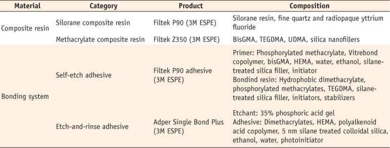
Occlusal surfaces of all teeth were thoroughly rinsed with water and dried with oil- and moisture-free compressed air. Experimental and control groups were further divided into four subgroups (n = 15) according to the adhesive system to be used (Figure 1). Information on the composite resins and adhesive systems used are summarized in Table 1.
  • Groups A and C: Filtek P90 (3M ESPE, St Paul, MN, USA) composite resin was used with its dedicated adhesive system (Filtek P90 adhesive). The self-etch primer was applied with gentle agitation using a microbrush for 20 seconds, gently air-thinned for 2 seconds, and polymerized for 10 seconds using a halogen light curing unit (Coltolux 2.5, Coltene AG, Feldwiesenstrasse Altstδtten, Switzerland) with 600 mW/cm2 output. The bonding resin was then applied and dispersed with a light stream of air, and light-cured for 20 seconds. P90 composite resin was built up in increments to a height of 5 mm to ensure sufficient bulk for the bond strength test. Each increment was light-cured for 40 seconds.

  • Groups B and D: Filtek Z350 (3M ESPE) composite resin and Adper Single Bond Plus adhesive system (3M ESPE) were used. Dentin surfaces were acid-etched using 35% phosphoric acid gel (3M ESPE) for 15 seconds and rinsed for 30 seconds. Excess moisture was removed with absorbent paper (Aria Paper & Film Industries Co., Tehran, Iran) and the surface kept moist. A single coat of adhesive was immediately applied onto the prepared dentin surface with agitation for 30 seconds and then gently air-dried for 3 seconds prior to light curing for 20 seconds. Filtek Z350 composite resin build-ups were placed as described in groups A and C.

Microtensile bond strength test

Specimens were stored in distilled water at 37℃ for 24 hours and then sectioned using a diamond saw under water lubrication (Isomet), perpendicular to the bonded interface. Two beams approximately 0.9 mm2 in diameter from the central area of each specimen were collected (n = 30 beams/group).17 Beams were mounted on a universal testing machine (Instron, Norwood, MA, USA) using cyanoacrylate adhesive (Loctite, Loctite Ltd., Dublin, Ireland). The samples were stressed to failure at a crosshead speed of 0.5 mm/min (Figure 2). Results were expressed in MPa by dividing the resultant force in Newton by the cross-sectional area of the specimen. The bond strength data were statistically analyzed by two-way ANOVA and post hoc Tukey test using SPSS 15 (α = 0.05).

Failure mode

Failure mode was determined with a stereomicroscope (Olympus, Tokyo, Japan) and classified as adhesive, adhesive failure between adhesive and dentin; cohesive, failure in composite resin or dentin; mixed, combination of adhesive and cohesive failures.18

Results

The mean MTBS values for all groups are presented in Table 2. Two-way ANOVA was conducted to examine the interaction between the effect of TAP application and the type of adhesive on MTBS of adhesives to coronal dentin. There was a statistically significant interaction between the effect of dentin treatment and adhesive on MTBS to coronal dentin (p = 0.003). It was shown that different types of adhesives significantly affected the MTBS (p < 0.001). However, TAP application showed no significant effect on the MTBS (p = 0.064). Control specimens in the Adper Single Bond Plus and TAP-treated specimens in the Filtek P90 groups showed the highest and the lowest bond strengths, respectively. Adper Single Bond Plus showed significantly higher mean MTBS than Filtek P90 adhesive in both experimental and control groups regardless of the dentin treatment (p < 0.05).
Failure modes are shown in Table 3. For Adper Single Bond Plus, most of the failures in both control and experimental groups were cohesive. There was no significant differences in the mode of failure between experimental and control groups (p > 0.05). For Filtek P90 adhesive, there was a significant difference between experimental and control groups (p = 0.038). The control and the experimental groups exhibited a majority of mixed and adhesive failures, respectively.

Discussion

The coronal seal plays a significant role on the final success of root-filled teeth. Lack of proper adhesion between restorative materials and access cavity in endodontically treated teeth may result in microleakage and compromise prognosis.19 Inevitable contact of endodontic materials during chemomechanical preparation of root canals may modify the dentin available for coronal restorations and jeopardize bonding procedures.4,20 Following increased use of TAP due to its sterilization efficacy in endodontic treatments, we assessed the effect of TAP application on the bond strength of two types of etch-and-rinse and self-adhesive dental adhesives to coronal dentin.2 Results showed no significant differences between control and experimental groups and, therefore, our null hypothesis was rejected.
TAP contains ciprofloxacin, metronidazole, and minocycline. Ciprofloxacin is a synthetic floroquinolone with rapid bactericidal action against gram-negative bacteria but with limited activity against gram-positive bacteria.21 For that reason, ciprofloxacin is often combined with metronidazole for treatment of mixed infections like root canal infections. Metronidazole is a nitroimidazole compound that exhibits broad spectrum of activity against protozoa and anaerobic bacteria.21 Minocycline exhibits broad spectrum of activity against gram positive and gram negative microorganisms. The optimal antimicrobial effect of TAP seems to occur between seven and 10 days, which was the application time chosen in our study.2
The demineralization effect of TAP has been confirmed by significant reduction in phosphate/amide ratio in TAP-treated dentin.5 The low pH (2.8) of this medicament and the ability of minocycline to chelate calcium and demineralize dental hard tissues may explain its significant demineralization effect.22 In the present study, although results are not significantly different, the MTBS of both etch-and-rinse and self-etch adhesives tended to be lower in the TAP-treated groups compared to the control groups. This tendency was more obvious in the self-etch group (22% reduction in mean MTBS) compared to the etch-and-rinse group (5.4% reduction in mean MTBS). Excessive demineralization of dentin prior to self-etch adhesive application may prevent optimal adhesive impregnation affecting MTBS as shown by Van Landuyt et al.23
In the present study, the etch-and-rinse adhesive system Adper Single Bond Plus showed significantly higher MTBS than that of the self-etch adhesive Filtek P90, irrespective of dentin treatment. Filtek P90 adhesive is a self-etch adhesive (pH = 2), which gently demineralizes the tooth structure resulting in a very thin micro-retentive pattern.24 This limits the ability of the bonding resin to penetrate the primed surface and might explain the inferior bond strengths of Filtek P90 adhesive compared to the etch-and-rinse adhesive in this study. Potential increased demineralization caused by TAP was not sufficient to increase the MTBS of Filtek P90 adhesive.
Mean MTBS in the present study ranged from 21.1 to 37.5 MPa. It is often not possible to differentiate between the strength of the adhesive and that of the composite resin or dentin when bond strengths exceed values of 18 - 20 MPa because of the increased likelihood of cohesive failure within the dentin.25 For the self-etch adhesive Filtek P90, the majority of the failures in the experimental group was adhesive with significant differences being noticed between experimental and control groups. Higher number of adhesive failures occurred in the TAP-treated group. The potential lack of proper dentin infiltration may explain the finding.26 Regarding Adper Single Bond Plus, a higher number of failures were cohesive suggesting that the bond strength between composite resin and dentin was greater than the cohesive strength of the composite resin or dentin itself.
Limitations of our study include the use of only one concentration and application time of TAP, the use of only ethanol/water-based adhesive systems, and the lack of aging of specimens. Furthermore the different characteristics of adhesive resins in the study (methacrylate-based versus silorane-based) could have affected the outcomes of the study. Also, the next session following TAP application may be the cleaning of the root canals with NaOCl and EDTA first. Considering the fact that no studies have investigated the possible interaction between NaOCl and EDTA with TAP, we used normal saline after 10 days to rinse the TAP. Regarding the substrate for bonding, we opted for using coronal dentin considering the similar results between radicular and coronal dentin in bonding studies.2,27,28 However, due to the fact that TAP might be directly applied in the canal below the orifice level, it seems rational to also consider radicular dentin in examining the possible effect of TAP on bond strength of adhesive resins when it comes to further studies.

Conclusions

The etch-and-rinse adhesive Adper Single Bond Plus presented significantly higher mean bond strengths than the self-etch adhesive Filtek P90, irrespective of the group. The superior bond performance of Adper Single Bond was confirmed by a fewer number of adhesive failures when compared to Filtek P90 adhesive. The influence of TAP in bond strength is insignificant.

Notes

No potential conflict of interest relevant to this article was reported.

References

1. Mohammadi Z. Antibiotics as intracanal medicaments: a review. J Calif Dent Assoc. 2009; 37:98–108. PMID: 19489526.
2. Ordinola-Zapata R, Bramante CM, Minotti PG, Cavenago BC, Garcia RB, Bernardineli N, Jaramillo DE, Hungaro Duarte MA. Antimicrobial activity of triantibiotic paste, 2% chlorhexidine gel, and calcium hydroxide on an intraoral-infected dentin biofilm model. J Endod. 2013; 39:115–118. PMID: 23228269.
crossref
3. Windley W 3rd, Teixeira F, Levin L, Sigurdsson A, Trope M. Disinfection of immature teeth with a triple antibiotic paste. J Endod. 2005; 31:439–443. PMID: 15917683.
4. Yurdagüven H, Tanalp J, Toydemir B, Mohseni K, Soyman M, Bayirli G. The effect of endodontic irrigants on the microtensile bond strength of dentin adhesives. J Endod. 2009; 35:1259–1263. PMID: 19720227.
crossref
5. Yassen GH, Chu TM, Eckert G, Platt JA. Effect of medicaments used in endodontic regeneration technique on the chemical structure of human immature radicular dentin: an in vitro study. J Endod. 2013; 39:269–273. PMID: 23321244.
6. Schwartz RS, Fransman R. Adhesive dentistry and endodontics: materials, clinical strategies and procedures for restoration of access cavities: a review. J Endod. 2005; 31:151–165. PMID: 15735460.
crossref
7. Van Meerbeek B, De Munck J, Yoshida Y, Inoue S, Vargas M, Vijay P, Van Landuyt K, Lambrechts P, Vanherle G. Buonocore memorial lecture. Adhesion to enamel and dentin: current status and future challenges. Oper Dent. 2003; 28:215–235. PMID: 12760693.
8. Sato I, Ando-Kurihara N, Kota K, Iwaku M, Hoshino E. Sterilization of infected root-canal dentine by topical application of a mixture of ciprofloxacin, metronidazole and minocycline in situ. Int Endod J. 1996; 29:118–124. PMID: 9206435.
9. Van Meerbeek B, Yoshihara K, Yoshida Y, Mine A, De Munck J, Van Landuyt KL. State of the art of self-etch adhesives. Dent Mater. 2011; 27:17–28. PMID: 21109301.
crossref
10. Tjäderhne L, Larjava H, Sorsa T, Uitto VJ, Larmas M, Salo T. The activation and function of host matrix metalloproteinases in dentin matrix breakdown in caries lesions. J Dent Res. 1998; 77:1622–1629. PMID: 9719036.
crossref
11. Manso AP, Marquezini L Jr, Silva SM, Pashley DH, Tay FR, Carvalho RM. Stability of wet versus dry bonding with different solvent-based adhesives. Dent Mater. 2008; 24:476–482. PMID: 17675145.
crossref
12. Maruyama H, Aoki A, Sasaki KM, Takasaki AA, Iwasaki K, Ichinose S, Oda S, Ishikawa I, Izumi Y. The effect of chemical and/or mechanical conditioning on the Er:YAG laser-treated root cementum: analysis of surface morphology and periodontal ligament fibroblast attachment. Lasers Surg Med. 2008; 40:211–222. PMID: 18366073.
crossref
13. Wattanawongpitak N, Nakajima M, Ikeda M, Foxton RM, Tagami J. Microtensile bond strength of etch-and-rinse and self-etching adhesives to intrapulpal dentin after endodontic irrigation and setting of root canal sealer. J Adhes Dent. 2009; 11:57–64. PMID: 19343928.
14. Santana FR, Pereira JC, Pereira CA, Fernandes Neto AJ, Soares CJ. Influence of method and period of storage on the microtensile bond strength of indirect composite resin restorations to dentine. Braz Oral Res. 2008; 22:352–357. PMID: 19148392.
crossref
15. Ozer F, Unlü N, Sengun A. Influence of dentinal regions on bond strengths of different adhesive systems. J Oral Rehabil. 2003; 30:659–663. PMID: 12787465.
16. Pashley DH, Ciucchi B, Sano H, Carvalho RM, Russell CM. Bond strength versus dentine structure: a modelling approach. Arch Oral Biol. 1995; 40:1109–1118. PMID: 8850649.
crossref
17. Hashimoto M, Ohno H, Kaga M, Sano H, Tay FR, Oguchi H, Araki Y, Kubota M. Over-etching effects on microtensile bond strength and failure patterns for two dentin bonding systems. J Dent. 2002; 30:99–105. PMID: 12381409.
crossref
18. Sano H, Shono T, Sonoda H, Takatsu T, Ciucchi B, Carvalho R, Pashley DH. Relationship between surface area for adhesion and tensile bond strength-evaluation of a micro-tensile bond test. Dent Mater. 1994; 10:236–240. PMID: 7664990.
19. Heling I, Gorfil C, Slutzky H, Kopolovic K, Zalkind M, Slutzky-Goldberg I. Endodontic failure caused by inadequate restorative procedures: review and treatment recommendations. J Prosthet Dent. 2002; 87:674–678. PMID: 12131891.
crossref
20. Cecchin D, Farina AP, Galafassi D, Barbizam JV, Corona SA, Carlini-Júnior B. Influence of sodium hypochlorite and edta on the microtensile bond strength of a self-etching adhesive system. J Appl Oral Sci. 2010; 18:385–389. PMID: 20835574.
crossref
21. Tripathi KD. Essentials of Medical Pharmacology. 5th ed. New Delhi: Jaypee Brothers Medical;1985. p. 710.
22. Minabe M, Takeuchi K, Kumada H, Umemoto T. The effect of root conditioning with minocycline HCl in removing endotoxin from the roots of periodontally-involved teeth. J Periodontol. 1994; 65:387–392. PMID: 8046553.
crossref
23. Van Landuyt KL, Kanumilli P, De Munck J, Peumans M, Lambrechts P, Van Meerbeek B. Bond strength of a mild self-etch adhesive with and without prior acid-etching. J Dent. 2006; 34:77–85. PMID: 15979226.
crossref
24. Van Landuyt KL, Peumans M, De Munck J, Lambrechts P, Van Meerbeek B. Extension of a one-step self-etch adhesive into a multi-step adhesive. Dent Mater. 2006; 22:533–544. PMID: 16300826.
crossref
25. Schreiner RF, Chappell RP, Glaros AG, Eick JD. Microtensile testing of dentin adhesives. Dent Mater. 1998; 14:194–201. PMID: 10196796.
crossref
26. Nakabayashi N, Watanabe A, Arao T. A tensile test to facilitate identification of defects in dentine bonded specimens. J Dent. 1998; 26:379–385. PMID: 9611944.
crossref
27. Walter R, Miguez PA, Pereira PN. Microtensile bond strength of luting materials to coronal and root dentin. J Esthet Restor Dent. 2005; 17:165–171. PMID: 15996387.
crossref
28. Abo-Hamar SE. Effect of endodontic irrigation and dressing procedures on the shear bond strength of composite to coronal dentin. J Adv Res. 2013; 4:61–67. PMID: 25685402.
crossref
Figure 1

Flow chart of the experimental groups and procedures.

rde-40-136-g001
Figure 2

Schematic diagram of the specimen preparation for the microtensile bond strength test. (a) Tooth decoronation below the cementoenamel junction; (b) Preparation of a flat dentin surface for bonding; (c) Dentin treatment; (d) Adhesive application; (e) Incremental resin composite buildup; (f) Cutting slab from the center of a bonded specimen after 24 h water storage; (g) A single slab containing the resin-dentin interface; (H) Beams for testing from the center slab; (i) attachment of a beam for bond strength evaluation. TAP, tri-antibiotic paste.

rde-40-136-g002
Table 1

Materials used in the study

rde-40-136-i001
Material Category Product Composition
Composite resin Silorane composite resin Filtek P90 (3M ESPE) Silorane resin, fine quartz and radiopaque yttrium fluoride
Methacrylate composite resin Filtek Z350 (3M ESPE) BisGMA, TEGDMA, UDMA, silica nanofillers
Bonding system Self-etch adhesive Filtek P90 adhesive (3M ESPE) Primer: Phosphorylated methacrylate, Vitrebond copolymer, bisGMA, HEMA, water, ethanol, silane-treated silica filler, initiator
Bondind resin: Hydrophobic dimethacrylate, phosphorylated methacrylates, TEGDMA, silane-treated silica filler, initiators, stabilizers
Etch-and-rinse adhesive Adper Single Bond Plus (3M ESPE) Etchant: 35% phosphoric acid gel
Adhesive: Dimethacrylates, HEMA, polyalkenoid acid copolymer, 5 nm silane treated colloidal silica, ethanol, water, photoinitiator

BisGMA, Bisphenol A-glycidyl methacrylate; TEGDMA, Triethyleneglycol-dimethacrylate; UDMA, Urethane dimethacrylate; HEMA, Hydroxylethyl methacrylate.

Table 2

Mean microtensile bond strength values and standard deviations expressed in MPa of different adhesive systems for experimental and control groups (n = 30)

rde-40-136-i002
Adhesive system Control group TAP-treated group
Adper Single Bond Plus + Filtek Z350 37.5 ± 4.2a 35.8 ± 4.5a
Filtek P90 system 27.0 ± 5.6b 21.1 ± 4.6c

TAP, tri-antibiotic paste.

Table 3

Mode of failure of adhesive systems bonded to saline- (control) and TAP-treated dentin (n = 30)

rde-40-136-i003
Failure mode Adper Single Bond Plus Filtek P90 adhesive
Control group TAP-treated group Control group TAP-treated group
Adhesive 3 3 6 14
Cohesive 17 16 12 9
Mixed 10 11 12 7

Adhesive, adhesive (interface) failure between adhesive and dentin; Cohesive, cohesive failure in composite resin or dentin; Mixed, mixed failure of adhesive and cohesive failures.

TAP, tri-antibiotic paste.

TOOLS
Similar articles