

 PDF
PDF ePub
ePub Citation
 Print
Print


Introduction
Nickel-Titanium (NiTi) rotary instruments create well-tapered and clean root canals with a low tendency of aberration, but are generally perceived as having a high risk of fracture during use.1,2,3 Numerous instruments have been designed and manufactured to reduce and eliminate the risk of instrument breakage. Additionally, a more convenient and effective alternative by clinicians is to make a glide path before inserting conventional NiTi rotary instruments into root canals, as this may reduce the risk of torsional failure and root canal transportation.4,5,6
Preparing glide paths using stainless steel K-files (size 10 or 15) is recommended as a practical way for shaping root canals.4,5,7,8 A K-file can be used in a watch-winding movement to remove any obstructing dentin in very narrow canals. This may later be followed by a vertical in-and-out movement, which is started with an amplitude of 1 mm and then gradually increased till sufficient dentin is worn away to allow the file to be advanced to the apex.8 Usually repeated push-and-pull movements are recommended until the next larger file in the sequence moves easily to the desired working length.9 A glide path of sufficient size ensures less torsional stress, thereby increasing the lifespan of the rotary instrument used for canal preparation. It also provides the clinician with greater confidence to handle more complex and challenging endodontic cases.6,10
Instrument systems, such as G-file (Micro-Mega, Besançon, France), ScoutRace (FKG Dentaire SA, La Chaux-de-Fonds, Switzerland) and PathFile (Dentsply Maillefer, Ballaigues, Switzerland), have been introduced for pre-flaring and glide path preparation procedures before the insertion of conventional rotary instruments for canal shaping. These systems are known to have a higher flexibility and cyclic fatigue resistance, and thus allow clinicians to make glide paths easily in a relatively short span of time.11 However, there are no guidelines on the optimal number of repetitive insertions at working length using these files to prepare a glide path efficiently with minimal risk of root canal transportation. This study evaluated the changes in canal diameters after repeated insertions of G-file at the working length for glide path preparation.
Materials and Methods
The G-file series (G1, size 12/0.03 taper; G2, size 17/0.03 taper) was used to prepare a glide path in J-shaped simulated resin canals (Dentsply Maillefer) with 35° curvature. The working length of the simulated canal, as defined by the tip of a size 10 K-file (Dentsply Maillefer) becoming visible at the apical foramen, was measured under an operating microscope (Leica M320 F12, Leica Microsystems, Wetzlar, Germany) at ×10 magnifications.
The rubber stops of G1 and G2 files were fixed with cyanoacrylate to ensure a stable working length. The G-files were operated with an endodontic motor (X-smart, Dentsply Maillefer), which was used at 400 rpm with torque set at 1.2 Ncm, as recommended by the manufacturer. First, G1 files were used to reach the working length twice in all canals, and then G2 files were inserted 1, 4, 7, and 10 times respectively using an up-and-down movement of 4 mm amplitude 'at the working length'. Ten simulated canals were prepared for each of the 4 experimental groups. The repetitive up-and-down movements at the working length were based on the current recommendations for glide path formation using stainless steel files.8,9 G1 files were discarded after use for 4 canals, 1 canal from each group; G2 files were used only for 1 canal before being discarded. Totally 10 G1 files were used for 40 samples and 40 G2 files were used for 40 samples.
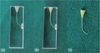
The simulated canals were filled with saline during instrumentation, and the debris was flushed out with copious irrigation (20 mL of saline) using a needle-syringe system and sonic vibration (EndoActivator, Dentsply Tulsa Dental Specialties, Tulsa, OK, USA). Then, the canals were subsequently lubricated with a separating gel medium (THE-Sep, MDCLUS, Cheongju, Korea) and injected with impression material (Imprint III Light Body, 3M Deutschland GmbH, Neuss, Germany) to make canal replicas. The replicas were removed after an hour of complete setting (Figure 1). The replicas of 5 new simulated canals were used as controls for measuring the diameters of un-instrumented canals, as well as to verify the accuracy of the replicas made of the impression material.
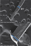
Canal replicas were evaluated under a scanning electron microscope (SEM, S-4800 II, Hitachi High Technologies, Pleasanton, CA, USA) 24 hours after the impression material had set at various magnifications (from ×50 to ×200). The apical preparation size (diameter) was measured at working length (D0) and at 1 mm level (D1) (Figure 2). Data was analysed by using one-way analysis of variance (ANOVA) and Duncan's post-hoc comparison to check for any differences between the groups at a significance level of 95%.
Results
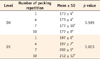
The diameter at D0 level did not showed significant difference by the different repetitions of pecking insertions at working length. However, at D1, the group in which the G2 file was inserted 10 times showed a significantly bigger canal diameter (p < 0.05, Table 1).
Discussion
The main goals of root canal preparation are to clean and shape the root canal system with minimal procedural errors while maintaining the original canal configuration.1,12 The NiTi rotary instruments have allowed all these goals to be achieved efficiently.1,12,13 However, NiTi rotary instruments are prone to torsional separation when exposed to high torsional stress occurred due to the contacts with canal wall, especially in the early stages of root canal preparation.2,14,15
The establishment of a glide path, a smooth tunnel from the orifice of the canal to the terminus of the root, is highly recommended to reduce the risk of instrument fracture.16 Blum et al. suggested that small flexible stainless steel hand files can be used to create a glide path, which allows sufficient space for rotary instruments to reach any part of the root canal.17 Berutti et al. also recommended manual pre-flaring of the root canal or glide path preparation before using NiTi rotary instruments.4 They also advocated that the diameter of glide path should be at least one size larger than the tip of the first rotary instrument to be used, in order to minimize torsional stresses on rotary instruments, which typically have a bigger taper than files that precede them. Manual pre-flaring and coronal enlargement have been shown to allow the safe use of NiTi rotary instruments by preventing torsional fracture and shaping aberrations.4,5,18,19
In recent years, many clinicians have used NiTi instruments for glide path preparation because of the high level of efficiency and convenience these instruments offer. However, no study has reported the optimal pecking times of repetitive file insertions and the relationship between the number of pecking times for glide path preparation and the resultant change of apical canal sizes. Therefore, this study evaluated the effect of repetitive pecking movements of G2 file at the working length during glide path preparation.
Under the conditions of this study, the number of G2 file insertion to working length made no significant difference to the apical diameter at D0, although 10 times of G2 file insertion resulted in significantly larger canal diameter at D1. Thus, it could be concluded that G2 files can be inserted to the working length up to 10 times in order to make an efficient glide path without any significant change of apical dimensions. The apical foramen diameter was not changed significantly by repetitive insertions of G2 file to the working length for up to 10 times, even in curved simulated canals that were used in this study.
In the present study, simulated canals in resin blocks and their replicas were used to minimise variations in observation. Even though the best way to evaluate clinical performance of NiTi rotary instrument is using the natural tooth, apical foramen size of most natural teeth is larger than 0.19 mm and/or varies.20 In this particular case, evaluating the performance of glide path file, the simulated resin canals offered the desired condition for experiment. A silicone impression material was used to make replicas of the instrumented resin canals. The replicas could be observed and measured in micrometre resolution under the SEM. The uninstrumented simulated canals that were used as controls showed the apical diameter of 0.15 mm at working length (the level of D0), which was confirmed by the replicas and SEM evaluation. These preliminary procedures supported that the replica did not have significant distortion.
Meanwhile, single file reciprocating systems, such as Reciproc (VDW, Munich, Germany) and WaveOne (Dentsply Maillefer), have been introduced recently. These systems have 25/0.08 sized instruments as their principal instruments, and the manufacturers claims that these instruments could shape the entire root canal, i.e. from orifice to apex, using only one file in common cases. However, if these files were indeed to be used in this manner, they would be expected to undergo significantly higher torsional stresses than the files in other systems that utilize multiple files of different sizes in sequence.21 In such cases, it is even more important to create a glide path to provide safe procedural conditions and a low risk of torsional failure. Stress generation during root canal preparations can be decreased by inserting each file to the working length only for the minimum possible number of times. As attained in this study, approximately 10 repetitive pecking motions at working length may give a proper lumen for these principal instruments to be used safely.
Glide path preparation using rotary instruments with a crown-down technique allows an early removal of pulp tissue and debris from the root canal and maintenance of the working length and patency, allows for an increased flow of irrigation solutions to the apical root canal, and also reduces clinicians' hand fatigue and save their chair time. In particular, the high flexibility of the 2% (e.g. PathFile; ScoutRace) or 3% (e.g. G-files) tapered instruments enables the clinician to follow the original canal anatomy and preserve it during glide path preparation, without ledge formation or root canal transportation. Under the condition of this study, 7 or 10 repetitive insertions of the G2 file to the working length enlarged the lumen sufficiently to 0.20 mm at D1 without significant differences at D0 level.
 XML Download