

 PDF
PDF ePub
ePub Citation
 Print
Print


Introduction
The success rate of endodontic treatment has been reported to be approximately 90%.1 However, clinicians often face conventional endodontic treatment failure, and in this situation, a surgical endodontic treatment is needed. Conventional surgical endodontic treatments have used a small round or an inverted cone bur for root-end preparation.2 This method has limitations due to the use of a large instrument in a limited surgical area. In the 1990s, ultrasonic devices were introduced to improve these limitations and have now become a standard of surgical endodontics.
Root-end preparation by using surgical burs with the traditional method has some problems. It is difficult to prepare a root-end cavity parallel to the long axis of the root; this can increase the possibility of perforation.3 The large size of the instrument results in a large amount of bone reduction, and this makes it difficult to prepare the appropriate depth.4 A long bevel is needed and this causes a large amount of dentinal tubule exposure.4,5 After the introduction of ultrasonic devices, many limitations with bur preparation have been solved.4,6,7 Ultrasonic devices have been used for surgical endodontic treatments since Bertrand et al. first used them for their surgical procedures.8 Many studies on the effectiveness of root-end preparation by using ultrasonic devices have been reported.7,8,9,10,11,12,13,14,15
A diamond-coated ultrasonic tip has been widely used for root-end preparation in conventional surgical endodontic procedures. It has been reported that its cutting efficiency is superior to that of a stainless steel tip, because while the stainless steel tip prepares a root-end cavity by a chipping effect, the diamond-coated tip prepares a cavity by using the side aspect of its diamond coating.9,10 A diamond-coated tip also shows less crack development and can eliminate a previously developed crack.10 The coating treatment of the ultrasonic tip may reduce the time required for root-end preparation by improving the cutting efficiency, but it sometimes causes severe abrasion of the canal wall and cracks. Saunders et al. first reported crack development in a root-end cavity when an ultrasonic device was used.11 Many other studies have reported that crack and microfracture developments increase upon the use of ultrasonic devices, but this is still controversial.2,12,13,14,15
Various ultrasonic tips have been introduced to improve the cutting efficiency and durability of ultrasonic devices. Recently, a microprojection tip, which has many microprojections on its stainless steel surface, has been introduced and may have the potential to replace the conventional diamond-coated tip, but few studies comparing the microprojection and diamond-coated tips have been reported thus far. Therefore, the purpose of this study was to compare the cutting efficiency of a newly developed microprojection tip and a diamond-coated tip under two different engine powers.
Materials and Methods
Forty freshly extracted single-rooted teeth were stored in a 2.5% sodium hypochloride solution for more than 24 hours to eliminate soft tissue debris. The apical 3 mm of each root was resected with a disk (Isomet Low-Speed Saw, Buehler, Lake Bluff, IL, USA) under copious sterile saline irrigation. All teeth were immersed in 1% methylene blue dye to check for any cracks or fractures. Any cracked teeth were excluded from the experiment.
The apical 3 mm of each root-end preparation was obtained with upward and downward pressure by using ultrasonic tips, KIS-1D (Obtura Spartan, Fenton, MO, USA) and JT-5B (B&L Biotech Ltd., Ansan, Korea). The ultrasonic engine (Obtura Spartan) was set to a power of 1 or 4. The 40 teeth were randomly divided into four groups: K1 (KIS-1D / Power 1), J1 (JT-5B / Power 1), K4 (KIS-1D / Power 4), and J4 (JT-5B / Power 4). The total time required for root-end preparations was recorded. The finishing time (end point) of preparation was decided when the ultrasonic tip reached the full working length of the cutting tip (3 mm).
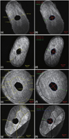
After root-end preparation, all roots were resected using a low-speed disk to make the apical specimens and examined with a confocal scanning microscope (LSM 5 Pascal Confocal Laser Scanning Microscopy, Carl Zeiss, Oberkochen, Germany). The confocal microscope examination was conducted at a magnification of ×20 for an analysis of the surface. The number of cracks and their length were recorded using a graphics tool (Zeiss LSM Image Examiner Ver. 3.1, Carl Zeiss). The size of the root-end cavity and remaining dentin thickness were measured with the assumption that the root-end cavity was oval in shape.
The cutting efficiency of the different tips under the two powers was statistically analyzed by two-way analysis of variance and a Mann-Whitney test using software (PASW Statistics 18, SPSS Inc., Chicago, IL, USA). Statistical significance was set at a confidence level of 95%.
Results
Cavity preparation time
Table 1 shows the root-end cavity preparation time for each group. There was no statistically significant difference in the time required between the instrument groups, but the Power-4 groups showed reduced preparation time for both instrument groups (p < 0.05).
Crack number and length
Table 1 also presents the number of cracks and their mean lengths. The K4 and J4 groups with a power of 4 showed significantly higher crack formation and longer cracks irrespective of the instruments used (p < 0.05). The K4 group with the KIS-1D tip used in the Power-4 experiments produced the development of significantly more cracks than the other groups (p < 0.05).
Minimum dentin thickness and preparation shape
The minimal remaining dentin wall thicknesses after cavity preparation are presented in Table 1, and the shape along with the cavity dimensions are presented in Table 2 and Figure 1. There was no statistically significant difference in the remaining dentin thickness or any of the other parameters after cavity preparation among all the groups.
Discussion
Diamond-coated ultrasonic tips have been used as a primary instrument for apical surgery. However, the disadvantage of a diamond-coated tip is the reduction of cutting efficiency over a period of time. When the cutting efficiency of an ultrasonic tip decreases, the time required for endodontic surgery increases. A diamond-coated tip easily loses its diamond particles when it is used in a canal smaller than its diameter; if this happens, the tip will not enter the small-diameter canal unless more force is applied.10 Recently, many diamond-coated tips having different shapes and surface textures have been introduced for maintaining durability and obtaining better efficiency. In this study, the newly introduced ultrasonic tips with multiple microprojections were compared with the conventional diamond-coated tips. The root-end preparations were performed with upward and downward pressure using ultrasonic devices as in other studies.10,13,15,16 The forces applied in these experiments were not mechanically constant, but the fact that these experiments mimicked actual clinical situations made the experiments meaningful.
In this study, the time required for the preparation of a 3-mm root-end cavity did not differ depending on the ultrasonic tip used but depended mainly on the power applied on the ultrasonic tip. The greater the power applied to the tip, the less was the time spent for the preparation. However, it led to the formation of a larger number of cracks and the longer length of the cracks, irrespective of the tip used.
This study was conducted using extracted teeth. Gray et al. reported that there was no difference in crack development between human cadaver teeth and extracted teeth.17 However, Calzonetti et al. reported that 45 teeth showed no crack development after root-end resection using ultrasonic devices in their cadaver study.18 Van Arx et al. reported that if the root-end preparation was performed in the presence of periodontal ligament, the stress to teeth would be reduced.16 Therefore, the crack development of the in vitro experiment might be overestimated.19
There is a report that the ease of detection of the number of cracks could be increased by more than two times when observed under a magnification of more than ×150, but in this study, a magnification of ×20 was sufficient to observe the cracks and microfractures.20 Engel and Steiman reported that cracks could develop not only in in vitro cavity preparation but also during tooth dehydration.21 Therefore, in the experimental procedures, dehydration was avoided. None of the experimental teeth were endodontically treated. Beling et al. reported that there was no difference in the number or type of cracks between endodontically treated teeth and those not treated.22 It was also reported that endodontic treatment might result in additional stress to or dehydration of the teeth.20
In the present study, the number and length of the cracks that developed were significantly different between power mode 1 and power mode 4. The types of ultrasonic tips did not show any differences, but a longer application time led to more cracks and longer cracks, which might be related to the cement cracks found in the J4 and K4 groups. The cement cracks developed radiating from the cementum to the cemento-dentinal junction. Because these cracks did not extend to the canal over the cemento-dentinal junction, it seems that they did not develop during root-end preparation. It can be assumed that these cracks developed due to extraction or aging.12
Layton et al. reported that crack occurrence during preparation was increased when maximum ultrasonic power was used.12 Taschieri et al. showed that crack development decreased when the ultrasonic device was set to moderate power.23 Thus, a moderate power of the ultrasonic device was recommended during cavity preparation.2 In the present study, when the power was increased from 1 to 4, there were significant increases in the number and the length of cracks with both the KIS-1D and the JT-5B tips. Therefore, if ultrasonic tips have the same cutting efficiencies, an increase in the ultrasonic power may not necessary. Considering the shortening of the preparation time by a power increase, it would be attractive to use a high power; however, avoiding a higher incidence of cracks and an increased crack length should be considered above all.
In this study, none of the cavity parameters after the cavity preparation showed any significant differences. The use of two different types of ultrasonic tips did not lead to any differences in the results. This could be attributed to the fact that the cavity preparation was performed alongside the original canal shape. Neither tip type resulted in any specific aberrations, including irregular cavity shapes. The minimum remaining dentin thickness is a factor influencing the longevity of tooth stability, and thus, it is important to conserve the tooth structure during cavity preparation. In the present study, the remaining distance from the cavity wall to the external tooth surface showed that there was no difference related to either a change in the power level or the two types of tips used. This implies that the remaining dentin thickness depends on more on the cavity shape or the tooth form than on the tip itself. In evaluating the relationship between the remaining dentin and the crack development after root-end preparation, Abedi et al. reported that 75% of the cracks developed when the wall was thinner than 1 mm after the root-end cavity preparation.13 In contrast, Khabbaz et al. observed that crack development was not related to the remaining dentinal thickness and claimed that the ultrasonic tip was a more important factor.16 In this study, 22 cases showed that the remaining dentinal thickness was less than 1 mm. Among them, 5 cases (23%) showed that cracks developed through the thinnest cavity wall. The remaining 18 cases showed a dentinal thickness of more than 1 mm and no signs of a crack. Future research will be valuable for developing another practical clinical guideline with more evidence for the use of these ultrasonic tips in apical preparation.


 XML Download