

 PDF
PDF ePub
ePub Citation
 Print
Print


Introduction
Paresthesia is defined as a sensory disturbance with clinical manifestations such as burning, prickling, tingling, numbness, itching or any deviation from normal sensation.1 Paresthesia of the inferior alveolar nerve can occur during various dental procedures like local anaesthetic injections, third molar surgery, orthognathic surgery, ablative surgery, implants, and endodontics.2,3
The possible etiologic factors for endodontics related paresthesia are periapical infection and iatrogenic injury to the nerve. Iatrogenic injury can be due to followings: mechanical trauma from over-instrumentation into the inferior alveolar canal; pressure exerted by the endodontic point or sealant within the inferior alveolar canal neurotoxicity due to the irrigants; intracanal medicaments and sealants which have gone past the apical foramen.3,4,5,6 Paresthesia due to periapical infections can be as a result of mechanical pressure on the mental nerve due to inflammatory edema.7 The periapical infectious process results in the release of the inflammatory products secondary to tissue damage and toxic metabolic products of bacteria leading to accumulation of purulent exudate in the mandibular bone. The associated edema or a subsequent hematoma can cause pressure on the nerve fibres and induce symptoms of paresthesia.7,8
The duration of paresthesia can vary from days to weeks or to several months and in some cases paresthesia might even become permanent. Permanent paresthesia can result from cases of actual irreversible nerve damage which may be due to laceration, prolonged pressure on the nerve or contact with toxic overfilled endodontic materials.9
The present paper describes a case of mental nerve paresthesia arising after the start of the endodontic therapy in left mandibular first molar.
Case report
A 40-year-old female patient was referred to the department of conservative dentistry and endodontics of Dr. Z. A Dental college, A.M.U Aligarh, India, with a chief complaint of severe pain associated with the left mandibular first molar and numbness in the left lower lip and chin. The patient reported that approximately 1 week earlier she had endodontic treatment initiated by a general dentist in her left mandibular first molar which had a carious exposure. On the next day after the initiation of endodontic treatment she developed severe pain in the left mandibular first molar and numbness in the left lower lip and chin. On reporting this to her dentist she was prescribed a combination of ofloxacin 200 mg and ornidazole 500 mg every 12 hours and aceclofenac potassium 100 mg and paracetamol 500 mg every 12 hours. The prescribed medication gave her relief from pain but no improvement in the feeling of numbness occurred and therefore her dentist made the referral.
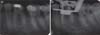
On examination with a dental probe, the area of numbness was found, extending from the mandibular midline to the left second premolar both intraorally and extraorally (Figures 1a and 1b). There was no deviation in sensory response of gingiva and tongue on probing. The left mandibular first molar showed unremoved proximal carious lesion (DO) and a temporary restoration placed in the access cavity with the tooth in occlusion. Intra-oral periapical radiograph revealed apical periodontal ligament widening in relation to both mesial and distal roots and slight apical root resorption in distal root (Figure 2a). After complete evaluation, diagnosis of acute apical periodontitis with mental nerve paresthesia was established and with the written informed consent of the patient it was decided to carry on the endodontic treatment along with the conservative management of paresthesia.
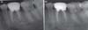
Local anesthesia was administered in the form of inferior alveolar nerve block and the involved tooth was isolated with a rubber dam. The temporary restoration was removed and the access cavity was prepared in a normal fashion. There was no active discharge from the canals. The canals were irrigated with 3% sodium hypochlorite (NaOCl) solution, and the instrumentation was done with stainless steel k-files (Dentsply-Maillefer, Ballaigues, Switzerland) and hand ProTaper files (Dentsply-Maillefer). The working lengths were established with an apex locator (Raypex-5, VDW, Munich, Germany) and confirmed radiographically (Figure 2b). The mesiobuccal and mesiolingual canals were prepared to an apical preparation size of F1 ProTaper whereas the distal canal was prepared to an apical preparation size of F2 ProTaper. The canals were then dried with sterile absorbent points, calcium hydroxide was placed as an intracanal medicament and the tooth was restored temporarily with a zinc oxide eugenol based intermediate restoration. The patient was prescribed with dexamethasone 0.5 mg every 12 hours for three days and was also prescribed with an oral methylcobalamin supplement (1,500 mcg once daily) owing to its role in enhancing proper neuronal functioning.10
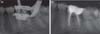
The patient was recalled after one week. Although the tooth had become asymptomatic, the feeling of numbness was still there. No intervention was done at this appointment and the patient was asked to continue methylcobalamin supplement and report after three weeks. The patient reported after three weeks with remarkable improvement in the feeling of paresthesia. The area of numbness was now reduced and was confined to the left lower lip region (Figure 3). The tooth was not tender on percussion or palpation but the obturation was still deferred to wait for the paresthesia to reduce further or to disappear completely. Two weeks later (i.e. 6 weeks after the initial visit) the paresthesia had mostly disappeared except for a small patch inside the left lower lip (Figure 4). The tooth was completely asymptomatic and therefore obturation was performed at this visit with laterally condensed gutta-percha and a zinc oxide eugenol based sealer (Figures 5a and 5b). The patient was seen again at 10 weeks from the initial visit as the symptoms of paresthesia had then subsided completely, and the patient was scheduled for restoration of the tooth. The tooth was restored with porcelain fused to metal full crown (Figure 6a). The tooth stays in function 1 year post-operatively with the area of paresthesia returned to normal sensation (Figure 6b).
Discussion
In the present case, the most probable cause of paresthesia seems to be periapical infection. Direct mechanical compression of the nerve or the release of toxic metabolites or both may have inhibited the normal function of mental nerve. Direct injury to the nerve trunk due to over instrumentation on the initial visit of endodontic treatment can also be a possibility. Paresthesia secondary to endodontic treatment may be caused by over instrumentation and/or overfill or the passage of various endodontic materials (root canal irrigants, sealers, and paraformaldehyde containing pastes) into the vicinity of the inferior alveolar nerve or its branches. During cleaning and shaping procedures, clinicians must strictly adhere to the accurate working length. Over-preparation of the canal and violation of apical foramen can lead to direct physical injury of the nerve or chemical nerve injuries from irrigating solutions and intracanal medicaments.11,12 Direct peripheral nerve injury has been classified by Seddon (1943)13 into three basic types: neurapraxia, axonotmesis and neurotmesis. Neurapraxia occurs due to a mild compression of the nerve trunk resulting in a temporary conduction block. Neurapraxia of the inferior alveolar nerve or mental nerve will usually manifest as a paresthesia or dysaesthesia of the lip and chin region.14 Axonotmesis refers to the actual degeneration of the afferent fibers as a result of internal/external irritation resulting in anesthesia.15 Neurotmesis is the complete severing of the nerve trunk, resulting in permanent paresthesia which can only be corrected by microsurgery and has a more guarded prognosis.13,14,15
The most likely form of injury in the present case seems to be neurapraxia due to either periapical infection or direct injury by over-instrumentation/inadvertent passage of the root canal irrigant or both. The tooth responded well to conservative treatment, and upon completion of the debridement and disinfection of the root canal, the symptoms of periapical infection subsided and paresthesia started to diminish. With the administration of the methylcobalamin, the symptoms of paresthesia further improved quickly and within 6 weeks there was a dramatic recovery with paresthesia reduced to a small patch inside the left lower lip. Methylcobalamin is the form of vitamin B12 that is active in the central nervous system. It is essential for cell growth and replication. Methylcobalamin may exert its neuroprotective effects through enhanced methylation, acceleration of nerve cell growth, or its ability to maintain already healthy homocysteine levels.16 Complete recovery of the case occurred within ten weeks.
Conclusions
Periapical infection and iatrogenic injury can be a cause of mental nerve paresthesia. During cleaning and shaping procedures, clinicians must strictly adhere to the accurate working length. Over-preparation of the canal and violation of apical foramen can lead to direct physical injury of the nerve. Irrigating solutions and intracanal medicaments can also lead to chemical nerve injuries. The best management of paresthesia secondary to endodontic treatment is prevention.


 XML Download