

 PDF
PDF ePub
ePub Citation
 Print
Print


Introduction
Physical sealing of the deep parts of pits and fissures has been established as a method for preventing occusal caries in children.1,2,3 The desired effects of sealant are to preserve retention and provide sufficient resin adaptation to the tooth walls.2,4 To achieve these goals different types of enamel surface pretreatments have been used such as cleaning the tooth surface by prophylactic brushing and enameloplasty by air polishing, air abrasion, burs or lasers.5,6,7,8,9,10
Enameloplasty by burs removes decalcified lesions from the tooth walls or debris from the bottom of the fissures, as well as providing better sealant penetration.7 However, concerns have been raised regarding sealant microleakage after using bur pretreatment. Some studies reported that burs led to less microleakage than conventional acid etch technique or air abrasion in sealed teeth.5,7,8,9 However, others reported opposite findings.10
Erbium yttrium-aluminum-garnet (Er:YAG) laser is another technique for cleaning or pretreatment of the enamel before sealant application.11 Compared to burs, laser ablation does not require local anesthesia and produces less noise and vibration, and is less painful and more comfortable for patients. However, laser ablation takes more time and requires expensive equipment, and safety considerations during laser use need to be taken into account.11,12 Pretreatment with Er:YAG laser alone prior to sealant application led to increased resin microleakage and less tensile bond strength of the sealant-to-enamel bond than acid etching.13,14,15,16,17,18 However, other studies found that the use of erbium laser associated with etching yielded similar microleakage and retention,18,19,20 or less microleakage, than conventional acid etching.21
The studies cited above were done with teeth in which the enamel was sound. However, dentists often seal deep groves and pits in teeth with fluorosis, and may be uncertain about the best way to proceed since the protocols1,2,3 for sealing defects in the normal dentition may need to be modified.22
Tooth fluorosis is a disturbance in the enamel structure that results from prolonged high levels of fluoride adsorption during tooth development. Teeth with fluorosis are categorized with the Thylstrup and Fejerskov Index (TFI) according to the clinical diagnosis and degree of tooth involvement as TFI 0, normal; 1-3, mild fluorosis; 4-5, moderate fluorosis; 6-9, severe fluorosis. Problems associated with tooth fluorosis include a hypermineralized outer layer followed by a hypomineralized enamel subsurface, increased microporosities on the enamel, enamel discoloration and unpleasant appearance.23 Over the years, effective bonding to fluorotic enamel may be compromised because of the hypermineralized outer layer, which is resistant to acid etching.23,24 Methods used to overcome the problem include applying dental adhesives, tooth surface pretreatment by grinding 0.5 mm of this layer with a bur or laser, and increasing the etching time.22,25,26,27 However, a recent study found that increasing the etching time led to decreased surface roughness.24 Also, no significant difference was observed in the depth of etching between teeth without fluorosis and teeth with mild flourosis; therefore, etching time may be similar under both circumstances.24,26 Loyola-Rodríguez et al. compared an acid etch technique to bur enameloplasty and sealing with a flowable composite in molar teeth with fluorosis. Their scanning electron microscopic findings showed that tooth surface pretreatment increased sealant penetration in teeth with fluorosis.22
Although several methods have been introduced for sealant pretreatment, the best method has yet to be established. To fill this gap in our knowledge of the effectiveness of different enamel pretreatments in molar teeth with fluorosis, the purpose of this in vitro study was to compare microleakage and sealant penetration in permanent molar teeth with fluorosis after different pretreatment methods.
Materials and methods
The research method was approved by the Human Ethics Review Committee of the School of Dentistry, Shiraz University of Medical Sciences. Permanent teeth were obtained during 4-month periods from dental clinics in three cities with reports of fluorosis located in two provinces (Fars and Bushehr). Immediately after extraction the specimens were stored in distilled water. The teeth were scaled, cleaned with fluoride-free pumice and a prophylaxis brush, and immersed in 0.1% chloramine T solution for 2 weeks for disinfection. Next, the teeth were stored in distilled water at 37℃. Finally after the occlusal surfaces were examined to discard any teeth with enamel abrasion, cracks, or caries, 120 third molars were selected with mild dental fluorosis (TFI 1 - 3).23
The teeth were randomly divided into 6 groups containing 20 teeth each, in which pretreatment procedures were performed on occlusal surfaces before fissure sealant (FS) application as follows:
Group 1: Phosphoric acid etch + FS (control group). First the occlusal surface was cleaned with a dry prophylactic brush and a low-speed hand piece for 10 seconds.9 Then 35% phosphoric acid (Scotchbond Etchant, 3M ESPE, St. Paul, MN, USA) were applied for 30 seconds. The tooth was rinsed and dried under a weak air stream. Then an unfilled FS (Clinpro, 3M ESPE) was applied and left for 20 seconds to ensure resin penetration. A halogen light curing unit (Coltolux, Coltène/Whaledent AG., Altstätten, Switzerland) with a power density of 550 mW/cm2 was used to cure the sealant for 20 seconds. To maximum curing depth the tip of the light curing instrument was placed as close as possible as to the occlusal surfaces. A tip of the explorer was used to ensure all the pits and fissure were sealed and to prevent void formation.
Group 2: Phosphoric acid etch + One-Step Plus + FS. A 2-step etch and rinse adhesive system with One-Step Plus (Bisco Inc, Schaumburg, IL, USA) was applied to the etched surface for 10 seconds, thinned by applying a weak air stream, and light-cured for 10 seconds. Then the teeth were sealed as described for group 1.
Group 3: Bur + phosphoric acid etch+ FS. Enameloplasty was done with a diamond bur (852/010, Dia Tessin, Vanetti, Gordevio, Switzerland) on the occlusal surface to wide fissures to a depth of 0.5 mm before enamel etching. Then the tooth surface was etched, rinsed and dried, and sealant was applied on the occlusal surfaces as in group 1.
Group 4: Bur + phosphoric acid etch + One-Step Plus + FS. All procedures were similar to group 3 except that an adhesive system (One-Step Plus) was used on the etched enamel before applying FS as described in group 2.
Group 5: Er:YAG laser + phosphoric acid etch + FS. The surface was pretreated with Er:YAG laser on the pit and fissure occlusal surface. Er:YAG laser (Fotona Fidelilis Plus III, Ljubljana, Slovenia) was used in enamel etch mode: 300 mJ, 20 Hz, 6 W, 140 µs, water 8, air 4, MSP mode (pulse duration 100 microseconds), R14 handpiece (100-degree), 0.8 mm fiber tip, 1 - 2 mm distance. Then the enamel margins were beveled with the same fiber tip at: 120 mJ, 10 Hz, 1.2 W, water 8, air 4, MSP mode. The occusal surface was sealed after etching the surfaces as described for the previous groups.
Group 6: Er:YAG + phosphoric acid etch + One-Step Plus + FS. The procedures were performed as for group 5 except that an adhesive system (One-Step Plus) was used before sealant placement.
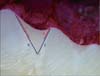
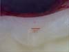
All teeth underwent thermal cycling for 1,000 cycles between 5℃ and 55℃ in a water bath, with a dwell time of 30 seconds and a 20-seconds transit time between the baths. The apices were sealed with sticky wax, and all tooth surfaces were covered with 2 layers of nail polish except for a 1-mm zone around the margins of each FS. To test for microleakage the samples were immersed in 0.5% basic fuchsin (Merck, Darmstadt, Germany) dye solution for 24 hours. The specimens were rinsed to remove excess dye and each tooth was sectioned bucolingually across the center of the sealant with a diamond saw (Letiz 1600, Leica, Wetzlar, Germany) and continuous water irrigation. Under blind conditions, two observers measured linear dye penetration in millimeters from the margin of the FS along the tooth and the sealant under a digital microscope (Dino Lite, Taipei, Taiwan) at ×50 magnification. The microscope was calibrated before evaluations. Consistency between examiners was ensured by measuring microleakage in 10 sectioned teeth. Proportion of microleakage (PM) was calculated by dividing the total length of dye penetration (c + d) by the total length of the enamel sealant interface (e + f, Figure 1). Penetration of the sealant was recorded as the unfilled area (mm2) divided by the total FS area (mm2) to obtain the proportion of unfilled area (PUA, a / (a + b), Figure 2).21,28 After the data were recorded, statistical analyses were done with the Kruskal-Wallis test to compare all groups together and the Mann-Whitney test for pairwise comparisons (p < 0.05).
Results
Table 1 shows the mean and standard deviation for the PM in all groups. Group 3 (bur + etching + FS) showed the greatest PM and group 6 (laser + etching + adhesive + FS) had the least PM. There were significant differences among all groups for the PM (p = 0.000). Pairwise comparisons between all groups revealed that only group 3 was significantly different from the other groups, except group 1. There were no significant differences among the other groups when compared with one another (p > 0.05, Table 2).
Table 1 shows the mean and standard deviation for the PUA in all groups. Bur pretreatment led to the greatest penetration into the fissures. Regarding penetration of the sealant into pits and fissures, our results showed no significant differences among groups (p = 0.124).
Discussion
Tooth surface pretreatment by enameloplasty has been used to improve fissure sealant success.6,29,30 The aims of this approach are to enhance enamel surface energy, increase surface area and increase resin penetration.5 This technique also removes debris and decalcified lesions as well as the prismless layer, which interferes with enamel etching and sealing ability of the sealant.5,31,32
Teeth with fluorosis, like other young permanent teeth with normal enamel, may have deep pits and fissures on the occlusal surfaces, which are susceptible areas for the development of dental caries.33 The use of resin-based materials is therefore recommended to reduce the likelihood of caries.1,2 Under these circumstances, dentists may prefer pretreatment of the enamel because of uncertainty about the complete removal of debris from deep parts of fissures with only a prophylactic brush, or doubts about the presence of decalcification on the enamel fissure walls or hypermineralization of the enamel outer layer.22,23 The present study was designed to address these potential situations.
All experimental groups in the present study had some degree of microleakage across sealant and tooth walls.5,6,9 There were no differences in PM between the control group (acid etching + FS) and pretreatment with a bur (groups 3 and 4). As in the present study, some earlier researches found no significant differences in sealant microleakage after acid etching compared to the use of a bur with etching.8,34 In contrast, some studies reported that bur pretreatment may reduce or increase sealant microleakage compared to acid etching.5,7,9,10 The differences between the results may be due to the use of different types of burs, methods to quantify microleakage, and types of teeth.7,15,16,21,28,29,30
In the present study, PM was the greatest in group 3 (bur + etch + FS). This result may be related to the formation of microfractures in the hydroxyapatite enamel crystals which may be more prominent in teeth with fluorosis because of the porosity in the enamel subsurface layer.10,23,25 In addition, bur preparation forms a smear layer which may affect resin adhesion to etched enamel and increase microleakage.5,16
There are two approaches to using laser pretreatment prior to sealant placement: laser ablation instead of acid etching and laser associated with etching. Although some believe that laser preparation alone may be as effective as acid etching,29,35 most studies recommended pretreatment with Er:YAG laser combined with acid etching, as used in the present study.13,15,16,20
Our results showed no significant differences in PM between the control group and either of the two laser groups (5 and 6). In agreement with our results, Lupi-Pégurier et al., Borsatto et al. and Manhart et al. found no significant differences in mircoleakage between Er:YAG laser with acid etching and acid etching alone before sealant application.15,17,19 In addition, Moshonvo et al. found no differences in sealant microleakage after Er;YAG laser preparation (without etching) and acid etching.36 The difference between these results may be related to differences in laser settings (characteristics), sealant materials and the type of teeth that were treated.13,20,21
We found that the PM in the laser-treated groups was lower than the other groups. In agreements with our findings, Baygin et al. and Khogli et al. showed that Er:YAG pretreatment followed by etching resulted in less microleakage than acid etching or bur pretreatment prior to sealant application.16,21 These results may reflect the fact that Er:YAG laser ablation leads to increased sealant adaption to the enamel walls by producing irregularities and thus increasing the roughness of the enamel surface and the available surface area more than acid etching alone. In addition, laser treatment can reach the deepest parts of fissures and remove debris, while avoiding the formation of a smear layer.28,29,37 It should be noted, however, that the use of laser preparation involves some difficulties for the operator: clinicians must use the correct angle and tip during preparation. To reduce errors a smaller tip may be useful; however, smaller tips are associated with problems such as the need to ensure an equal level of ablation at the same speed, which requires more time and energy compared to larger tips.28 Thus operator experience and expertise are important for the success of laser pretreatment.
In the present study an acetone-based one-bottle adhesive was used in three groups (groups 2, 4 and 6). The use of bonding agents in these groups reduced mean PM compared to groups 1, 3 and 5. Using an adhesive before the FS is applied enhances acid-etched tooth surface wetting, which may be more prominent in teeth with fluorosis. The adhesive may also reduce microleakage, enhance resin flow into the fissures, and thus improve adhesion and clinical success.38,39,40 For example, Lygidakis et al. reported that the use of a one-bottle adhesive system increased sealant retention in hypomineralized molars.38
Penetration of the sealant into the depth of pits and fissures is a factor that may affect sealant retention.41 The degree of penetration may be influenced by fissure morphology and pretreatment of the enamel surface. In agreement with our results, Khogli et al., found no significant differences in sealant penetration between bur, laser and conventional acid etching techniques.21 However, at least one study found that bur preparation increased sealant penetration.30 The differences between studies may be due to differences in the quantitative methods used to determine the proportion of the unfilled area.21,28 An advantage of the present study is that mean sealant penetration was compared in samples without pretreatment (groups 1 and 2) and samples prepared with an adhesive (groups 4 and 6). The application of an adhesive may be associated with air entrapment or excessive thickness of the bonding agent, which can interfere with complete penetration of the resin.29 Sealant penetration in our group 3 (bur preparation) was better than other groups - a finding consistent with results published by Salama and Al-Hamad.30 Widening the fissures with a bur may create smooth tooth walls and increase resin flow; however, it is an invasive procedure.5,16
Micoleakage is a laboratory phenomenon and may not precisely reflect the clinical situation. Our method aimed to evaluate the adhesion of different materials to the tooth surface; however, microleakage test results may be influenced by factors such as temperature changes. To control for this possible effect, we used thermocycling in the present study. Also among the potential limitations of this in vitro study are the effects of functional loading and pH cycling, which were not considered here.42 A further possible limitation is the effect of age of the teeth with fluorosis, which we did not consider in our analysis.25 Additional clinical studies should be designed to compare different types of enameloplasty before sealant application in other types of teeth with fluorosis.
Conclusions
Based on our results, there was no difference in the proportion of microleakage between conventional acid etching and bur or laser surface pretreatment before sealant resin was applied in teeth with fluorosis. According to the results for proportions of mircoleakage and resin penetration, Er:YAG laser pretreatment may offer an alternative method before fissure sealant application in teeth with mild fluorosis.


 XML Download