

 PDF
PDF ePub
ePub Citation
 Print
Print


Introduction
The anatomy of the tooth has long been an unresolved mystery, and complexity of root canal morphology has been commonly found. The inability to recognize the presence of canals and treat them adequately results in unfavourable outcomes from root canal treatment.1-4 Maxillary molar morphology has been extensively studied with a specific emphasis on the mesiobuccal root because of its complexity.1,5-9 Hess and Zurcher showed that ramifications are prominent features of the mesiobuccal root of maxillary molars, and the presence of such anatomic complexities makes it difficult for clinicians to achieve endodontic treatment goals.10-12 One of the rare complexities is the presence of a third canal in the mesiobuccal root of maxillary second molar. Incidences varying from 1.6% to 16% have been reported in in vitro studies.13,14 Clinically, a third mesiobuccal canal was first reported by Ferguson et al.15 in the maxillary first molar. Ozcan et al. were first to report a third mesiobuccal canal in the maxillary second molar in a case report.16 Various canal configurations have been reported in mesiobuccal roots of maxillary second molars using different techniques in vitro.8,17-20 The canal configuration for the mesiobuccal root in the case reported by Ozcan et al. was 3-2, which is included in the classification given by Gulabivala et al. as an addition to the Vertucci classification.16,19,21 Presented here is a case where three canals were located in the mesiobuccal root of a maxillary second molar with a configuration of 3-2-1. To the best of our knowledge, the above canal configuration for the mesiobuccal root of a maxillary second molar has not been reported to date in any in vivo or in vitro studies. It was possible to confirm the internal anatomy of the mesiobuccal root and treat it adequately with the help of clinical, radiographic, and multi-detector computed tomography (MDCT) scan findings.
Case report
A 36-year-old female came to the department with the chief complaint of dull spontaneous pain in the upper left posterior tooth for the previous 2 weeks. She reported a fractured restoration on the same tooth few months back. On examination, an open cavity with a dislodged restoration and exposed mesiobuccal pulp horn was seen with respect to the maxillary left second molar. An intraoral radiograph revealed the presence of radiolucency encroaching on the pulp horn with an apparently normal periapex. Thermal sensitivity tests elicited a lingering pain in the patient. Based on the clinical and radiographic findings, a diagnosis of irreversible pulpitis was formulated.
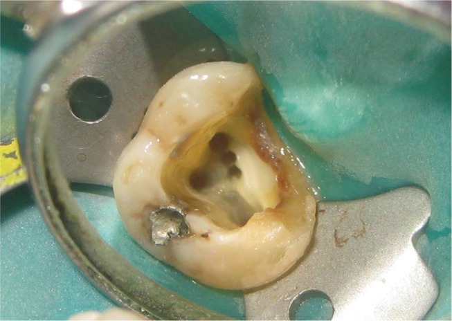
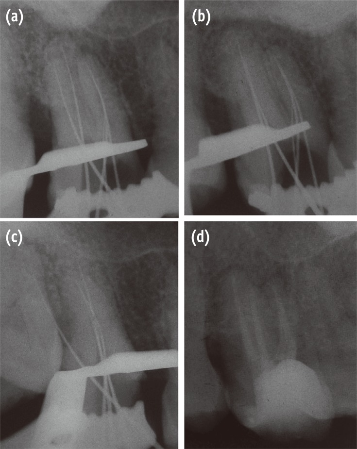
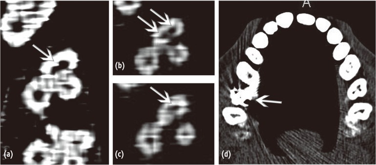
Subsequent to anesthesia and rubber dam isolation, access cavity preparation was initiated and the mesiobuccal, distobuccal, and palatal canals were located. Anticipating the presence of extra canals, further exploration was carried out deliberately and two sticky points were located. This led to finding of two additional canals in the mesiobuccal root on the line connecting the mesiobuccal and palatal canal orifice (Figure 1). Magnifying loupes (2.5X, ST250, STAC Dental Instruments Inc., Brampton, Canada), a DG 16 endodontic explorer, and sodium hypochlorite were the adjuncts used to locate the canals. After locating the orifices, an attempt was made to understand the internal anatomy of the mesiobuccal root using K files and intraoral periapical radiographs. While negotiating the canals individually in the mesiobuccal root, there was no hindrance in the movement of the #10 K file in any of the three canals. However, simultaneous insertion posed obstruction in the vertical movement of the files in the 2 extra canals. The above finding raised an element of doubt regarding the configuration of the canals. Intraoral periapical radiographs were taken from three different horizontal angulations (Figures 2a, 2b and 2c). However, all of the radiographs suffered from overlapping of the endodontic files radiopacity. Only a mesially angulated radiograph revealed limited information on where files associated with the second and third mesiobuccal canals were seen to be meeting in the middle third of the root approximately and continue to merge with the file in first mesiobuccal canal (Figure 2b). To avoid any missed canals, and for better understanding of the tooth's internal anatomy, a MDCT (Brilliance CT 64-channel, Philips Healthcare, Andover, MA, USA) scan was suggested because cone-beam computed tomography (CBCT) was not available in the set up or in the vicinity. The interpretation regarding the number of separate orifices and canals present in the coronal third or at the orifice level was inconsistent with the clinical or radiographic findings. In contrast to the 3 orifices and canals, clinically and radiographically, MDCT revealed only 1 large canal (Figure 3a). In the middle and apical third, 2 canals and 1 canal respectively could be distinguished in both radiographic and MDCT images (Figures 3b and 3c).
The configuration of the canals in the mesiobuccal root was perceived to be 3-2-1 (Figure 4) after correlating and computing the clinical, radiographic, and MDCT scan findings. Three separate coronal orifices extending apically to three separate canals were identified as mesiobuccal (MB), middle mesiobuccal (m-MB), and palatal mesiobuccal (p-MB) based on their anatomic positions. The m-MB and p-MB merged into one canal at a distance of approximately 5 mm from the orifice level. Following this, the canal joined the MB canal at a distance of approximately 9 mm from the orifice level. Finally, the canal ended with a single apical foramen at the apex. The distances mentioned above were calculated using the thickness of each slice of the MDCT scan images, which was 0.7 mm obtained using DentaScan software (GE Healthcare, Milwaukee, WI, USA). After understanding the anatomy of the mesiobuccal root and confirming the absence of extra canals in other roots, the treatment proceeded. The biomechanical preparation of MB, distobuccal, and palatal canals was completed using ProTaper NiTi rotary files (Dentsply Maillefer, Ballaigues, Switzerland). m-MB and p-MB canals were prepared with K-files using the step-back technique. Following one week of intracanal medication with calcium hydroxide, the canals were obturated using the cold lateral compaction technique (Figure 2d).
Discussion
Despite the reports of three mesiobuccal canals in the mesiobuccal root of the maxillary second molar, there have been very few reports of such cases. Based on the literature, many reasons can be listed for the paucity of reports. One specific reason could be the anatomic complexity of the tooth itself, making it impossible to locate additional canals. Indirect or non-specific reasons could be radiographic insensitivity, lack of visual aids during setup, or lack of practitioner's commitment.
Anatomically, the openings of canals situated palatal to the MB canal are difficult to locate because of their smaller size. As explained by Eskoz et al., initially the canal in the MB root is the shape of a kidney bean.5 With continued deposition of secondary dentin, the isthmus between the poles becomes narrower and eventually may even close, resulting in multiple canals. Because the mesiolingual segment of the canal surrounds the smaller of the poles of the kidney bean, it will close off leaving a small space, thus making it more difficult to locate.
The small size and superimposition over another root canal account for the difficulty in the location of extra canals.25 The information acquired using radiographs is valuable, but it suffers from insensitivity and lack of reliability when it comes to assessment of the number of root canals present.26,27 However, with the increasing use of visual aids, especially the endodontic microscope, locating and finding canals has become easier.28,29 Loupes have also been proven to be equally effective as the endodontic microscope in locating the second mesiobuccal canal.30 Apart from the factors mentioned above, careful clinical examination and the practitioner's commitment also play a major role in the detection of extra canals.27,31
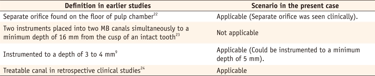
The literature has not been able to provide a clear definition of what constitutes a canal. After comparing the present case with some of the definitions given earlier, it was reasonable to assume that a third canal was present in the mesiobuccal root in the present case (Table 1).
Confusion has prevailed over the nomenclature of canals, especially in the maxillary molars. For instance, the second mesiobuccal canal has been termed the MB2, or the mesiopalatal or mesiolingual canal.32-34 Ozcan et al. termed the third mesiobuccal canal the MB3.16 However, the numbers do not reveal the anatomic location but only the presence of additional canals.35 In a recent clinical study, a new nomenclature was suggested for maxillary molars, based on which the canals in the present case were named.35 The proposed nomenclature is easy to understand and communicate, and is self-explanatory.
In the above case, a mesially angulated radiograph uncovered the internal anatomy, but because of inherent disadvantages of intraoral periapical radiographs, application of an advanced imaging modality such as CBCT was found to be valid, given its ability to localize and describe the internal and external anatomy in three dimensions.36,37 However, due to the non-availability of a CBCT unit in the vicinity, an MDCT scan was recommended. Although the MDCT provides a better image quality compared to conventional CT systems, CBCT has been proven to be better for imaging hard tissues in the maxillofacial region.38,39 In the present case, the inability of the MDCT scan to reveal three separate orifices seen clinically could be attributed to various causes. Firstly, the MDCT used here had a spatial resolution of 0.32 mm, which could be larger than the fine septa dividing the canals. Secondly, the larger voxel size could have had a negative impact. As proved by Bauman et al., detection of MB2 canals increased from 60.1% at a 0.4 mm voxel size (CBCT) to 93.3% at a 0.125 mm voxel size, and the voxel size of the MDCT used here was 0.23 mm with a slice thickness of 0.7 mm.40 Thirdly, in the present case an artifact was seen to be hindering the clarity of the MDCT images (Figure 3d). As proven before, metal objects including dental restorations in the scan field can lead to severe streaking artifacts.41-42 In the present case, an amalgam restoration on the right maxillary second molar caused a streaking artifact that distorted the images obtained at the level of the orifice and coronal third of canals from left maxillary second molar. It has been shown that MDCT suffers more from metal artifacts, which is another reason to opt for CBCT over MDCT.43
Caution was taken to avoid over-preparation of m-MB since the "danger zone" in the MB root exists across most of the distal surface of the MB root anatomically near to where the root joins the crown of the tooth.13 Henceforth, K files were used for biomechanical preparation to avoid aggressive preparation.
The present case is the first report of three canals in the mesiobuccal root of the maxillary second molars in an Indian population. No other case report on an Indian population could be found in Pubmed Search. Additionally, in an in vitro study using CBCT in an Indian population, none of the maxillary second molars were reported to have three canals in the mesiobuccal root.12 The root canal configuration of 3-2-1 encountered here is not included in the Vertucci classification or its modification, and a similar configuration was only found in the maxillary first molar in an in vitro study on a Turkish population.44

 XML Download