

 PDF
PDF ePub
ePub Citation
 Print
Print


Introduction
In most of its clinical applications, one or more surfaces of mineral trioxide aggregate (MTA) are exposed directly to blood which may also penetrate into body of the MTA slurry. Blood contamination of unset MTA adversely affects the setting reaction and mechanical strength of the material.1 Vanderweele et al. reported that following blood contamination of MTA in perforation sites, resistance to displacement decreased.2 In addition, Nekoofar et al. demonstrated that blood contamination influenced the development of MTA crystals and decreased its surface microhardness and compressive strength.3-5 It is well known that another disadvantage of MTA is its extended setting time.6-10
While MTA is a derivative of Type 1 Portland cement (PC), addition of setting accelerator admixtures such as calcium chloride (CaCl2) and disodium hydrogen phosphate (Na2HPO4) have been suggested to decrease the setting time of MTA.6,7,11-13 They also accelerate the early strength of PC.14 Therefore, they might be able to protect MTA against adverse effects of blood contamination by reducing the possibility of infusion of the blood into the material whilst at the same time improving its early strength. To date, no studies have reported the effects of blood contamination on rapid-setting MTA materials.
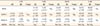
Root MTA (RMTA, Lotfi research group, Tabriz, Iran) is one of several commercially available types of MTA. Several cytological, animal and clinical studies have shown similar characteristics of RMTA and ProRoot White MTA (Dentsply Tulsa Dental, Tulsa, OK, USA).15-23 Scanning electron microscopic (SEM) analysis has shown that both ProRoot MTA and RMTA contained a complex mixture of mineral phases with highly visible randomly distributed particles of bismuth oxide.24 In these materials, the size of crystalline particles and bismuth oxide particles were the same.24 In addition, electron probe microanalysis has shown that their major components such as Lime (CaO), Silica (SiO2), Bismuth Oxide (Bi2O3), Aluminum Oxide (Al2O3) and Magnesium Oxide (MgO) are similar (Table 1).24
The purpose of this study was to evaluate the effect of blood contamination on the compressive strength of RMTA, as a model of MTA-like materials, which was modified by adding CaCl2 and Na2HPO4, in order to investigate the ability of these admixtures to prevent adverse effects of blood contamination.
Materials and Methods
The materials and groups evaluated were Group1, RMTA (n = 15); Group 2, RMTA modified with CaCl2 (RMTA-C) (n = 20); Group 3, RMTA modified with Na2HPO4 (RMTA-N) (n = 20); Group 4, RMTA contaminated with blood (n = 15); Group 5, RMTA-C contaminated with blood (n = 20); Group 6, RMTA-N contaminated with blood (n = 20). The compressive strength of specimens was evaluated after 3 hours, 24 hours, and 1 week (5 specimens at each time interval). In addition, in the modified groups (2, 3, 5 and 6) the compressive strength of an additional 5 specimens in each group was evaluated after 1 hour to evaluate their initial strength.
Specimen preparation technique
Stainless steel cylindrical split molds with a height of 6 mm and an internal diameter of 4 mm were used according to ISO 9917-1.25
Group 1 (RMTA): RMTA powder was mixed with sterile water in a 3/1 ratio. The powder was weighed using a digital scale model PL303 with 0.001 g accuracy (Mettler Toledo Inc., Columbus, OH, USA). The volume of sterile water was determined using a transferpette with 0.001 mL accuracy (BRAND GMBH + Co KG, Wertheim, Germany). The powder and liquid were poured into plastic capsules and were mixed mechanically for 30 seconds using an amalgamator (Farazmehr Co., Esfahan, Iran) at 4,500 rpm. The mixed MTA slurries were then placed into the metallic moulds according to method described by Nekoofar et al.4
Group 2 (RMTA-C): The specimens were prepared in the same way as Group 1 but instead of sterile water, a 5% CaCl2 solution was used. To prepare this solution 5.0 g of CaCl2 (Merck, Darmstadt, Germany) was dissolved in 100 mL of sterile water.
Group 3 (RMTA-N): In this group the RMTA powder was mixed with 2.5% (weight) Na2HPO4 powder (Merck). The specimens were prepared in the same way as Group 1.
Groups 4, 5, and 6 (blood contaminated specimens): The specimens in groups 4, 5, and 6 were prepared in the same way as Groups 1, 2, and 3 respectively. The only difference being that before MTA placement the moulds were contaminated with blood by filling them with whole fresh human blood that was then removed by aspirating with a syringe to leave a coating of blood on the internal walls of the moulds. The fresh blood was obtained by a trained medical nurse from a volunteer member of the research group. The procedure was approved by the ethics committee of the Zahedan University of Medical Sciences.
All specimens were incubated at 37℃ in a fully saturated humidity. At each time interval the specimens were removed from the molds and assessed for the presence of voids or chipped edges and damaged specimens were excluded and replaced with new specimens.
The compressive strength of the specimens was then evaluated using a universal testing machine Model H5K5 (Haunsfield Test Equipment, Redhill, UK). The compressive load was recorded until the loading failure point was reached. Loading failure was used to calculate the compressive strength of the specimens in terms of megapascal (MPa) using the following equation: C = 4P/πD2 where P (N) is loading failure, and D (mm) is the diameter of the specimen.
The effects of blood contamination on compressive strength of the specimens at different time intervals were analyzed using T-tests using the Statistical Package for the Social Sciences version 16 (SPSS Inc., Chicago, IL, USA). Also the effects of setting accelerators themselves and time on compressive strength were analyzed with twoway analysis of variance (ANOVA) and post hoc Tamhane's. The level of significance for the data analysis was 95%. In order to assess the normality of the data, the error terms of all experimental groups calculated. Then the normal distribution of error terms were analyzed by one-sample kolmogorov-smirnov test which showed that the differences were not statistically significant (p < 0.05).
Results
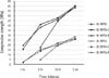
A summary of the compressive strength (Mean ± SD) of experimental groups is shown in Table 2. The highest mean compressive strength value was recorded for the original RMTA group after 1 week (62.64 ± 3.2 MPa) and the lowest mean compressive strength value was recorded for the blood contaminated RMTA-C group after 1 hour (2.08 ± 0.23 MPa).
Effect of blood contamination
In blood contaminated specimens the compressive strength was significantly lower than that of specimens without blood contamination at all time intervals for all experimental materials (p < 0.05, Figure 1).
Effect of accelerators
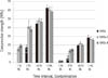
After 3 hours, the compressive strength of specimens in the RMTA groups (with or without blood contamination) was significantly lower than that of the same RMTA-C and RMTA-N groups (*1 and *2, p < 0.05, Figure 2). The difference was not significant at other time intervals (Figure 2). There was no significant difference between the compressive strength of the RMTA groups after 3 hours (with or without blood contamination) and that of the RMTA-C and RMTA-N groups after 1 hour (+1 and +2, Figure 2).
Effect of time
In all groups, the compressive strength of specimens increased significantly over time (p < 0.05, Figure 1).
Discussion
MTA is a hydraulic cement that consists mainly of dicalcium silicate and tricalcium silicate that produces calcium silicate hydrate (CSH) gel and calcium hydroxide during the hydration process.26,27 The setting and strength of hydraulic cements depends on the formation of CSH gel and ettringate (hydrated calcium sulfoaluminate) on nucleation sites of calcium hydroxide crystals.5 Because of the various clinical applications of MTA, such as pulp capping, furcal perforation repair etc., sufficient strength is necessary for MTA to withstand compressive pressures. Also, compressive strength is an indicator of the setting and hydration processes.3,4,28-30 Therefore, in the present study the compressive strength was evaluated to assess the effects of blood contamination on features of accelerated RMTA.
In the present study, blood contamination resulted in lower compressive strength values for the original RMTA specimens at each time interval (Figure 1). This finding was similar to previous studies that concluded that blood contamination adversely affected the physical properties of MTA.2-4 Lack of acicular crystals that are indicative of ettringate formation have been reported in blood contaminated MTA by Nekoofar et al.3-5 In addition, it has been stated that the 'air entrainment' features of blood proteins affect the microstructure of cements and increases their porosity. An increase in the porosity of hydraulic cements such as MTA is associated with a decrease in compressive strength.31 However, in the present study the same phenomenon was also observed in the modified groups, which means that addition of CaCl2 and Na2HPO4 did not prevent the negative effects of blood on compressive strength.
Kogan et al. reported that 3% and 5% CaCl2 solutions reduced the compressive strength of MTA.6 Following incorporation of 10% CaCl2 to MTA powder, similar results were reported by Lee et al. during the initial phase of setting; however, the final compressive strength did not change.32 In the present study, the compressive strength of uncontaminated RMTA specimens modified with CaCl2 was higher than specimens in the original RMTA group after 3 hours (*1, Figure 2), which can be explained by a reduction in porosity following CaCl2 addition, as reported by Hong et al.8 However, such a difference was not observed at the later time intervals. In addition, the compressive strength of RMTA specimens modified with CaCl2 after 1 hour was comparable to the compressive strength of RMTA specimens after 3 hours (+1, Figure 2). Therefore, it can be assumed that the initial increase in the compressive strength of RMTA was related to the accelerated setting reaction induced by CaCl2. The same trend was observed in specimens modified by Na2HPO4 (*1 and +1, Figure 2). Therefore, it can be concluded that (as for CaCl2) Na2HPO4 only accelerated the setting reaction of RMTA and did not improve its hydration process and physical characteristics. Liu et al. reported a similar trend with an increase in initial compressive strength of tricalcium silicate (a major constituent of MTA) following incorporation of tricalcium aluminate.31 Comparison of blood contaminated specimens of all experimented materials also revealed the same pattern as uncontaminated ones (Figure 1; *2 and +2, Figure 2).
These findings could explain the inability of accelerator admixtures to prevent adverse effects of blood contamination. While, blood penetrates into MTA slurries immediately after material placement, its adverse effects on compressive strength occur in the initial (early) phase of setting and subsequently are not prevented by setting accelerators. Protecting MTA from blood contamination may be the best strategy to preventing these adverse effects.
In the present study, the compressive strength of all specimens increased significantly over time (Figure 1), which has been also demonstrated in previous studies.2,3,30,31,33 The highest reported compressive strength of ProRoot MTA after 72 hours to 28 days was 71.36 - 86.02 MPa.4,28,29,33 In the present study the compressive strength of uncontaminated specimens of Root MTA after 7 days (58.64 - 62.64 MPa) was close to the reported range. However, despite improvement over time the compressive strengths of blood contaminated specimens did not reach the aforementioned level even after 1 week (Figure 1).

 XML Download