

 PDF
PDF ePub
ePub Citation
 Print
Print


Abstract
Objectives
The purpose of this study was to determine the effect of resin infiltration technique on color and surface hardness of white spot lesion (WSL) with various degrees of demineralization.
Materials and Methods
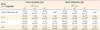
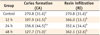
Ten human upper premolars were cut and divided into quarters with a 3 × 4 mm window on the enamel surface. Each specimens were separated into four groups (n = 10) and immersed in demineralization solution to create WSL: control, no treatment (baseline); 12 h, 12 hr demineralization; 24 h, 24 hr demineralization; 48 h, 48 hr demineralization. Resin infiltration was performed to the specimens using Icon (DMG). CIEL*a*b* color parameters of the enamel-dentin complex were determined using a spectroradiometer at baseline, after caries formation and after resin infiltration. Surface hardness was measured by Vickers Micro Hardness Tester (Shimadzu, HMV-2). The differences in color and hardness among the groups were analyzed with ANOVA followed by Tukey test.
Figures and Tables
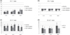 | Figure 1(a) Mean values of CIE L* for three groups (12 h, 24 h, and 48 h demineralization time) measured at baseline, after careis formation, and after resin infiltration; (b) Mean values of CIE a* for three groups; (c) Mean values of CIE b* for three groups; (d) Mean values of color change (CIE ΔE*) after caries formation (BL-CA) and after resin infiltration (BL-RI) for three groups.
|
References
1. Kielbassa AM, Muller J, Gernhardt CR. Closing the gap between oral hygiene and minimally invasive dentistry: a review on the resin infiltration technique of incipient (proximal) enamel lesions. Quintessence Int. 2009. 40:663–681.
2. Featherstone JD. Prevention and reversal of dental caries: role of low level fluoride. Community Dent Oral Epidemiol. 1999. 27:31–40.

3. Fontana M, Young DA, Wolff MS, Pitts NB, Longbottom C. Defining dental caries for 2010 and beyond. Dent Clin North Am. 2010. 54:423–440.
4. Kim ME, Jung IY, Kum KY, Lee CY, Roh BD. In vivo quantitative analysis of remineralization effect of remineralization solution 'R' of incipient enamel dental caries. J Korean Acad Conserv Dent. 2002. 27:175–182.
5. Kim S, Kim EY, Jeong TS, Kim JW. The evaluation of resin infiltration for masking labial enamel white spot lesions. Int J Paediatr Dent. 2011. 21:241–248.
6. Tüfekçi E, Merrill TE, Pintado MR, Beyer JP, Brantley WA. Enamel loss associated with orthodontic adhesive removal on teeth with white spot lesions: an in vitro study. Am J Orthod Dentofacial Orthop. 2004. 125:733–739.
7. Gwinnett AJ, Ceen RF. Plaque distribution on bonded brackets: a scanning microscope study. Am J Orthod. 1979. 75:667–677.
8. Rocha Gomes Torres C, Borges AB, Torres LM, Gomes IS, de Oliveira RS. Effect of caries infiltration technique and fluoride therapy on the colour masking of white spot lesions. J Dent. 2011. 39:202–207.
9. Meyer-Lueckel H, Paris S, Kielbassa AM. Surface layer erosion of natural caries lesions with phosphoric and hydrochloric acid gels in preparation for resin infiltration. Caries Res. 2007. 41:223–230.
10. ten Cate JM, Buijs MJ, Miller CC, Exterkate RA. Elevated fluoride products enhance remineralization of advanced enamel lesions. J Dent Res. 2008. 87:943–947.
11. Son HJ, Kim WC, Jun SH, Kim YS, Ju SW, Ahn JS. Influence of dentin porcelain thickness on layered all-ceramic restoration color. J Dent. 2010. 38:Suppl 2. e71–e77.
12. Zandoná AF, Zero DT. Diagnostic tools for early caries detection. J Am Dent Assoc. 2006. 137:1675–1684.
13. Bishara SE, Ostby AW. White Spot Lesions: formation, prevention, and treatment. Semin Orthod. 2008. 14:174–182.
14. Cochrane NJ, Cai F, Huq NL, Burrow MF, Reynolds EC. New approaches to enhanced remineralization of tooth enamel. J Dent Res. 2010. 89:1187–1197.
15. Son JH, Hur B, Kim HC, Park JK. Management of white spots: resin infiltration technique and microabrasion. J Korean Acad Conserv Dent. 2011. 36:66–71.
16. Joiner A, Hopkinson I, Deng Y, Westland S. A review of tooth colour and whiteness. J Dent. 2008. 36:Suppl 1. S2–S7.
17. Ko CC, Tantbirojn D, Wang T, Douglas WH. Optical scattering power for characterization of mineral loss. J Dent Res. 2000. 79:1584–1589.
18. Iwami Y, Hayashi N, Takeshige F, Ebisu S. Relationship between the color of carious dentin with varying lesion activity, and bacterial detection. J Dent. 2008. 36:143–151.
19. Lim HN, Yu B, Lee YK. Spectroradiometric and spectrophotometric translucency of ceramic materials. J Prosthet Dent. 2010. 104:239–246.
20. Lim HN, Yu B, Lim JI, Lee YK. Correlations between spectroradiometric and spectrophotometric colors of all-ceramic materials. Dent Mater. 2010. 26:1052–1058.
21. Douglas RD, Steinhauer TJ, Wee AG. Intraoral determination of the tolerance of dentists for perceptibility and acceptability of shade mismatch. J Prosthet Dent. 2007. 97:200–208.
22. Kim Y, Son HH, Yi K, Kim HY, Ahn J, Chang J. The color change in artificial white spot lesions measured using a spectroradiometer. Clin Oral Investig. 2012. 01. 27. (Epub ahead of print).
23. Knösel M, Attin R, Becker K, Attin T. External bleaching effect on the color and luminosity of inactive white-spot lesions after fixed orthodontic appliances. Angle Orthod. 2007. 77:646–652.
 XML Download