

 PDF
PDF ePub
ePub Citation
 Print
Print


Abstract
Objectives
This study analyzed the difference in color caused by different thickness in enamel layer of composite resins when applied with single and layering placement technique, and evaluated if the results agreed with the shade guide from the manufacturers to verify reliability of the color matching process of the manufacturers.
Materials and Methods
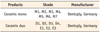
For single composite resin samples, 6 mm diameter and 4 mm thickness cylindrical samples were fabricated using Ceram-X mono (DENTSPLY DeTrey) and CIE L*a*b* values were measured with spectrophotometer. Same process was done for layering composite resin samples, making 3 dentinal shade samples, 4 mm thickness, for each shade using Ceram-X duo (DENTSPLY DeTrey) and enamel shade resins were layered in 2 mm thickness and CIE L*a*b* values were measured. These samples were ground to 0.2 mm thickness each time, and CIE L*a*b* values were measured to 1 mm thickness of enamel shade resin.
Results
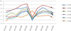
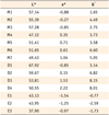
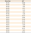
Color difference (ΔE*) between single and layering composite resin was 1.37 minimum and 10.53 maximum when layering thicknesses were between 1 mm and 2 mm and 6 out of 10 same shade groups suggested by manufacturer showed remarkable color difference at any thickness (ΔE* > 3.3).
Conclusion
When using Ceram-X mono and duo for composite resin restoration, following the manufacturer's instructions for choosing the shade is not appropriate, and more accurate information for Ceram-X duo is needed on the variation and expression of the shades depending on the thickness of the enamel.
Figures and Tables
Figure 1
Color differences between single composite and double composite according to the thickness of the double composite.

References
1. Hwang IN, Lee KW. Translucency of light cured composite resins depends on thickness & its influence on color of restorations. J Korean Acad Conserv Dent. 1999. 24:585–603.
2. Miller LL. Shade matching. J Esthet Dent. 1993. 5:143–153.

3. Sproull RC. Color matching in dentistry. 3. Color control. J Prosthet Dent. 1974. 31:146–154.
4. Grajower R, Revah A, Sorin S. Reflectance spectra of natural and acrylic resin teeth. J Prosthet Dent. 1976. 36:570–579.
5. Macentee M, Lakowski R. Instrumental colour measurement of vital and extracted human teeth. J Oral Rehabil. 1981. 8:203–208.
6. Goodkind RJ, Schwabacher WB. Use of a fiber-optic colorimeter for in vivo color measurements of 2830 anterior teeth. J Prosthet Dent. 1987. 58:535–542.
7. Cho KM, Shin DH. Color analysis of the natural teeth with a modified intraoral spectrophotometer. J Korean Acad Conserv Dent. 1998. 23:223–235.
8. O'Brien WJ, Groh CL, Boenke KM. A one-dimensional color order system for dental shade guides. Dent Mater. 1989. 5:371–374.
9. Hwang IN, Park SJ, Kim SW, Kim TG, Youm CM, Cho SJ, Hwang YC, Park YJ, Oh WM. The influence of layering placement of different shade composite resins on surface color. J Korea Res Soc Dent Mater. 2003. 30:325–335.
10. Hwang IN, Oh WM. Colorimetric analysis of extracted human teeth and five shade guides. J Korean Acad Conserv Dent. 1997. 22:769–781.
11. Preston JD. Current status of shade selection and color matching. Quintessence Int. 1985. 16:47–58.
12. Goodkind RJ, Keenan KM, Schwabacher WB. A comparision of Chromascan and spectrophotometric color measurements of 100 natural teeth. J Prosthet Dent. 1985. 53:105–109.
13. Brodbelt RH, O'Brien WJ, Fan PL. Translucency of dental porcelains. J Dent Res. 1980. 59:70–75.
14. Brodbelt RH, O'Brien WJ, Fan PL, Frazer-Dib JG, Yu R. Translucency of human dental enamel. J Dent Res. 1981. 60:1749–1753.
15. Cook WD, McAree DC. Optical properties of esthetic restorative materials and natural dentition. J Biomed Mater Res. 1985. 19:469–488.
16. Grajower R, Wozniak WT, Lindsay JM. Optical properties of composite resins. J Oral Rehabil. 1982. 9:389–399.
17. Yeh CL, Miyagawa Y, Powers JM. Optical properties of composites of selected shades. J Dent Res. 1982. 61:797–801.
18. Miyagawa Y, Powers JM. Prediction of color of an esthetic restorative material. J Dent Res. 1983. 62:581–584.
19. Kubelka P. New contributions to the optics of intensely light-scattering materials. J Opt Soc Am. 1948. 38:448–457.
20. Ishikawa-Nagai S, Sato R, Furukawa K, Ishibashi K. Using a computer color-matching system in color reproduction of porcelain restorations. Part 1: Application of CCM to the opaque layer. Int J Prosthodont. 1992. 5:495–502.
21. Crisp S, Abel G, Wilson AD. The quantitative measurement of the opacity of aesthetic dental filling materials. J Dent Res. 1979. 58:1585–1596.
22. Powers JM, Dennison JB, Lepeak PJ. Parameters that affect the color of direct restorative resins. J Dent Res. 1978. 57:876–880.
 XML Download