

 PDF
PDF ePub
ePub Citation
 Print
Print


Abstract
Objectives
The aim of this study was to test the hypothesis, that the effectiveness of irrigation in removing smear layer in the apical third of root canal system is dependent on the depth of placement of the irrigation needle into the root canal and the enlargement size of the canal.
Materials and Methods
Eighty sound human lower incisors were divided into eight groups according to the enlargement size (#25, #30, #35 and #40) and the needle penetration depth (3 mm from working length, WL-3 mm and 9 mm from working length, WL-9 mm). Each canal was enlarged to working length with Profile.06 Rotary Ni-Ti files and irrigated with 5.25% NaOCl. Then, each canal received a final irrigation with 3 mL of 3% EDTA for 4 min, followed by 5 mL of 5.25% NaOCl at different level (WL-3 mm and WL-9 mm) from working length. Each specimen was prepared for the scanning electron microscope (SEM). Photographs of the 3mm area from the apical constriction of each canal with a magnification of ×250, ×500, ×1,000, ×2,500 were taken for the final evaluation.
Figures and Tables
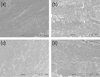 | Figure 1Representative photograph in the WL-3 mm groups. (a) #25, WL-3 mm; (b) #30, WL-3 mm; (c) #35, WL-3 mm; (d) #40, WL-3 mm. (a) and (b) presence of the smear layer on the surface, ×1,000; (c) presence of debris in the dentinal tubules, ×1,000; (d) removal of the smear layer from the surface, ×1,000.
|
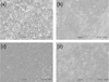 | Figure 2Representative photograph in the WL-9 mm groups. (a) #25, WL-9 mm; (b) #30, WL-9 mm; (c) #35, WL-9 mm; (d) #40, WL-9 mm. (a) and (b) smear layer is not removed, and tubule apertures are totally obliterated, ×1,000; (c) smear layer is thin as evidenced by crack over tubule aperture, ×1,000; (d) dentinal tubules are exposed, but some are blocked by smear layer, ×1,000.
|


References
1. Baugh D, Wallace J. The role of apical instrumentation in root canal treatment: a review of the literature. J Endod. 2005. 31:333–340.

2. Schilder H. Cleaning and shaping the root canal. Dent Clin North Am. 1974. 18:269–296.
3. Vertucci FJ. Root canal anatomy of the human permanent teeth. Oral Surg Oral Med Oral Pathol. 1984. 58:589–599.
4. Verma P, Love RM. A Micro CT study of the mesiobuccal root canal morphology of the maxillary first molar tooth. Int Endod J. 2011. 44:210–217.
5. Zehnder M. Root canal irrigants. J Endod. 2006. 32:389–398.
6. Rutala WA, Weber DJ. Uses of inorganic hypochlorite (bleach) in health-care facilities. Clin Microbiol Rev. 1997. 10:597–610.
7. Bystrom A, Sundqvist G. The antibacterial action of sodium hypochlorite and EDTA in 60 cases of endodontic therapy. Int Endod J. 1985. 18:35–40.
8. Yang SE, Bae KS. SEM study on the anaerobic bacterial adhesion to the dentin of root canal. J Korean Acad Conserv Dent. 2001. 26:350–359.
9. Chow TW. Mechanical effectiveness of root canal irrigation. J Endod. 1983. 9:475–479.
10. Abou-Rass M, Piccinino MV. The effectiveness of four clinical irrigation methods on the removal of root canal debris. Oral Surg Oral Med Oral Pathol. 1982. 54:323–328.
11. Kahn FH, Rosenberg PA, Gliksberg J. An in vitro evaluation of the irrigating characteristics of ultrasonic and subsonic handpieces and irrigating needles and probes. J Endod. 1995. 21:277–280.
12. Sedgley CM, Nagel AC, Hall D, Applegate B. Influence of irrigant needle depth in removing bioluminescent bacteria inoculated into instrumented root canals using real-time imaging in vitro. Int Endod J. 2005. 38:97–104.
13. Boutsioukis C, Lambrianidis T, Vasiliadis L. Clinical relevance of standardization of endodontic irrigation needle dimensions according to the ISO 9,626:1991 and 9,626:1991/Amd 1:2001 specification. Int Endod J. 2007. 40:700–706.
14. Boutsioukis C, Lambrianidis T, Kastrinakis E. Irrigant flow within a prepared root canal using various flow rates: a computational fluid dynamics study. Int Endod J. 2009. 42:144–155.
15. Boutsioukis C, Lambrianidis T, Kastrinakis E, Bekiaroglou P. Measurement of pressure and flow rates during irrigation of a root canal ex vivo with three endodontic needles. Int Endod J. 2007. 40:504–513.
16. Boutsioukis C, Gogos C, Verhaagen B, Versluis M, Kastrinakis E, Van der Sluis LW. The effect of apical preparation size on irrigant flow in root canals evaluated using an unsteady computational fluid dynamics model. Int Endod J. 2010. 43:874–881.
17. Lendini M, Alemanno E, Migliaretti G, Berutti E. The effect of highXMLLink_XYZfrequency electrical pulses on organic tissue in root canals. Int Endod J. 2005. 38:531–538.
18. Calvo Pérez V, Medina Cárdenas ME, Sánchez Planells U. The possible role of pH changes during EDTA demineralization of teeth. Oral Surg Oral Med Oral Pathol. 1989. 68:220–222.
19. Gulabivala K, Ng YL, Gilbertson M, Eames I. The fluid mechanics of root canal irrigation. Physiol Meas. 2010. 31:R49–R84.
20. Aktener BO, Bilkay U. Smear layer removal with different concentrations of EDTA-ethylenediamine mixtures. J Endod. 1993. 19:228–231.
21. Perez F, Rouqueyrol-Pourcel N. Effect of a low-concentration EDTA solution on root canal walls: a scanning electron microscopic study. Oral Surg Oral Med Oral Pathol Oral Radiol Endod. 2005. 99:383–387.
22. Torabinejad M, Khademi AA, Babagoli J, Cho Y, Johnson WB, Bozhilov K, Kim J, Shabahang S. A new solution for the removal of the smear layer. J Endod. 2003. 29:170–175.
23. Nakashima K, Terata R. Effect of pH modified EDTA solution to the properties of dentin. J Endod. 2005. 31:47–49.
24. Ram Z. Effectiveness of root canal irrigation. Oral Surg Oral Med Oral Pathol. 1977. 44:306–312.
25. Orstavik D, Kerekes K, Molven O. Effects of extensive apical reaming and calcium hydroxide dressing on bacterial infection during treatment of apical periodontitis: a pilot study. Int Endod J. 1991. 24:1–7.
26. Hoskinson SE, Ng YL, Hoskinson AE, Moles DR, Gulabivala K. A retrospective comparison of outcome of root canal treatment using two different protocols. Oral Surg Oral Med Oral Pathol Oral Radiol Endod. 2002. 93:705–715.
27. Kerekes K, Tronstad L. Long-term results of endodontic treatment performed with a standardized technique. J Endod. 1979. 5:83–90.
 XML Download