

 PDF
PDF ePub
ePub Citation
 Print
Print


Introduction
Since portable dental X-ray machine was developed to be used in military field, it has been used for forensics at disaster areas and for patients who cannot access care in dental offices, i.e. those who are home-bound or handicapped.1-6 Recently, portable dental X-rays have been used for identification of the dead bodies by Indonesia's tsunami damage or by hurricane in Louisiana, USA.7 In Korea, portable dental X-ray machines are sometimes used for implant surgery and endodontic treatment and even in general diagnosis, since it is convenient. Some American states including Michigan, Ohio, and Washington allow the limited use of portable dental X-ray machines and define the conditions of use for these machines.8-10 But, Korea doesn't have rules or guidelines on radiation protection for the portable dental X-ray machine.
The operator leakage and scattered radiation from portable dental radiography were greater than those from fixed dental radiography because it is handheld.11 According to National Institute of Food and Drug Safety Evaluation (NIFDS) regulation in Korea, when portable dental X-ray machine is used at places other than the operating room, the emergency room and the intensive care room, it is imperative to wear the lead apron and to use radiation protective partition.12 This inspecting standard is mostly about patient radiation protection, not for the operator who directly handles the equipment. The operator was prohibited from holding of the tube housing or cone with his or her hand to reduce the chance of occupational exposure. Moreover the operator should stand 6 feet from the X-ray tube during the exposure. Dentists or assistants using portable dental X-ray unit usually pay little attention on radiation protection due to their little understanding of operator exposure.
Therefore this study was done to evaluate the efficiency of various remedies for reduction of the operator' radiation dose in dental radiography by portable dental X-ray machines.
Materials and Methods
1. Materials
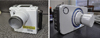
Dose measurements were conducted using a Human skull DXTRR III (Dentsply Rinn, Elgin, IL, USA) (Figure 1) and were recorded using an ionization chamber (Medical X-ray system test equipments Model 9015RM with 1,800 chamber, RadCal Corp., Monrovia, CA, USA) (Figure 2).
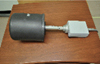
Two kinds of portable dental X-ray machines were included in this study. One (DX3000, Dexcowin, Seoul, Korea) was operated at 60 kVp, 1 mA and could be equipped with a removable external lead shield in place at the end of the cone. The other (Rextar, Posdion, Seoul, Korea) could be attached with two types of cones, 6 cm and 14 cm in length and operated at 70 kVp, 2 mA (Figure 3).
2. Methods
1) Operator's radiation dose measurement with the use of the backscatter shield
(1) At the operator's hands level
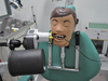
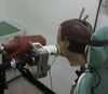
Dose measurement was carried out at the operator's hand level during the exposure of lower anterior and posterior teeth, with and without a lead backscatter shield attached at the end of cone. The setting for exposure was 60 kVp, 1 mA and 0.4 seconds for the lower anterior teeth, 0.8 seconds for the lower posterior teeth using digital sensor (Figures 4 and 5).
(2) At the operator's chest and waist levels
The scattered radiation was measured at the operator's chest and waist levels at a horizontal distance of 20 cm away from the X-ray machine during the exposure of lower posterior teeth, with and without a lead backscatter shield attached at the end of cone. A vertical distance of 130 cm from the floor was used as the chest height and 70 cm from the floor as the waist height (Figure 6).
2) Operator's radiation dose measurement with the use of the lead gloves
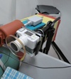
The doses at the hand level were measured in cases of wearing the lead gloves and not wearing them when human skull DXTTR III was exposed to portable X-ray unit. The lead sleeves which have lead equivalence 0.23 mm Pb at 60 kVp were used instead of the lead gloves due to size and form of ionization chamber (Figure 7).
3) Operator's radiation dose measurements according to the cone length of tubehead
Rextar X-ray machine (Posdion, Seoul, Korea) could attach two kinds of cone, 6 cm and 14 cm in length. Operator's radiation dose at the hand level were measured with both of them. Each setting time for anterior and posterior teeth exposure was 0.1 and 0.2 second in a 6 cm length cone, respectively. In a 14 cm cone, exposure time was 0.2 and 0.43 second, respectively (Figure 8).
Results
1. Operator's radiation dose with the use of the backscatter shield
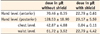
When a lower periapical radiograph was taken using a portable dental X-ray machine, the backscatter shield to the end of cone reduced operator's radiation dose to 32% (posterior teeth) and 23% (anterior teeth) at the hand level, 0.1% at the chest level and 37% at the waist level (Table 1).
2. Operator's radiation dose with the use of the lead gloves
When the lower anterior and posterior periapical radiography was taken using a portable dental X-ray machine, the lead gloves decreased operator's radiation dose at the hand level to 26% and 31% respectively (Table 2).
3. Operator's radiation dose according to the cone length of tubehead
When the lower anterior and posterior periapical radiography was taken using portable dental X-ray machine, the long cone (14 cm length cone) reduced operator's radiation dose at the hand level to 48% and 52% respectively (Table 3).
Discussion
The portable dental X-ray machine has the advantage of free movement outside the X-ray room. For this reason, the use of this equipment is on the increase in the dental clinics. Though its convenience and utility are accepted in the disaster area, the weak point is that the operator has the equipment in his/her hands. The operator has the possibility of direct exposure to leakage radiation from the tube head and scattered radiation from the patient.
Some authors reported that operator exposure due to leakage and scattered radiation using the hand-held dental X-ray system are well below established occupation exposure limits and insignificant compared with established radiation safety guidelines of 50 mSv per year.7,13-15 Even so, operator exposure to radiation should be reduced to keep the ALARA (as low as reasonably achievable) principle of dose optimization. Methods of dose reduction that can be used in portable dental radiography were offered. The focus in this study is not about the risk of the operators but the effect of dose reduction methods like backscatter shield, lead gloves and long cone. In further study, it is recommended to do dose measurements using TLD dosimetry to estimate operator's biologic effect.
Operator's radiation dose is mitigated by the use of shielding devices to reduce leakage exposure and to minimize backscatter from the patient.16 NOMAD (Arbiex, Orem, UT, USA) has an external lead-acrylic backscatter shield permanently attached at the end of the tube, with which the low operator exposure is described by the manufacturer.17 The use of the NOMAD presented no significant scattered radiation risk to any member of the operators.15,18 When three portable X-ray machines were tested for the occupational dosimetry, the dose at the operator's hand with protective shielding was the lowest.18 In this study, we found that the dose at the operator's hand level of the machine provided with the circular lead shields was reduced to 23 - 32%, in comparison to the case with the shields.
Most of portable dental X-ray machines, recently available in the Korea market, have no radio-protective shielding on the cone. Also, there is no legal standard for the additional reduction in radiation exposure to the operator. According to the results, we intend to provide a guideline for safe use of portable dental X-ray machines so that operators are able to use the appropriate shielding device while taking radiographs using a portable dental X-ray unit.
Conclusions
Use of a backscatter shield reduced the operator's radiation dose at hand level of X-ray tubehead to 23 - 32%, the lead gloves to 26 - 31%, and long cone of 14 cm to 48 - 52%. When portable dental X-ray systems are used, it is recommended to select the X-ray machine attached with a backscatter shield and a longer cone, and to wear the lead gloves.


 XML Download