

 PDF
PDF ePub
ePub Citation
 Print
Print


Abstract
Objectives
The purpose of this study was to evaluate the buccolingual curvature at the apical one third in type II mesial canals of mandibular molars using the radius and angle of curvature.
Materials and Methods
Total 100 mandibular molars were selected. Following an endodontic access in the teeth, their distal roots were removed. #15 H- or K-files (Dentsply Maillefer) were inserted into the mesiobuccal and mesiolingual canals of the teeth. Radiographs of the teeth were taken for the proximal view. Among them, type II canals were selected and divided into two subgroups, IIa and IIb. In type IIa, two separate canals merged into one canal before reaching the apex and in type IIb, two separate canals merged into one canal within the apical foramen. The radius and angle of curvature of specimens were examined.
Results
In type II, mean radius of curvature in mesiolingual and mesiobuccal canals were 2.82 mm and 3.58 mm, respectively. The radius of the curvature of mesiolingual canals were significantly smaller than that of mesiobuccal canals in type II, and especially in type IIa. However, there were no statistically significant differences in radius of curvature between mesiobuccal and mesiolingual canals in type IIb and there were no significant differences in angle of curvature between type IIa and IIb.
Figures and Tables
Figure 1
The proximal radiographic views of type II mesial canals of mandibular molar. (a) It shows type IIa mesial canals of mandibular molar; (b) It shows type IIb mesial canals of mandibular molar.
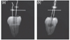
Figure 2
Technique used for determining the radius and angle of curvature in the proximal view. (a) Technique used for determining the radius and angle of curvature in type IIa mesial canals of mandibular molars; (b) Technique used for determining the radius and angle of curvature in type IIb mesial canals of mandibular molars. To determine these parameters, a straight line (l1 or l'1) is drawn along the long axis of coronal portion of the canal. A second line (l2 or l'2) is drawn along the long axis of the apical portion of the canal. There is a point on each of these lines at which the canal deviates to begin (point a1 or a2) or end (point b1 or b2) the canal curvature. The curved portion of the canal is represented by a sector with tangents at points a1 (or a2) and b1 (or b2). The angle of curvature (α1 and α2) is angle formed by perpendicular lines drawn from the points a1 (or a2) and b1 (or b2) that intersect at the center of sector (c1 or c2). The radius of curvature (γ1 and γ2) is the length of the radius of the sector measured in millimeters.
a1 or a2, a point on each of lines at which the canal deviates to begin; b1 or b2, a point on each of lines at which the canal deviates to end; c1 or c2, center of sector; l1 or l'1, line along the long axis of coronal portion of the canal; l2 or l'2, line along the long axis of the apical portion of the canal; α1 and α2, the angle of curvature; γ1 and γ2, the radius of curvature. This figure was partly modified with permission from the original one of Pruett et al.18
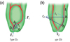
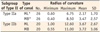
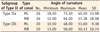
Figure 3
Radius and angle of curvature in Type IIa and IIb. In type IIa, radius of curvature of MLs were significantly smaller than MBs (p < 0.05). However, there were no statistically significant differences in radius of curvature between mesiobuccal and mesiolingual canals in type IIb and there were no significant differences in angle of curvature in type IIa, IIb.
ML, mesiolingual canal; MB, mesiobuccal canal.
*A significant difference was found between radius of ML and MB (p < 0.05).


References
1. Cunningham CJ, Senia ES. A three-dimensional study of canal curvatures in the mesial roots of mandibular molars. J Endod. 1992. 18:294–300.

2. Pineda F, Kuttler Y. Mesiodistal and buccolingual roentgenographic investigation of 7,275 root canals. Oral Surg Oral Med Oral Pathol. 1972. 33:101–110.
3. Cimilli H, Mumcu G, Cimilli T, Kartal N, Wasselink P. The correlation between root canal patterns and interorificial distance in mandibular first molars. Oral Surg Oral Med Oral Pathol Oral Radiol Endod. 2006. 102:e16–e21.
4. Peters OA. Current challenges and concepts in the preparation of root canal systems: a review. J Endod. 2004. 30:559–567.
5. Tzanetakis GN, Kontakiotis EG, Maurikou DV, Marzelou MP. Prevalence and management of instrument fracture in the postgraduate endodontic program at the Dental School of Athens: a five-year retrospective clinical study. J Endod. 2008. 34:675–678.
6. Iqbal MK, Kohli MR, Kim JS. A retrospective clinical study of incidence of root canal instrument separation in an endodontics graduate program: a PennEndo database study. J Endod. 2006. 32:1048–1052.
7. Vertucci FJ. Root canal anatomy of the human permanet teeth. Oral Surg Oral Med Oral Pathol. 1984. 58:589–599.
8. Sattapan B, Nervo GJ, Palamara JE, Messer HH. Defects in rotary nickel-titanium files after clinical use. J Endod. 2000. 26:161–165.
9. Sotokawa T. An analysis of clinical breakage of root canal instruments. J Endod. 1988. 14:75–82.
10. Parashos P, Messer HH. Rotary NiTi instrument fracture and its consequence. J Endod. 2006. 32:1031–1043.
11. Wolcott S, Wolcott J, Ishley D, Kennedy W, Johnson S, Minnich S, Meyers J. Separation incidence of protaper rotary instruments: a large cohort clinical evaluation. J Endod. 2006. 32:1139–1141.
12. Kartal N, Cimilli HK. The degrees and configurations of mesial canal curvatures of mandibular first molars. J Endod. 1997. 23:358–362.
13. Schneider SW. A comparison of canal preparations in straight and curved root canals. Oral Surg Oral Med Oral Pathol. 1971. 32:271–275.
14. Weine FS. Endodnotic therapy. 1996. 5th ed. St. Louis: Mosby;356–357.
15. Kyomen SM, Caputo AA, White SN. Critical analysis of the balanced force technique in endodontics. J Endod. 1994. 20:332–337.
16. Hankins PJ, ElDeeb ME. An evaluation of the canal master, balanced force, and step-back techniques. J Endod. 1996. 22:123–130.
17. Günday M, Sazak H, Garip Y. A comparative study of three different root canal curvature measurement techniques and measuring the canal access angle in curved canals. J Endod. 2005. 31:796–798.
18. Pruett JP, Clement DJ, Carnes DL Jr. Cyclic fatigue testing of nickel-titanium endodontic instruments. J Endod. 1997. 23:77–85.
19. Jeong H, Park SJ, Park SH, Choi GW. Morphology of the apical root canal system in Korean mandibular first molar. J Korean Acad Conserv Dent. 2009. 34:137–144.
20. Zelada G, Varela P, Martín B, Bahíllo JG, Magán F, Ahn S. The effect of rotational speed and the curvature of root canals on the breakage of rotary endodontic instruments. J Endod. 2002. 28:540–542.
21. Alomairy KH. Evaluating two techniques on removal of fractured rotary nickel-titanium endodontic instruments from root canals: an in vitro study. J Endod. 2009. 35:559–562.
22. Kum KY, Kim SY, Kim ES, Kim HC, Baek SH, Cho YB, Hwang HK. Practice and application of NiTi rotary systems in clinical endodontics. 2006. 1st ed. Seoul: DaehanNarae Publishing Inc.;18–37.
23. Lee DG, Park JM, Hwang HK. A study on the c-shaped root canal system of mandibular second molar. J Korean Acad Conserv Dent. 2007. 32:335–342.
24. Slowey RR. Root canal anatomy. Road map to successful endodontics. Dent Clin North Am. 1979. 23:555–573.
25. Haïkel Y, Serfaty R, Bateman G, Senger B, Allemann C. Dynamic and cyclic fatigue of engine-driven rotary nickel-titanium endodontic instruments. J Endod. 1999. 25:434–440.
26. Schäfer E, Diez C, Hoppe W, Tepel J. Roentgenographic investigation of frequency and degree of canal curvatures in human permanent teeth. J Endod. 2002. 28:211–216.
 XML Download