

 PDF
PDF ePub
ePub Citation
 Print
Print


Abstract
Background:
Studies on the relationship between vitamin D and visceral fat area (VFA; intra-abdominal fat area) have been actively conducted. But, there is a few Korean population-based studies about the association between serum vitamin D level and VFA. The aim of our study was to explore the correlation between serum 25-hydroxyvitamin D (25[OH]D) levels and VFA measured using bioelectrical impedance analysis (BIA; electric impedance) in healthy Korean adults.
Methods:
This cross-sectional study involved 1,945 adults aged 20-70 years who visited a health promotion center. All subjects underwent the BIA to estimate the VFA. Serum 25(OH)D level was measured using chemiluminescent immunoassay. Multiple regression analysis was performed to identify independent correlation of VFA and serum 25(OH)D level.
Results:
The prevalence of vitamin D deficiency (25[OH]D: 20-29 ng/mL) and insufficiency (25[OH]D <20 ng/mL) were 54.4% and 38.1%, respectively. After having adjusted age and season, VFA were negatively associated with serum 25(OH)D levels in both men (P<0.001) and women (P<0.001). The obese group with VFA ≥ 100 cm2 had significant lower serum 25(OH)D level in men (P<0.001) and women (P=0.0034).
Go to : 
REFERENCES
1.Lips P. Vitamin D deficiency and secondary hyperparathyroidism in the elderly: consequences for bone loss and fractures and therapeutic implications. Endocr Rev. 2001. 22(4):477–501.

2.Ahn J., Albanes D., Berndt SI., Peters U., Chatterjee N., Freedman ND, et al. Vitamin D-related genes, serum vitamin D concentrations and prostate cancer risk. Carcinogenesis. 2009. 30(5):769–76.
3.Cardoso AT., Nanji L., Costa J., Vaz-Carneiro A. Analysis of the cochrane review: vitamin D supplementation for prevention of cancer in adults. Cochrane Database Syst Rev. 2014, 6: CD007469. Acta Med Port. 2014. 27(4):411–3.
4.Strange RC., Shipman KE., Ramachandran S. Metabolic syndrome: a review of the role of vitamin D in mediating susceptibility and outcome. World J Diabetes. 2015. 6(7):896–911.
5.Yao Y., Zhu L., He L., Duan Y., Liang W., Nie Z, et al. A meta-analysis of the relationship between vitamin D deficiency and obesity. Int J Clin Exp Med. 2015. 8(9):14977–84.
6.Zittermann A., Ernst JB., Gummert JF., Börgermann J. Vitamin D supplementation, body weight and human serum 25-hydrox-yvitamin D response: a systematic review. Eur J Nutr. 2014. 53(2):367–74.
7.Fox CS., Massaro JM., Hoffmann U., Pou KM., Mauro-vich-Horvat P., Liu CY, et al. Abdominal visceral and subcutaneous adipose tissue compartments: association with metabolic risk factors in the Framingham Heart Study. Circulation. 2007. 116(1):39–48.
8.Holick MF. Vitamin D deficiency. N Engl J Med. 2007. 357(3):266–81.
9.Organization WH. The Asia-Pacific perspective: redefining obesity and its treatment. Sydney: Health Communications Australia. 2000.
10.Sim SJ., Park HS. The cut-off values of body fat to identify cardiovascular risk among Korean adults. J Korean Soc Study Obes. 2004. 13(1):14–21.
11.Choi HJ., Yoon K., Han KH., Kim SH. The cut-off values of the visceral fat area to identify metabolic syndrome among Korean adults: using visceral fat area presented by body composition analyzer, inbody 4.0. Korean J Obes. 2006. 15(4):219–26.
12.Lips P., Hosking D., Lippuner K., Norquist JM., Wehren L., Maalouf G, et al. The prevalence of vitamin D inadequacy amongst women with osteoporosis: an international epidemiological investigation. J Intern Med. 2006. 260(3):245–54.
13.Arunabh S., Pollack S., Yeh J., Aloia JF. Body fat content and 25-hydroxyvitamin D levels in healthy women. J Clin Endocrinol Metab. 2003. 88(1):157–61.
14.Snijder MB., van Dam RM., Visser M., Deeg DJ., Dekker JM., Bouter LM, et al. Adiposity in relation to vitamin D status and parathyroid hormone levels: a population-based study in older men and women. J Clin Endocrinol Metab. 2005. 90(7):4119–23.
15.Jorde R., Sneve M., Emaus N., Figenschau Y., Grimnes G. Cross-sectional and longitudinal relation between serum 25-hydroxyvitamin D and body mass index: the Tromsø study. Eur J Nutr. 2010. 49(7):401–7.
16.Sun X., Cao ZB., Tanisawa K., Ito T., Oshima S., Ishimi Y, et al. Associations between the serum 25(OH)D concentration and lipid profiles in Japanese men. J Atheroscler Thromb. 2015. 22(4):355–62.
17.Shin SR., Han AL., Park SH. Vitamin D status and its relation with abdominal adiposity and cardiovascular risk factors of Korean adults in certain areas. Korean J Obes. 2015. 24(1):30–5.
18.Ross AC., Manson JE., Abrams SA., Aloia JF., Brannon PM., Clinton SK, et al. The 2011 report on dietary reference intakes for calcium and vitamin D from the Institute of Medicine: what clinicians need to know. J Clin Endocrinol Metab. 2011. 96(1):53–8.
19.Vanlint S. Vitamin D and obesity. Nutrients. 2013. 5(3):949–56.
20.Hjelmesaeth J., Hofsø D., Aasheim ET., Jenssen T., Moan J., Hager H, et al. Parathyroid hormone, but not vitamin D, is associated with the metabolic syndrome in morbidly obese women and men: a cross-sectional study. Cardiovasc Diabetol. 2009. 8:7.
21.Kremer R., Campbell PP., Reinhardt T., Gilsanz V. Vitamin D status and its relationship to body fat, final height, and peak bone mass in young women. J Clin Endocrinol Metab. 2009. 94(1):67–73.
22.Mai XM., Chen Y., Camargo CA Jr., Langhammer A. Cross-sectional and prospective cohort study of serum 25-hydroxyvitamin D level and obesity in adults: the HUNT study. Am J Epidemiol. 2012. 175(10):1029–36.
23.Pathak K., Soares MJ., Calton EK., Zhao Y., Hallett J. Vitamin D supplementation and body weight status: a systematic review and meta-analysis of randomized controlled trials. Obes Rev. 2014. 15(6):528–37.
24.Chandler PD., Wang L., Zhang X., Sesso HD., Moorthy MV., Obi O, et al. Effect of vitamin D supplementation alone or with calcium on adiposity measures: a systematic review and meta-analysis of randomized controlled trials. Nutr Rev. 2015. 73(9):577–93.
25.Pannu PK., Zhao Y., Soares MJ. Reductions in body weight and percent fat mass increase the vitamin D status of obese subjects: a systematic review and metaregression analysis. Nutr Res. 2016. 36(3):201–13.
26.Kim D., Kim J. Association between serum 25-hydroxyvitamin D levels and adiposity measurements in the general Korean population. Nutr Res Pract. 2016. 10(2):206–11.
27.Nagai M., Komiya H., Mori Y., Ohta T., Kasahara Y., Ikeda Y. Estimating visceral fat area by multifrequency bioelectrical impedance. Diabetes Care. 2010. 33(5):1077–9.
28.Xu L., Cheng X., Wang J., Cao Q., Sato T., Wang M, et al. Comparisons of body-composition prediction accuracy: a study of 2 bioelectric impedance consumer devices in healthy Chinese persons using DXA and MRI as criteria methods. J Clin Densitom. 2011. 14(4):458–64.
29.Ryo M., Maeda K., Onda T., Katashima M., Okumiya A., Nishida M, et al. A new simple method for the measurement of visceral fat accumulation by bioelectrical impedance. Diabetes Care. 2005. 28(2):451–3.
30.Zhang M., Li P., Zhu Y., Chang H., Wang X., Liu W, et al. Higher visceral fat area increases the risk of vitamin D insufficiency and deficiency in Chinese adults. Nutr Metab (Lond). 2015. 12:50.
Go to :
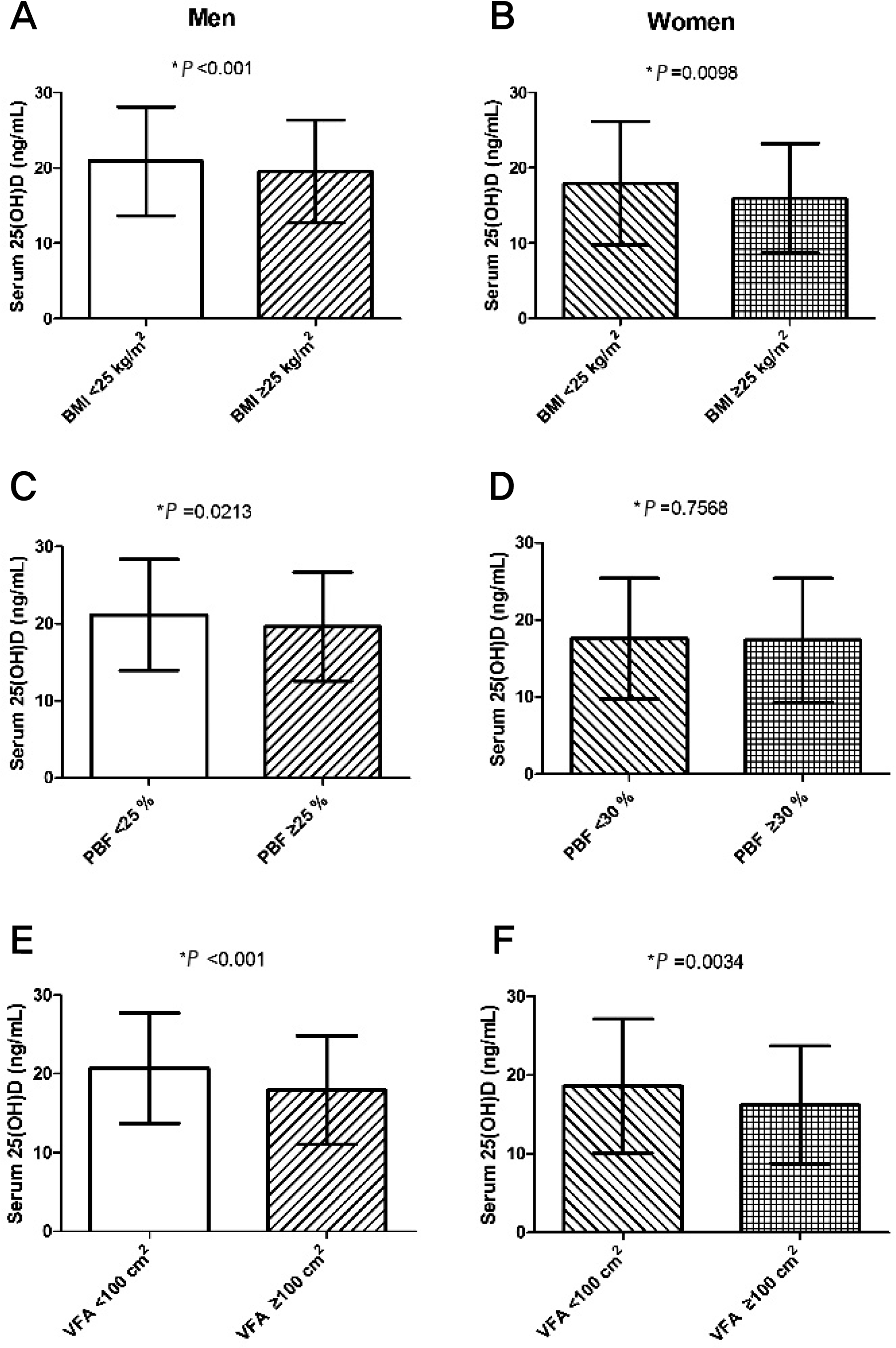 | Figure 1.Differential serum 25-hydroxyvitamin D (25[OH]D) concentrations in subjects with or without obesity. (A, B) Comparison of serum 25-hydroxyvitamin D (25[OH]D) levels according to body mass index (BMI) in men and women. Subjects with a BMI ≥25 kg/m2 were classified as obese. (C, D) Comparison of serum 25(OH)D levels according to percentage body fat (PBF) in men and women. Men with a PBF ≥25 % and women with a PBF ≥30 % were classified as obese. (E, F) Comparison of serum 25[OH]D levels according to visceral fat area (VFA) in men and women. Subjects with a VFA ≥100 cm2 were classified as obese. Data are presented as mean±standard deviation. ∗P value from an independent-sample t-test. |
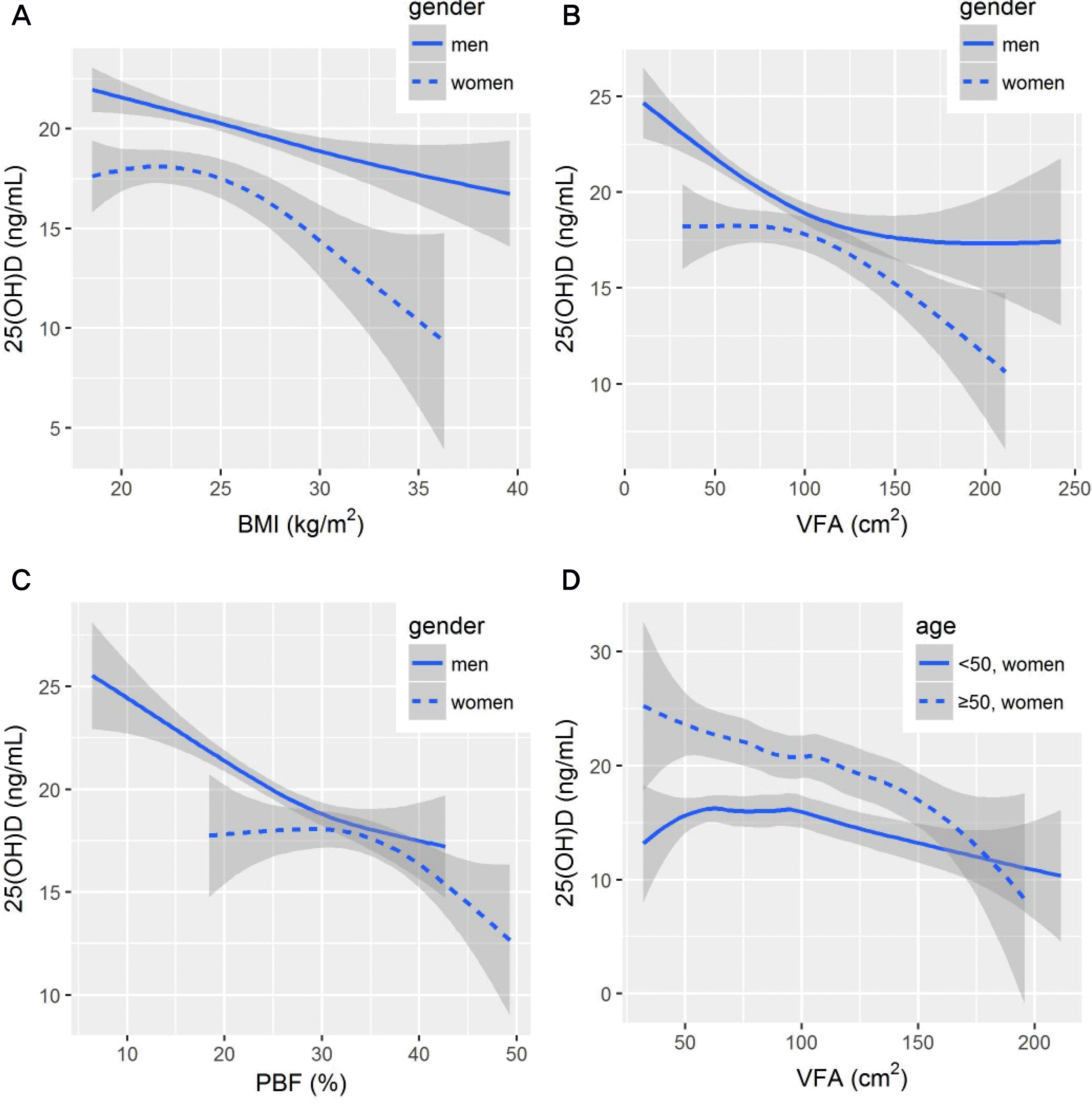 | Figure 2.Correlation between 25(OH)D and fatness indices in men and women (A-C); in women aged ≥50 and <50 years (D). Smoothing curve with 95% confidence interval (shaded areas) are fitted by a Loess curve. |
Table 1.
Characteristics of study subjects
| Total (n=1,945) | Men (n=1,395) | Women (n=550) | Pa | |
|---|---|---|---|---|
| Age (y) | 46±9.5 | 46.3±9.1 | 47±10.4 | 0.204 |
| BMI (kg/m2) | 24.6±3.2 | 25±3 | 23.3±3.2 | <0.001 |
| Normal (18.5-22.9) | 639 (32.9) | 346 (24.8) | 293 (53.3) | <0.001 |
| Overweight (23-24.9) Obese (≥25) | 517 (26.6) 789 (40.6) | 401 (28.7) 648 (46.5) | 116 (21.1) 141 (25.6) | 0.001 <0.001 |
| Percentage body fat (%) | 27±8 | 24.4±5.4 | 33.1±5.8 | <0.001 |
| Non-obese (<25 in males, <30 in females) | 939 (48.3) | 786 (56.3) | 153 (27.8) | <0.001 |
| Obese (≥25 in males, ≥30 in females) | 1,006 (51.7) | 619 (43.7) | 397 (72.2) | <0.001 |
| VFA (cm2) | 82.5±31.7 | 78±29 | 94.1±10.4 | <0.001 |
| Normal (<100) High-VFA (≥100) | 1,501 (77.2) 444 (22.8) | 1,161 (83.2) 234 (16.8) | 340 (61.8) 210 (38.2) | <0.001 <0.001 |
| Blood pressure (mmHg) | ||||
| Systolic | 118.5±13.9 | 120.4±13.2 | 113.4±14.4 | <0.001 |
| Diastolic | 78.4±11.3 | 80.1±11.1 | 74.3±10.9 | <0.001 |
| 25(OH)D (ng/mL) | 19.5±7.4 | 20.3±7 | 17.3±8 | <0.001 |
| Normal (≥30) | 144 (7.4) | 103 (7.4) | 41 (7.5) | 0.957 |
| Insufficiency (20-29.9) Deficiency (<20) | 742 (38.1) 1,059 (54.4) | 596 (42.7) 696 (49.9) | 146 (26.5) 363 (66) | <0.001 <0.001 |
| Total cholesterol (mg/dL) | 193.4±34.8 | 193.4±35 | 193.6±34.4 | 0.916 |
| Triglyceride (mg/dL) | 138.9±91 | 153.1±98.3 | 102.8±54 | <0.001 |
| HDL cholesterol (mg/dL) | 50.7±11.8 | 48.2±10.3 | 57.1±13.1 | <0.001 |
| LDL cholesterol (mg/dL) | 119.2±25.6 | 120.4±25.7 | 116.2±25.3 | 0.001 |
| Fasting glucose (mg/dL) | 87.2±10.4 | 88.1±10.6 | 84.8±9.8 | <0.001 |
Table 2.
Correlation between serum 25(OH)D and variables
| Men | Women | |||
|---|---|---|---|---|
| ra | Pb | r | P | |
| Age | 0.142 | <0.001 | 0.305 | <0.001 |
| Body mass index | -0.127 | <0.001 | -0.134 | 0.002 |
| Percentage body fat | -0.195 | <0.001 | -0.096 | 0.025 |
| Visceral fat area | -0.185 | <0.001 | -0.153 | <0.001 |
| Systolic blood pressure | -0.079 | 0.003 | -0.038 | 0.379 |
| Diastolic blood pressure Total cholesterol | -0.115 -0.087 | <0.001 0.001 | -0.064 0.060 | 0.133 0.160 |
| Triglyceride | -0.312 | <0.001 | -0.163 | <0.001 |
| High density lipoprotein cholesterol | 0.058 | 0.031 | 0.006 | 0.892 |
| Low density lipoprotein cholesterol | -0.029 | 0.284 | 0.105 | 0.014 |
| Fasting glucose | -0.062 | 0.020 | -0.029 | 0.493 |
Table 3.
Regression analysis of relationship between vitamin D and obesity indices
| Men | Women | |||
|---|---|---|---|---|
| β±SE | Pa | β±SE | P | |
| Model 1b | ||||
| Body mass index | -0.004±0.064 | 0.950 | -0.342±0.111 | 0.002 |
| Percentage body fat | -0.142±0.035 | <0.001 | -0.158±0.059 | 0.008 |
| Visceral fat area | -0.018±0.007 | 0.008 | -0.034±0.010 | -0.149 |
| Model 2c | ||||
| Body mass index Percentage body fat | -0.188±0.060 -0.211±0.033 | 0.002 <0.001 | -0.492±0.100 -0.225±0.056 | <0.001 <0.001 |
| Visceral fat area | -0.032±0.006 | <0.001 | -0.046±0.009 | <0.001 |
 XML Download