ABSTRACT
Middle aortic syndrome (MAS) is very uncommon vascular pathology characterized by a long segmental narrowing or obstruction of the abdominal and/or distal thoracic aorta, commonly involving with the visceral and renal arteries. This syndrome may be presented with various physical signs of coarctation of the aorta, including resistant hypertension, renal insufficiency and/or mesenteric ischemia. Here, we report a case of a 64-year-old man with severe hypertension. He was diagnosed with MAS associated with stenosis of visceral and renal vessels by use of computed tomography and magnetic resonance angiography.
References
1. Celik T, Kursaklioglu H, Iyisoy A, Turhan H, Amasyali B, Kocaoglu M, et al. Hypoplasia of the descending thoracic and abdominal aorta: a case report and review of literature. J Thorac Imaging. 2006; 21:296–9.

2. Terramani TT, Salim A, Hood DB, Rowe VL, Weaver FA. Hypoplasia of the descending thoracic and abdominal aorta: a report of two cases and review of the literature. J Vasc Surg. 2002; 36:844–8.

3. Sen PK, Kinare SG, Engineer SD, Parulkar GB. The middle aortic syndrome. Br Heart J. 1963; 25:610–8.

5. Delis KT, Gloviczki P. Middle aortic syndrome: from presentation to contemporary open surgical and endovascular treatment. Perspect Vasc Surg Endovasc Ther. 2005; 17:187–203.

6. Ilica AT, Bilici A, Ilhan A, Kara M, Gur S. Thoracoabdominal aorta coarctation with bilateral renal artery involvement: diagnosis with multidetector CT angiography (MDCTA). Int J Cardiovasc Imaging. 2007; 23:645–8.

7. Tosun O, Sanlidilek U, Cetin H, Ozdemir O, Kurt A, Sakarya ME, et al. Unusual congenital aortic anomaly with rare common celiamesenteric trunk variation: MR angiography and digital substraction angiography findings. Cardiovasc Intervent Radiol. 2007; 30:1061–4.

8. Kim SJ, Ha J. Middle aortic syndrome: report of 4 cases. Korean J Vasc Endovasc Surg. 1995; 11:230–7.
9. Kim DK, Kim YJ, Ryu JS, Eom WS, Cho JH, Jeong YT. Middle aortic syndrome diagnosed at 51 years of age. Korean J Med. 2004; 66:293–7.
10. Kim JA, Kim DB, Jang SW, Kwon BJ, Cho EJ, Song JH, et al. Hypertensive heart failure with severe arteriosclerotic stenosis of the descending aorta. Korean Circ J. 2007; 37:590–3.

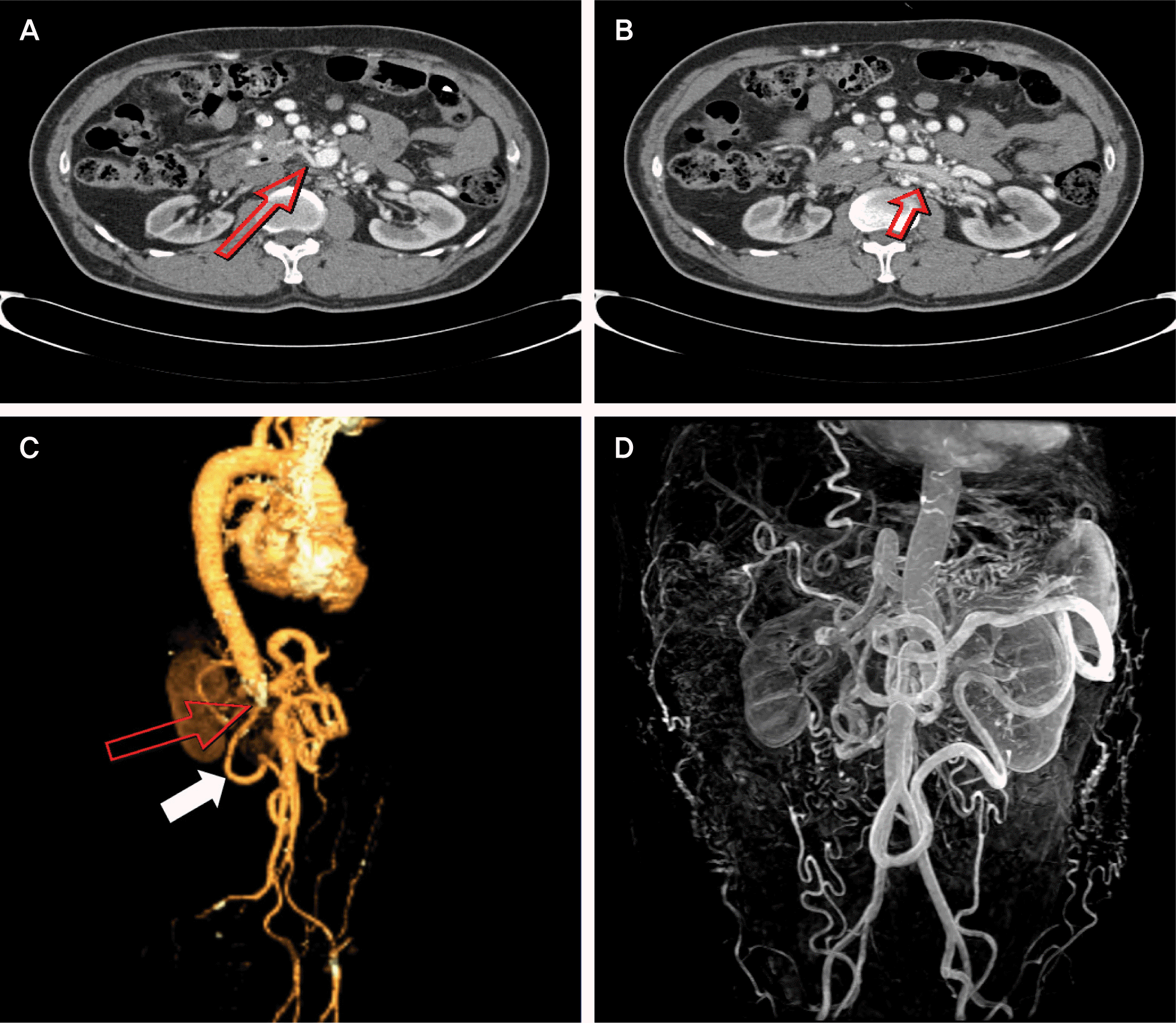
Fig. 1.
Imaging of computed tomography (CT) and magnetic resonance angiography (MRA). (A) Origin of right renal artery (arrow) was not found around aorta, supplied by branch vessels arose from distal superior mesenteric artery and left renal artery. (B) Left renal artery (arrow) originating from hypoplastic abdominal aorta was severely calcified and stenosed. (C) Abrupt luminal obstruction of abdominal aorta just below the celiac trunk (Upper arrow). Superior mesenteric artery is supplied by the arc of Riolan (Lower arrow). Both iliac arteries are normal. (D) MRA showing similar but more detailed vasculature as compared with CT.


 PDF
PDF ePub
ePub Citation
 Print
Print


 XML Download