Journal List > Electrolyte Blood Press > v.6(1) > 1089757

Kim: The Role of Angiopoietin-1 in Kidney Disease

Abstract

Injury to the renal microvasculature and inflammatory process may be major factors in the progression of renal disease, therefore, protection of the renal endothelial cell and regulation of inflammatory process may be an important therapeutic target of renal disease. Thus, we evaluated the protective effect of cartilage oligomeric matrix protein-angiopoietin-1 (COMP-Ang1) in unilateral ureteral obstruction (UUO)-induced renal fibrosis, cyclosporine A (CsA)-induced renal injury, and the diabetic nephropathy model. In the UUO model, morphologic examination indicated less tubular injury and tubulointerstitial fibrosis in mice that received COMP-Ang1 compared to vehicle-treated mice. Interstitial type I collagen, myofibroblast accumulation, renal surface microvasculature and renal blood flow were higher after treatment with COMP-Ang1 compared to vehicle-treated mice. COMP-Ang1 treatment decreased monocyte/macrophage infiltration, tissue levels of transforming growth factor β1, and Smad 2/3 phosphorylation and increased Smad 7 in the obstructed kidney. In CsA-induced renal injury, histologic examination showed significantly decreased CsA-induced tubular damage and tubulointerstitial fibrosis in COMP-Ang1 treated mice. COMP-Ang1 administration also decreased increased macrophage infiltration, adhesion molecule expression, TGF-β1, and Smad 2/3 levels in CsA-treated kidneys, while increasing Smad 7 levels. Laser-Doppler sonographic findings and endothelial factor VIII staining revealed that COMP-Ang1 had a preservative effect on peritubular vasculature. In the diabetic nephropathy model, COMP-Ang1 reduced albuminuria and decreased mesangial expansion, thickening of the glomerular basement membrane and podocyte foot process broadening and effacement. COMP-Ang1 may delay the fibrotic changes in the kidney of diabetic db/db mice through its anti-inflammatory or metabolic effects. In conclusion, COMP-Ang1 may be an endothelium-specific and anti-inflammatory therapeutic modality in fibrotic renal disease.

Introduction

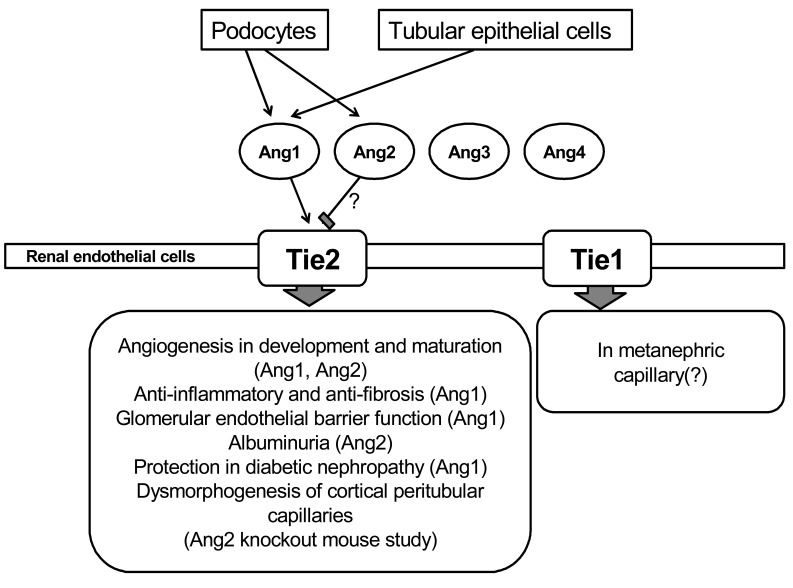
Angiopoietins are a family of growth factors which act on the Tie receptor on endothelial cells. The angiopoietin family consists of 4 angiopoietins, i.e., angiopoietin-1 (Ang1), angiopoietin-2 (Ang2), angiopoietin-3 (Ang3), and angiopoietin-4 (Ang4)1-3). Ang1 is known as an angiogenic factor4), however, the function of Ang2, Ang3 and Ang4 is not well known. Angiopoietins' receptor system consists of two receptor tyrosine kinases, Tie1 and Tie2. The Tie receptors are exclusively expressed by endothelial cells and hematopoietic cells5). It has been also reported that Tie2 is also expressed in macrophages, eosinophils, and hematopoietic stem cells5, 6). Although Tie1 and Tie2 share a similar structure consisting of an extracellular domain with 33% similarity and an intracellular tyrosine kinase domain with 76% similarity, the exact role of Tie1 is not well known 7). Ang1 and Ang2 bind to the same site in Tie2 with similar affinities. Ang1 is a widely expressed ligand for the Tie2 tyrosine kinase receptor that is expressed on endothelial cells, and it regulates vascular growth, development, maturation, and permeability8, 9). Ang1-induced Tie2 phosphorylation in endothelial cells can activate the protein kinase B/Akt pathway that conducts endothelial survival10-12). Although Ang1 is known as an angiogenic factor, it has been also demonstrated that Ang1 has an anti-inflammatory effect13, 14). Although the exact role of Ang2 is not well known, it has been reported that Ang2 functions as an antagonist ligand of Tie215). Ang1 has potential therapeutic applications in inducing angiogenesis, enhancing endothelial cells survival, and preventing vascular leakage12, 16-20) (Fig. 1).
In this section, I focus on the therapeutic effect of Ang1 in kidney disease

Angiopoietin-1 and Tie2 expression in kidney

Ang1 is also associated with physiologic or pathophysiologic conditions in the kidney21). In kidney development, Tie2 is detected in capillaries of the nephrogenic cortex, glomerular tufts, cortical interstitium, and medulla including vessels in the vasa recta22, 23). Thus, Ang1 and Tie2 play roles in the maturation of glomeruli and the vasa rectae. Ang1 protein is detected in podocytes in normal glomeruli and was highly expressed in podocyte foot processes24). Tie2 has been demonstrated on glomerular capillary endothelial cells and peritubular capillary endothelial cells23). Thus, circulating Ang1 may have an effect on renal Tie2 in the endothelial cell and renal Ang1 may also have paracrine effect on renal Tie225, 26).
Disturbance of endothelial integrity in the kidney may cause a reduction in glomerular filtration rate or proteinuria. Ang1 in the glomerulus or renal epithelial cells may have a role in maintenance of the glomerular or peritubular endothelium in the kidney. In pathophysiologic conditions, folic acid-induced nephropathy is associated with increased Ang1 protein expression in renal epithelia and arteries27). It was also reported that loss of glomerular capillaries during the course of anti-glomerular basement membrane glomerulonephritis in mice was temporally associated with decreases in endothelial survival molecules Ang128). Therefore, Ang1 may have an important role in maintenance of normal kidney development of the glomerular or peritubular endothelium and various kidney disease conditions.

Cartilage oligomeric matrix protein-angiopoietin-1 in renal fibrosis

Most forms of chronic kidney disease finally progress to interstitial fibrosis. The severity of tubulointerstitial changes is the best indicator of the progression of renal dysfunction29, 30). Renal tubulointerstitial fibrosis is an important feature in unilateral ureteral obstruction (UUO). In humans, chronic and acute ureteral obstruction can occur in various clinical situations such as ureteral stone or ureteral carcinoma. Injury to the renal microvasculature and inflammatory condition may be major factors in the process of kidney disease31). In particular, injury to the peritubular capillary endothelium of the kidney may be a factor in tubulointerstitial disease32-34). It has been reported that impaired angiogenesis may occur in the diseased kidney and can contribute to renal scarring33). Renal ischemia that is caused by vascular obliteration can be a major contributor to renal scarring. Therefore, endothelial cells play an important role in renal disease progression in the unilateral ureteral obstructed kidney. Because renal microvasculature injury involves a critical process in renal fibrosis, a growth factor or cytokine with an endothelial protective or angiogenic effect may have a protective role in renal fibrosis in the UUO model35-37).
Recently, a variant of Ang1, cartilage oligomeric matrix protein (COMP)-Ang1 was recently developed. COMP-Ang1 is soluble and more potent than native Ang1 in phosphorylating Tie2 and signaling via Akt in primary cultured endothelial cells13, 38). Thus, I evaluated the protective effect of COMP-Ang1 in the UUO-induced renal fibrosis model39).
In the UUO model, COMP-Ang1 preserved the renal platelet-endothelial cell adhesion molecule-1- and Tie2-positive endothelial cell. Morphologic examination demonstrated less tubular injury and renal interstitial fibrosis in mice that received COMP-Ang1 than in vehicle-treated mice. Type I collagen and myofibroblast accumulation were significantly decreased by COMP-Ang1 treatment. COMP-Ang1 stimulated Tie2 and Akt phosphorylation in ureteral obstructed kidneys. Renal surface microvasculature and renal blood flow were higher after treatment with COMP-Ang1 than in vehicle-treated mice. COMP-Ang1 treatment suppressed monocyte/macrophage infiltration, tissue levels of transforming growth factor β1 (TGF-β1), and Smad 2/3 phosphorylation and increased Smad 7 in the obstructed kidney. These results suggest that COMP-Ang1 treatment can prevent the progression of renal fibrosis in UUO.

Cartilage oligomeric matrix protein-angiopoietin-1 in diabetic nephropathy model

Inflammatory processes have been recently seen as the important factor in the pathogenesis of diabetic nephropathy. In this study, db/db mice were treated with adenovirus expressing either COMP-Ang1 or LacZ40). To evaluate histology, inflammatory, metabolic, and fibrotic parameters and signalling pathways, we used diabetic nephropathy model.
COMP-Ang1 reduced albuminuria in 24 hour urine analysis and decreased mesangial expansion, thickening of the glomerular basement membrane, and podocyte foot process broadening and effacement in histologic examination. COMP-Ang1 decreased both kidney expression of the adhesion molecule and the number of f4/80-positive macrophage infiltration in diabetic db/db mice. In addition, COMP-Ang1 significantly decreased fasting blood glucose level, epididymal fat weight to body weight ratio, and epididymal adipocyte size in diabetic db/db mice. COMP-Ang1 also ameliorated the fibrotic process in the kidney of diabetic db/db mice through its anti-inflammatory and metabolic effects. COMP-Ang1 suppressed renal levels of TGF-β1, α-smooth muscle actin, fibronectin, as well as Smad 2/3, but increased Smad 7. In human umbilical vein endothelial cells, COMP-Ang1 treatment decreased high glucose-induced nuclear factor-κB (NF-κB) activation. COMP-Ang1 mediated inhibition of increased NF-κB-DNA binding activity in nuclear extracts from human umbilical vein endothelial cells grown in high glucose was significantly blocked by soluble Tie2 receptor-Fc. The side effect of COMP-Ang1 is not well known. However, there are several conditions associated with COMP-Ang1 such as slight increased redness in the nose and increased vascular density in the trachea. These results suggest that COMP-Ang1 ameliorated the fibrotic processes in the kidney of diabetic db/db mice through its anti-inflammatory or metabolic effects.

Cartilage oligomeric matrix protein-angiopoietin-1 in cyclosporine-induced renal injury

In cyclosporine (CsA)-induced renal injury, COMP-Ang1 significantly decreased CsA-induced tubular damage and tubulointerstitial fibrosis in histologic examination. COMP-Ang1 also reduced the number of macrophage infiltration and expression of adhesion molecules in the CsA-induced renal injury model. COMP-Ang1 administration decreased CsA-induced increase of TGF-β1 and Smad 2/3 in kidneys, while increasing Smad 7 levels. Laser-Doppler sonographic findings and endothelial factor VIII staining revealed that COMP-Ang1 had a protective effect on peritubular vasculature and intrarenal hemodynamic alteration in CsA-induced renal injury.

Conclusion

The protective effect of COMP-Ang1 on of the renal endothelial cell and regulation of inflammatory process may be an important therapeutic target of renal disease. Therefore, COMP-Ang1 may be an endothelium-specific therapeutic modality in various renal diseases.

References

1. Davis S, Yancopoulos GD. The angiopoietins: Yin and Yang in angiogenesis. Curr Top Microbiol Immunol. 1999; 237:173–185. PMID: 9893351.
crossref
2. Valenzuela DM, Griffiths JA, Rojas J, Aldrich TH, Jones PF, Zhou H, et al. Angiopoietins 3 and 4: diverging gene counterparts in mice and humans. Proc Natl Acad Sci U S A. 1999; 96:1904–1909. PMID: 10051567.
crossref
3. Nishimura M, Miki T, Yashima R, Yokoi N, Yano H, Sato Y, et al. Angiopoietin-3, a novel member of the angiopoietin family. FEBS Lett. 1999; 448:254–256. PMID: 10218486.
crossref
4. Koblizek TI, Weiss C, Yancopoulos GD, Deutsch U, Risau W. Angiopoietin-1 induces sprouting angiogenesis in vitro. Curr Biol. 1998; 8:529–532. PMID: 9560344.
crossref
5. Kukk E, Wartiovaara U, Gunji Y, Kaukonen J, Buhring HJ, Rappold I, et al. Analysis of Tie receptor tyrosine kinase in haemopoietic progenitor and leukaemia cells. Br J Haematol. 1997; 98:195–203. PMID: 9233584.
crossref
6. Suda T. Function of TIE2/angiopoietin in the angiogenesis and hematopoiesis. Rinsho Ketsueki. 2000; 41:251–255. PMID: 10846448.
7. Schnurch H, Risau W. Expression of tie-2, a member of a novel family of receptor tyrosine kinases, in the endothelial cell lineage. Development. 1993; 119:957–968. PMID: 8187650.
crossref
8. Breier G, Damert A, Plate KH, Risau W. Angiogenesis in embryos and ischemic diseases. Thromb Haemost. 1997; 78:678–683. PMID: 9198238.
crossref
9. Breier G. Angiogenesis in embryonic development--a review. Placenta. 2000; 21(Suppl A):S11–S15. PMID: 10831116.
crossref
10. Kwak HJ, So JN, Lee SJ, Kim I, Koh GY. Angiopoietin-1 is an apoptosis survival factor for endothelial cells. FEBS Lett. 1999; 448:249–253. PMID: 10218485.
crossref
11. Kwak HJ, Lee SJ, Lee YH, Ryu CH, Koh KN, Choi HY, et al. Angiopoietin-1 inhibits irradiation- and mannitol-induced apoptosis in endothelial cells. Circulation. 2000; 101:2317–2324. PMID: 10811601.
12. Kim I, Kim HG, So JN, Kim JH, Kwak HJ, Koh GY. Angiopoietin-1 regulates endothelial cell survival through the phosphatidylinositol 3'-Kinase/Akt signal transduction pathway. Circ Res. 2000; 86:24–29. PMID: 10625301.
crossref
13. Cho CH, Kammerer RA, Lee HJ, Yasunaga K, Kim KT, Choi HH, et al. Designed angiopoietin-1 variant, COMP-Ang1, protects against radiation-induced endothelial cell apoptosis. Proc Natl Acad Sci U S A. 2004; 101:5553–5558. PMID: 15060280.
crossref
14. Kim I, Kim HG, Moon SO, Chae SW, So JN, Koh KN, et al. Angiopoietin-1 induces endothelial cell sprouting through the activation of focal adhesion kinase and plasmin secretion. Circ Res. 2000; 86:952–959. PMID: 10807867.
crossref
15. Maisonpierre PC, Suri C, Jones PF, Bartunkova S, Wiegand SJ, Radziejewski C, et al. Angiopoietin-2, a natural antagonist for Tie2 that disrupts in vivo angiogenesis. Science. 1997; 277:55–60. PMID: 9204896.
crossref
16. Marti HH, Risau W. Angiogenesis in ischemic disease. Thromb Haemost. 1999; 82(Suppl 1):44–52. PMID: 10695485.
crossref
17. Jain RK, Munn LL. Leaky vessels? Call Ang1! Nat Med. 2000; 6:131–132. PMID: 10655092.
crossref
18. Thurston G, Suri C, Smith K, McClain J, Sato TN, Yancopoulos GD, et al. Leakage-resistant blood vessels in mice transgenically overexpressing angiopoietin-1. Science. 1999; 286:2511–2514. PMID: 10617467.
crossref
19. Fujikawa K, de Aos Scherpenseel I, Jain SK, Presman E, Christensen RA, Varticovski L. Role of PI 3-kinase in angiopoietin-1-mediated migration and attachment-dependent survival of endothelial cells. Exp Cell Res. 1999; 253:663–672. PMID: 10585289.
crossref
20. Thurston G, Rudge JS, Ioffe E, Zhou H, Ross L, Croll SD, et al. Angiopoietin-1 protects the adult vasculature against plasma leakage. Nat Med. 2000; 6:460–463. PMID: 10742156.
crossref
21. Satchell SC, Mathieson PW. Angiopoietins: microvascular modulators with potential roles in glomerular pathophysiology. J Nephrol. 2003; 16:168–178. PMID: 12768063.
22. Yuan HT, Suri C, Yancopoulos GD, Woolf AS. Expression of angiopoietin-1, angiopoietin-2, and the Tie-2 receptor tyrosine kinase during mouse kidney maturation. J Am Soc Nephrol. 1999; 10:1722–1736. PMID: 10446940.
crossref
23. Woolf AS, Yuan HT. Angiopoietin growth factors and Tie receptor tyrosine kinases in renal vascular development. Pediatr Nephrol. 2001; 16:177–184. PMID: 11261688.
crossref
24. Satchell SC, Harper SJ, Tooke JE, Kerjaschki D, Saleem MA, Mathieson PW. Human podocytes express angiopoietin 1, a potential regulator of glomerular vascular endothelial growth factor. J Am Soc Nephrol. 2002; 13:544–550. PMID: 11805186.
crossref
25. Lu YH, Deng AG, Li N, Song MN, Yang X, Liu JS. Changes in angiopoietin expression in glomeruli involved in glomerulosclerosis in rats with daunorubicin-induced nephrosis. Acta Pharmacol Sin. 2006; 27:579–587. PMID: 16626513.
26. Satchell SC, Anderson KL, Mathieson PW. Angiopoietin 1 and vascular endothelial growth factor modulate human glomerular endothelial cell barrier properties. J Am Soc Nephrol. 2004; 15:566–574. PMID: 14978158.
crossref
27. Long DA, Woolf AS, Suda T, Yuan HT. Increased renal angiopoietin-1 expression in folic acid-induced nephrotoxicity in mice. J Am Soc Nephrol. 2001; 12:2721–2731. PMID: 11729241.
crossref
28. Yuan HT, Tipping PG, Li XZ, Long DA, Woolf AS. Angiopoietin correlates with glomerular capillary loss in anti-glomerular basement membrane glomerulonephritis. Kidney Int. 2002; 61:2078–2089. PMID: 12028448.
crossref
29. Nath KA. Tubulointerstitial changes as a major determinant in the progression of renal damage. Am J Kidney Dis. 1992; 20:1–17. PMID: 1621674.
crossref
30. Risdon RA, Sloper JC, De Wardener HE. Relationship between renal function and histological changes found in renal-biopsy specimens from patients with persistent glomerular nephritis. Lancet. 1968; 2:363–366. PMID: 4173786.
crossref
31. Bohle A, Mackensen-Haen S, Wehrmann M. Significance of postglomerular capillaries in the pathogenesis of chronic renal failure. Kidney Blood Press Res. 1996; 19:191–195. PMID: 8887259.
crossref
32. Ohashi R, Kitamura H, Yamanaka N. Peritubular capillary injury during the progression of experimental glomerulonephritis in rats. J Am Soc Nephrol. 2000; 11:47–56. PMID: 10616839.
crossref
33. Kang DH, Joly AH, Oh SW, Hugo C, Kerjaschki D, Gordon KL, et al. Impaired angiogenesis in the remnant kidney model: I. Potential role of vascular endothelial growth factor and thrombospondin-1. J Am Soc Nephrol. 2001; 12:1434–1447. PMID: 11423572.
crossref
34. Ohashi R, Shimizu A, Masuda Y, Kitamura H, Ishizaki M, Sugisaki Y, et al. Peritubular capillary regression during the progression of experimental obstructive nephropathy. J Am Soc Nephrol. 2002; 13:1795–1805. PMID: 12089375.
crossref
35. Yang J, Liu Y. Blockage of tubular epithelial to myofibroblast transition by hepatocyte growth factor prevents renal interstitial fibrosis. J Am Soc Nephrol. 2002; 13:96–107. PMID: 11752026.
crossref
36. Morrissey J, Hruska K, Guo G, Wang S, Chen Q, Klahr S. Bone morphogenetic protein-7 improves renal fibrosis and accelerates the return of renal function. J Am Soc Nephrol. 2002; 13(Suppl 1):S14–S21. PMID: 11792757.
crossref
37. Chevalier RL, Klahr S. Therapeutic approaches in obstructive uropathy. Semin Nephrol. 1998; 18:652–658. PMID: 9819156.
38. Cho CH, Kammerer RA, Lee HJ, Steinmetz MO, Ryu YS, Lee SH, et al. COMP-Ang1: a designed angiopoietin-1 variant with nonleaky angiogenic activity. Proc Natl Acad Sci U S A. 2004; 101:5547–5552. PMID: 15060279.
crossref
39. Kim W, Moon SO, Lee SY, Jang KY, Cho CH, Koh GY, et al. COMP-angiopoietin-1 ameliorates renal fibrosis in a unilateral ureteral obstruction model. J Am Soc Nephrol. 2006; 17:2474–2483. PMID: 16885409.
crossref
40. Lee S, Kim W, Moon SO, Sung MJ, Kim DH, Kang KP, et al. Renoprotective effect of COMP-angiopoietin-1 in db/db mice with type 2 diabetes. Nephrol Dial Transplant. 2007; 22:396–408. PMID: 17085463.
crossref
Fig. 1
The function of Angiopoietins (Angs) in kidney development and disease conditions. Tie, angiopoietin receptor.
ebp-6-22-g001
TOOLS
Similar articles