

 PDF
PDF ePub
ePub Citation
 Print
Print


INTRODUCTION
Pyogenic liver abscess (PLA) has an estimated annual incidence of 2.3 cases per 100,000 populations.1 PLA can be classified by size, etiologic microorganism, and gas formation. Gas-forming PLA (GFPLA) is defined as the presence of gas within the abscess. GFPLA was first described in 1944 by Smith.2 Scientific understanding and distinct behavior of GFPLA remain elusive. Associations of GFPLA with diabetes mellitus (DM), Klebsiella pneumoniae infection, and higher mortality have been reported.34 Due to advances in management of hepatocellular carcinoma (HCC) with radiofrequency ablation (RFA) and transarterial chemoembolization (TACE), GFPLA is increasingly reported.56 However, majority of studies on PLA did not report GFPLA separately. There are very few reports with distinct emphasis on GFPLA. Heterogeneity and small sample size of reported studies restrict their clinical impact. In addition, majority of GFPLA reports are restricted to case reports of rare associations. In the era of multimodal care, the outcomes of PLA have improved and are not really determined by aetiologic microorganism7 or size of abscess.8 The mere presence of gas within the abscess should not be a determinant of GFPLA outcome. Hence, it is important to study biologic characteristics of GFPLA that make it a distinct clinical entity. Factors contributing to its increased mortality risk associated with gas formation should also be determined. The paucity of GFPLA reports does not permit a high quality meta-analysis or a systematic review. Hence, we performed a qualitative review to consolidate evidences and enhance our understanding of GFPLA.
MATERIALS AND METHODS
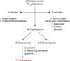
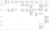
A review of English literature in PubMed was done using MeSH terms “gas forming” AND “Liver abscess, pyogenic”, “gas” AND “Liver abscess, pyogenic”, “gas” AND “Liver abscess”, “gas forming” AND “Liver abscess”. References of these articles were then searched to include additional studies. The search was restricted to publications on or after 1960, yielding 79 publications up to June 2016. Six Japanese, six Korean, and one Spanish language publications were excluded. Seven English language publications were deemed irrelevant (e.g., animal studies) and thus excluded. One textbook chapter was also excluded. The remaining 58 publications consisted of 15 case series and 43 case reports. Two authors reported on the same patients in their second publications. Only the most recent publication was considered.9101112 Finally, 13 case series including 2,630 patients yielded 313 GFPLA patients. These patients formed the study group. A flow chart of the literature search and studies included is shown in Fig. 1.
Age, gender, diabetes mellitus (DM), bacteriology, etiology, clinical profile, investigation results (biochemical and radiological), management, operative indications, outcomes, and predictors of mortality were reviewed. No statistical analysis was performed due to data heterogeneity.
RESULTS
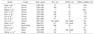
Table 1 lists the 13 case series reporting GFPLA. Eight reports described that the mean incidence of GFPLA was 13.2% (range, 5.6% to 31.8%). Six out of these eight studies were from Taiwan. Yang et al.3 and Chou et al.10 compared the age of patients with GFPLA vs. non-GFPLA. Mean ages of GFPLA patients in these two studies were 56.2±9.6 and 57±10.5 years, respectively, while those of non-GFPLA patients were 55.6±12.3 and 55.1±13.7 years, respectively. Both studies concluded that there were no significant difference in age between the two groups. Yang et al.3 and Chou et al.10 also compared gender preponderance between GFPLA and non-GFPLA patients. The male to female ratio of patients with GFPLA was 2.3 or 1.5 in the two studies. For non-GFPLA patients, the male to female ratio was 2.2 or 1.3. Both studies concluded that there was no significant gender difference between GFPLA and non-GFPLA patients.
Yang et al.3 have compared symptoms of GFPLA and non-GFPLA patients and found no significant difference. However, Chou et al.10 found that the duration of symptoms was shorter (p<0.05) in GFPLA patients (5.2±5.3 days) compared to that in non-GFPLA patients (7.6±10 days). Four studies have reported that GFPLA is mostly (75–91.9%) cryptogenic.341013 Five studies have described the association of DM with GFPLA.34101314 DM was diagnosed in 83.5% (range, 74.2–95%) of GFPLA patients. However, only 38.3% (range, 27.5–45.6%) of non-GFPLA patients had DM. Chou et al.10 have demonstrated that GFPLA patients have significantly higher DM rates compared to non-GFPLA patients (71 in 83 GFPLA patients (85.5%) vs. 113 in 341 non-GFPLA patients (33.1%), p<0.01). There were conflicting data regarding association of poorly controlled DM with GFPLA. Yang et al.3 demonstrated that there was no significant difference in mean blood sugar level between GFPLA and non-GFPLA patients (357±107 mg/dl in GFPLA group vs. 333±149 mg/dl in non-GFPLA group). On the other hand, Chou et al.10 demonstrated that GFPLA patients had higher mean blood sugar level compared to non-GFPLA patients (383.0±167.7 mg/dl vs. 262.6±168.0 mg/dl, p<0.01). Furthermore, Lin et al.15 showed that all GFPLA patients in their study had HbA1c ≥7.0%. However, these levels were not significantly different from those in non-GFPLA patients.15
Five authors have studied GFPLA bacteriology.34101316 They all described that monomicrobial Klebsiella pneumoniae (K. pneumoniae) was the most common bacterium found in patients with GFPLA. Klebsiella pneumoniae was found in 85.9% (range, 80.6–100%) of positive liver pus cultures in GFPLA patients versus 67.7% (range, 65.8–85.7%) in non-GFPLA patients. Only Chou et al.10 demonstrated that there was significant (p<0.01) difference in liver pus aspirate yield of K. pneumoniae between GFPLA and non-GFPLA patients. Similarly, 90.1% (range, 87–100%) of GFPLA patients were positive for blood cultures of K. pneumoniae whereas only 63.2% of non-GFPLA patients were culture positive for K. pneumoniae. Only Chou et al.10 demonstrated significant (p<0.01) difference in blood culture yield of K. pneumoniae between GFPLA and non-GFPLA patients. Additionally, Chou et al.10 described that, of 83 liver pus cultures and 83 blood cultures, only 64 (77.1%) liver pus cultures and 54 (65.1%) blood cultures were positive for K. pneumoniae in the GFPLA group.
Regarding laboratory investigations, Yang et al.3 described that there were no significant differences in percentages of those with albumin level ≤2.5 g/dl, alkaline phosphatase ≥150 IU/L, bilirubin ≥2 mg/dl, or leukocytes ≥20,000/mm3 between GFPLA and non-GFPLA patients. In their study, the percentage of patients who had aspartate aminotransferase (AST) level ≥100 IU/L was higher (p<0.05) in GFPLA patients than that in non-GFPLA patients.3 However, Chou et al.10 described that GFPLA patients had higher levels of blood glucose (21.3±9.3 mmol/L vs. 14.6±9.3 mmol/L, p<0.01), AST (176±191 U/L vs. 84±109 U/L, p<0.01), alkaline phosphatase (ALP) (275±187 U/L vs. 215±141.8 U/L, p<0.01), and serum urea nitrogen (9.6±6.8 mmol/L vs. 7.4±6.6 mmol/L, p<0.01) at hospital admission. Albumin levels in GFPLA patients were significantly lower than those in non-GFPLA patients (2.3±0.5 g/L vs. 2.6±0.6 g/L, p<0.01). However, there wase no significant difference in hemoglobin, bilirubin, or leukocyte level between the two groups of patients.10
Regarding radiological investigation, GFPLA can be most reliably diagnosed via ultrasound (US) or computerized tomography (CT) scan. Lee et al.13 reported 100% detection rate with either US or CT. Plain radiographs are less reliable. Foo et al.14 reported a sensitivity of 25.7% with plain radiographs. Three studies have described radiological findings of GFPLA.3410 On ultrasound, Yang et al.3 described GFPLA as ‘diffuse hyperechoic spots with acoustic shadowing’ while Chou et al.10 described it as ‘hyperechoic lesions with reverberation’. On CT scan, GFPLA was described as ‘low attenuation area with Hounsfield units similar to that of the lungs’ by Yang et al.3 whereas Chou et al.10 described that GFPLA ‘was easily diagnosed by the presence of gas’. Lee et al.4 have studied radiographic features associated with poor prognosis. This will be discussed later in the manuscript.
Chou et al.10 compared the singularity/multiplicity between GFPLA and non-GFPLA without finding significant difference between the two (p=0.14). In GFPLA group of 83 patients, 53 (63.9%) had solitary abscess while 30 (36.1%) had multiple abscesses In the non-GFPLA group of 341 patients, 246 (72.1%) had solitary abscess while 95 (27.9%) had multiple abscesses. Chou et al.10 compared size differences and concluded that GFPLA were larger (p<0.01). Abscess size >5 cm was seen in 67.5% patients with GFPLA vs. 45.7% in non-GFPLA patients.10 Lee et al.13 reported a mean GFPLA size of 6.5 cm (range, 1.5–11 cm). Chou et al.10 compared the location of abscess (right, left, or both lobes) without find any significant difference (p=0.08).
Four studies have reported complications of GFPLA.3101517 Chou et al.10 studied septic shock, bacteremia, pleural effusion, rupture, acute renal failure, respiratory failure, endophthalmitis, wound infection, gastrointestinal tract bleeding, multiple organ failure, empyema, recurrent abscess, meningitis, pericardial effusion, and length of hospital stay. Only septic shock (32.5% vs. 11.7%, p<0.01) and bacteremia (65.1% vs. 39%, p<0.01) were significantly higher in GFPLA. Jun CH et al. studied spontaneous rupture and showed that GFPLA had significantly increased risk.17 Out of 23 patients with rupture, five (21.7%) were patients with GFPLA. Of 579 patients without rupture, 29 (5%) were patients with GFPLA. Using multivariate analysis, GFPLA was found to be an independent risk factor for spontaneous rupture (OR: 3.65, 95% CI: 1.17–11.5, p=0.026). Yang et al.3 studied sub-hepatic abscess, empyema, rupture, acute renal failure, pleural effusion, septic shock, and bacteremia. They did not detect any significant difference for these complications (p>0.05 for all). Lastly, Lin et al.15 studied the risk of patients with GFPLA for metastatic infection and found no significant difference: 12.2% of patients without metastatic infection vs. 8.3% of patients with metastatic infection (p=0.693).
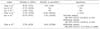
Six studies have reported the mortality rate of GFPLA, ranging from 25.7 to 37.1% (mean, 77 out of 254 patients, 30.3%) (Table 2).3410121314 These rates are higher in comparison with those of non-GFPLA patients (mean, 87 out of 971 patients (9%); range, 4.1–14.4%).3101214 Via univariate analysis, four studies demonstrated that GFPLA patients had significantly higher mortality rates: 30.4% (7 out of 23 patients),3 27.7% (23 out of 83 patients),10 25.7% (9 out of 35 patients),14 and 27.5% (8 out of 29 patients).12 Two authors further demonstrated difference in mortality on multivariate analysis, with OR of 35.7 (95% CI: 7.2–178.4, p<0.0001) and 9.4 (95% CI: 3.0–24.5, p<0.01), respectively.1214
Lee et al.4 described clinical and radiologic predictors of mortality among patients with GFPLA. Elevated serum levels of glucose and/or creatinine as well as more acute clinical presentations predicted mortality. When non-survivors and survivors were compared, serum levels of glucose and creatinine were higher in non-survivors (glucose: 467.1±175.5 mg/dl vs. 314.2±144.1 mg/dl, p<0.01; creatinine: 2.47±0.85 mg/dl vs. 1.23±0.53 mg/dl, p<0.001).4 Regarding radiologic factors, alveolar gas pattern and pneumoperitoneum on plain radiograph were found to be significant predictors for mortality.4 Thirteen (68.4%) of 19 patients with GFPLA died when any of these two findings was involved.4 Additionally, 52.2% (12 of 23) of patients that died had abscesses with alveolar gas pattern whereas 15.4% (6 out of 39) of those that survived (p<0.01) had abscesses with alveolar gas pattern.4 Also, 21.7% of those that died (5 out of 23 patients) had pneumoperitoneum whereas none of those that survived (p<0.05) had pneumoperitoneum.4 On CT scan, globular configuration (87.5% vs. 16.7%, p<0.001), shaggy margins (53.3% vs. 17.2%, p<0.05), alveolar internal structure (57.1% vs. 16.7%, p<0.05), and total gas content (50% vs. 15.4%, p<0.05) were found to be mortality predictors.4 However, bilobar involvement, multiple abscesses, or maximal diameter of abscess was not a significant predictor of mortality.4
Transarterial embolization/transarterial chemoembolization (TAE/TACE) is increasingly reported to be an aetiology of GFPLA.618 Chen et al.18 reported 80% (4 out of 5 PLA cases) of TAE/TACE-related GFPLA and Huang et al.6 reported 100% (7 out of 7) of TAE/TACE-related GFPLA. However, the incidence of GFPLA as a complication of TAE/TACE remained low (at 0.88%18 and 0.27%,6 respectively).
DISCUSSION
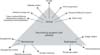
GFPLA was described as early as 1944 by Smith.2 It has been reported that patients with GFPLA have high mortality risk. However, there is a paucity of data on GFPLA and its distinct biologic characteristics. For the first time, an attempt to consolidate current GFPLA literature is done in a systematic manner here (Table 3). This review has shown that GFPLA is associated with DM and Klebsiella pneumoniae infection. It presents more often with septic shock and bacteraemia. It has higher mortality. Fig. 2 shows adversaries associated with GFPLA.
It has been reported that prompt resuscitation, parenteral antibiotics according to local antibiogram, and proactive percutaneous drainage procedures can improve outcome of PLA.7821 Chemaly et al.22 have reported that blood cultures from PLA patients fail to yield a bacterial isolate in more than 60% of patients. Similarly, we have reported that pus cultures from only 60% of PLA patients yield bacteria.2223 Hence, a clinician should be aware of local antibiogram and treat PLA with empirical antibiotics accordingly. In a recent international task force report from 79 countries, world's leading experts have raised awareness of healthcare workers to improve prescribing behavior in treating intra-abdominal infections and recommended that empiric antimicrobial therapy should be broad enough to cover all likely organisms because inappropriate therapy is associated with poor outcomes and the development of bacterial resistance.24
In our previous study of 264 patients treated for culture negative PLA (CNPLA), we have found that empiric treatment according to local antibiogram is adequate, with outcomes similar to patients with Klebsiella pneumoniae PLA (KPPLA).21 Unfortunately, due to absence of uniform reporting of bacterial isolates and rates of negative cultures, we are unable to derive conclusions for GFPLA. The current study concludes that GFPLA is more likely due to Klebsiella pneumoniae. However, it remains unclear if this association is reflective of the overall global trend of increasing incidence of KPPLA. In earlier reports, Streptococcus milleri and Escherichia coli have been found to be common monomicrobial causative agents.25 We have previously found that KPPLA is more likely to be associated with systemic organ dysfunction compared to CNPLA.21 It is likely that the increased incidence of septic shock and organ dysfunction as evidenced by elevated creatinine are multifactorial in patients with GFPLA instead of being solely due to Klebsiella pneumoniae infection.
In a multicenter observational study including 4533 patients with mean age of 51.2 years and diverse etiologies of sepsis, Sartelli et al.26 have reported that age >70 years can predict mortality. Mean age of patients with GFPLA has been reported to be lower than mean age of KPPLA patients.7 Hence, old age cannot be attributed for organ dysfunction or mortality.
In patients with KPPLA and ECPLA, the incidence of DM is approximately 40% based on our previous study.7 The current review shows that majority (83.5%) of GFPLA patients have DM. Septic shock and organ dysfunction might be related to poor glycemic control or DM-related end-organ dysfunction. High serum glucose also impairs leukocyte function and compromises immune defense mechanisms. It is a risk for mortality.4 However, only one study has reported that GFPLA patients have higher blood glucose levels on admission than non-GFPLA patients.10 More data are needed to clarify the relationship between blood glucose level and GFPLA.
Our review also shows that GFPLA is associated with mono-microbial infection and raised AST levels. In a comparative study on CNPLA and KPPLA, we have found that patients with KPPLA have elevated AST levels and larger PLA size.21 However, majority of studies included in our review are retrospective. We are unable to conclude if AST elevation is due to Klebsiella pneumoniae sepsis or if it is a determinant of PLA size.
Our study shows that GFPLA is associated with larger PLA size. PLA size is a predictor of rupture and rupture necessitates surgical intervention. Reported series on GFPLA are limited in samples. Hence, no clear trend is observed with regards to the need for surgical intervention. We have found that even a giant PLA can be managed by percutaneous drainage. The need for surgery is rare.8 However, failure of non-operative management or rupture with peritonitis remains clear indication for surgery.3410 Regarding complications, GFPLA is more likely to be associated with spontaneous rupture, septic shock, and bacteremia. It is possible that the presence of gas raises intracavitary tension. Hence, GFPLA has higher likelihood of both systemic bacteremia and spontaneous rupture. In the presence of compromised immunity, patients often deteriorate rapidly with endotoxemia and Gram-negative sepsis.
We have reported that Escherichia coli associated PLA patients treated with percutaneous drainage have higher mortality compared to KPPLA patients and postulated that this could be due to difference in timing of percutaneous drainage.7 A distinction between a ‘proactive’ versus ‘salvage’ percutaneous drainage was proposed. Due to retrospective nature of studies, it remains unclear if a ‘proactive’ policy of percutaneous drainage may impact clinical outcomes and reduce mortality associated with GFPLA. It remains to be determined if outcomes could be improved by proactive percutaneous drainage. It is our policy to drain all GFPLA patients regardless of size or clinical response to antibiotic treatment.78 A study by Lee et al. have identified that raised serum creatinine, raised serum glucose, and more acute clinical presentations are three predictors of mortality in GFPLA.4 Radiological findings such as alveolar gas pattern, pneumoperitoneum, globular configuration, shaggy margins, and total gas content can also predict mortality.4
The retrospective nature and small sample size of published reports are limitations of our study. However, we believe that this summative review will raise awareness about GFPLA. It also provides a benchmark for future reporting. As PLA is common in Asia, understandably majority of reports are from Asia. Due to geographic diversity in overall management, there might be possible bias. However, widespread adoption and implementation of sepsis campaign guidelines with multimodal care can reduce the impact of such limitation. The higher mortality of GFPLA may be due to old reports included in the review. Hence, high volume centers need to report their experience in managing GFPLA. Furthermore, it remains to be determined if the mere presence of gas in PLA impacts outcomes or if the presence of gas is a marker of immune compromise which is a determinant of outcomes. These conclusions can only be derived from prospective studies.
 XML Download