Abstract
Pancreaticoenteric anastomosis is the origin of postoperative pancreatic fistula (POPF). Although a variety of methods have been proposed to decrease the POPF rate, randomized controlled trials performed so far have failed to demonstrate superiority of any particular method to the others. Cattell-Warren duct-to-mucosa pancreaticojejunostomy (PJ) is a widely practiced procedure. Their method is challenging, especially when the pancreatic duct is small. We assumed that the difficulty resides in the pancreatic duct becoming difficult to access when the posterior row is tied before suturing the anterior row. We have modified the duct-to-mucosa PJ so that the entire circumference of the inner layer can be sutured and tied in one-step by anchoring and retracting the anterior row. The jejunal roux-limb and pancreatic stump are positioned spatially apart, allowing enough space for free needle work. During a 13-year period, 151 patients underwent pancreaticoduodenectomy with this method, and the cumulative POPF and mortality rates were 37.1% and 4.6%, respectively. These rates were stable throughout the study period, implicating a relative independence from surgeons' experience. We believe that our method is intuitive, easy to grasp, and can be readily adopted even by surgeons not accustomed to pancreaticoduodenectomy.
Pancreaticoduodenectomy (PD) is the standard method used for the management of diverse periampullary pathologies, both benign and malignant.12 Despite recent improvements in surgical techniques and perioperative management protocols, the procedure still carries high morbidity and mortality rates (up to 65% and ~5%, respectively).3
The major complications of PD originate from anastomotic failure of pancreaticoenterostomy, which is one of the most compromised and unavoidable procedures.34 To overcome this dilemma, a variety of methods of pancreaticoenteric anastomoses have been proposed but they failed.4 Pancreaticoenterostomy can be classified as pancreaticojejunostomy (PJ) or pancreaticogastrostomy (PG). Despite claims that PG is superior to PJ due to its lower anastomotic failure rate, randomized controlled studies could not prove the advantage of one method over the other.456 In turn, PJ can be further classified into three categories; end-to-side, duct-to-mucosa, and end-to-end invagination (dunking).7 Numerous minor modifications to pancreatic ductal stenting and suture techniques have been suggested for each of these categories, but no one particular method has been demonstrated to be superior to the others.8910 Currently, the adoption of one particular PJ method seems to depend on the surgeon's preference and his or her experience.
If various pancreaticoenteric reconstruction methods have similar complication rates, the next factor that should be considered while choosing a PJ method would be reproducibility or feasibility because PJ leakage rates are shown to be related to the individual operator's experience.11 One of the most widely practiced PJ technique is duct-to-mucosa anastomosis, which was originally described by Warren and Cattell.12 This technique has the following advantages: it is easy to grasp, is applicable to other anastomoses such as hepaticojejunostomy, has a long history, and many surgeons are familiar with it. However, the Cattell-Warren PJ can be technically challenging when the pancreatic duct has a small diameter. The working space between the pancreatic stump and the jejunal opening is too narrow once the posterior layer of the anastomosis has been completed. It is possible that this technical difficulty contributes to the higher anastomotic failure rate reported in patients with a small pancreatic duct.13
In this study, we introduced a slight modification of the Cattell-Warren PJ technique which resolves the insufficient space problem as the whole duct circumference is completed in one step compared to that in the commonly practiced ‘posterior-first’ technique.14 We believe that the method is intuitive and can be performed by beginners. In addition, we have presented a case of a patient who had to undergo a reoperation 50 months after PD due to locoregional recurrence. This case provided an example of how healing occurs after the current PJ method.
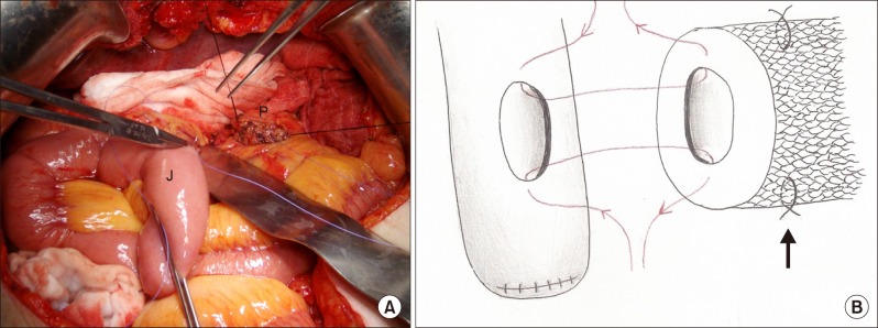
Resection of PD is performed in the generally accepted manner. The decision whether or not to preserve the pylorus is made prior to resection; we usually perform pylorus-preserving PD, except in cases of advanced pancreatic head cancer. When the portal vein has not been invaded by the tumor, the pancreas is resected anterior to the superior mesenteric vein (about 5 mm of a pancreatic stump is sufficient for future PJ anastomosis). Prior to pancreatic resection, two stay sutures are placed on both superior and inferior margins of the remnant pancreatic stump close to the expected resection line (to control the dorsal pancreatic artery branches and the inferior pancreatic artery) (Fig. 1). The pancreatic resection margin is sent for frozen section examination for assessing possible tumor invasion. If the pancreatic resection margin has been infiltrated by the tumor, PJ is abandoned and PG is considered instead. Further dissection and resection of the remnant pancreas makes PJ technically challenging because of the compromised working field. A jejunal Roux-limb is prepared in an antecolic or retrocolic manner, so as to provide tension-free PJ and hepaticojejunostomy. Blood supply to the jejunal stump can be confirmed by direct palpation of the arterial pulse or by the absence of discoloration. A small puncture is made at the antimesenteric border of the Roux-limb by electrocautery, and four tagging sutures are placed through the entire jejunal wall. This prevents inadvertent separation of the jejunal mucosa and the serosa during the course of duct-to-mucosa anastomosis. In addition, a plastic tube stent is prepared by cutting the tube into a 2 to 3 cm segment, with one end slanted to facilitate intubation into the pancreatic duct. The stent should fit loosely; when there is any doubt regarding its position and duct patency, gentle milking of the remnant pancreas should result in the flow of clear pancreatic juice through the stent.
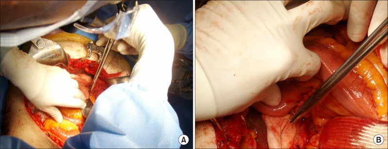
The jejunum and the pancreatic stump are located such that they face each other, leaving sufficient space for the needle work (Fig. 1A). Two 4-0 polyglactin (Vicryl®) sutures are performed at the inferior (3 o'clock) and superior (9 o'clock) edges of the pancreatic duct, and fixed in situ with small hemostatic clamps (Fig. 1B). An out-in-in-out needle movement starting from the jejunal side is recommended to ensure accurate pancreatic duct puncture. Also, the needle entering through the pancreatic duct wall should go as deep as possible from the duct margin and it should be perpendicular to the duct wall, to avoid inadvertent tearing while knot making. The exit of the needle is rather unimportant; just following the needle curvature is enough.
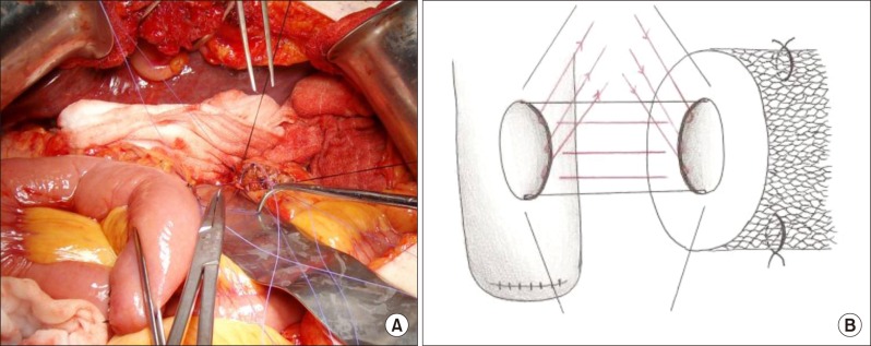
Suturing of the posterior semicircle of duct-to-mucosa anastomosis is started at the center (6 o'clock), and then to the either side (4:30 and 7:30 o'clock). The needle movement is conducted in the same manner as the anchoring sutures described above; except that it goes in the in-out-out-in direction, and it is started from the pancreatic duct to provide secure needle placement. Occasionally, the pancreatic parenchymal thickness posterior to the duct is so thin that the pancreatic duct is nearly exposed. It should be fine unless the knot is too tight at the time of tying and will tear the posterior pancreatic duct wall. The loose ends of the threads are clamped and retracted cephalad (Fig. 2A and B).
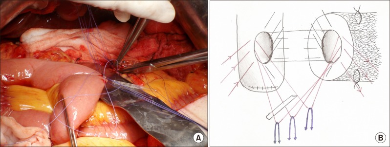
First, the anterior (12 o'clock) stitch is placed in an out-in-stent-in-out order starting from the jejunum. The stent is punctured so that the pancreatic duct side is slightly shorter than the jejunal side; this reduces the possibility of stent migration into the pancreatic duct and subsequent pancreatitis. A thread is then anchored to retract the suture loop, and the free ends of all suture materials are held together with a hemostat (Fig. 3A). On either side of the central suture (1:30 and 10:30), two additional sutures are placed in the same manner as anchoring, and all anterior row sutures are retracted caudad (Fig. 3B).
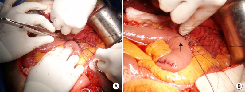
Using atraumatic forceps, the assistant approximates the jejunal Roux-limb to the pancreatic stump. Suture materials are gently tightened during the approximation to prevent dog-ear formation. Knot tying starts at the superior (9 o'clock) and inferior (3 o'clock) sutures. Alternative gentle traction of the loose ends of the suture material prevents dog-ear formation and loosening. There should be sufficient working space for knot-tying because the anterior row has been anchored and retracted (Fig. 4A). Once tied, both superior and inferior sutures are held with hemostats and retracted cephalad and caudad, respectively. The posterior row sutures are then tied in the same manner and cut. At this stage, the posterior circumference is complete.
A small probe is then introduced into the pancreatic duct to identify the duct orifice. After removing the anchoring thread (12 o'clock), the stent is inserted all the way into the pancreatic duct. There should be little resistance if the stent is placed correctly in the duct, and gentle milking of the remnant pancreas can confirm its proper location by the flow of pancreatic juice. After aligning the jejunal opening with the pancreatic duct orifice, traction applied to both ends of the 12 o'clock suture will cause the stent to pop-up, so as to insert the blunt end of the stent automatically into the jejunum. The suture is tied in situ after confirming the stent location by palpating its end through the jejunal wall (Fig. 4B). Sutures are tied on either side of the stent (10:30 and 1:30) after removing the anchoring thread. The whole circumference of the inner layer of duct-to-mucosa anastomosis is thus completed.
The outer seromuscular layer is sutured with non-absorbable suture material using an atraumatic curved needle and 3-0 black silk (Mersilk®). An interrupted simple suture is started at the inferior and superior borders of the PJ. The needle should enter from pancreatic parenchyma and it should be perpendicular to the pancreatic surface with a rounding motion compatible with the needle's curvature (Fig. 5A). The posterior row is placed in the same manner; the posteroinferior space can be exposed by lifting the PJ a little anteriorly, and the posterosuperior space can be approached from the patient's right-upper side by lifting the PJ anteriorly (Fig. 5B). Care should be taken not to puncture the stent when needle resistance is felt. When tying, the knot should be pushed to the side of the pancreas and it should not be tied too tightly to avoid pancreatic parenchymal tearing. Suturing of the anterior row of the seromuscular layer is straightforward. The entire PJ anastomosis is now complete.
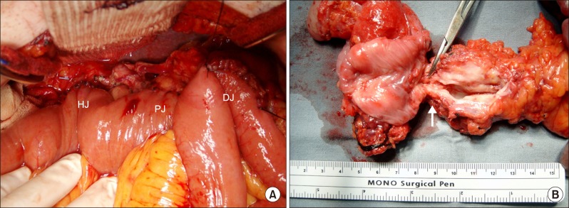
Hepaticojejunostomy is performed at the natural location proximal to the PJ, and gastro- or duodenojejunostomy is performed ~40 cm proximal to the hepaticojejunostomy (Fig. 6A). We usually drain the PJ area with two occlusive suction drains (Jackson-Pratt drain) inserted along the superior and inferior borders of PJ, respectively.
A 58-year-old male patient underwent pylorus-preserving PD due to T3N0M0 distal common bile duct cancer, only to suffer local recurrence 50 months later. He was in a good general condition and eager to undergo a reoperation. Total pancreatectomy, splenectomy, and subtotal gastrectomy were performed. The specimen showed locally recurrent adenocarcinoma at the previous PJ site which was clear at the initial operation, with patent pancreatic duct to jejunal mucosa continuity (Fig. 6B). Despite making an uneventful recovery, the patient died of multiple intra- and retroperitoneal recurrences 18 months after the reoperation.
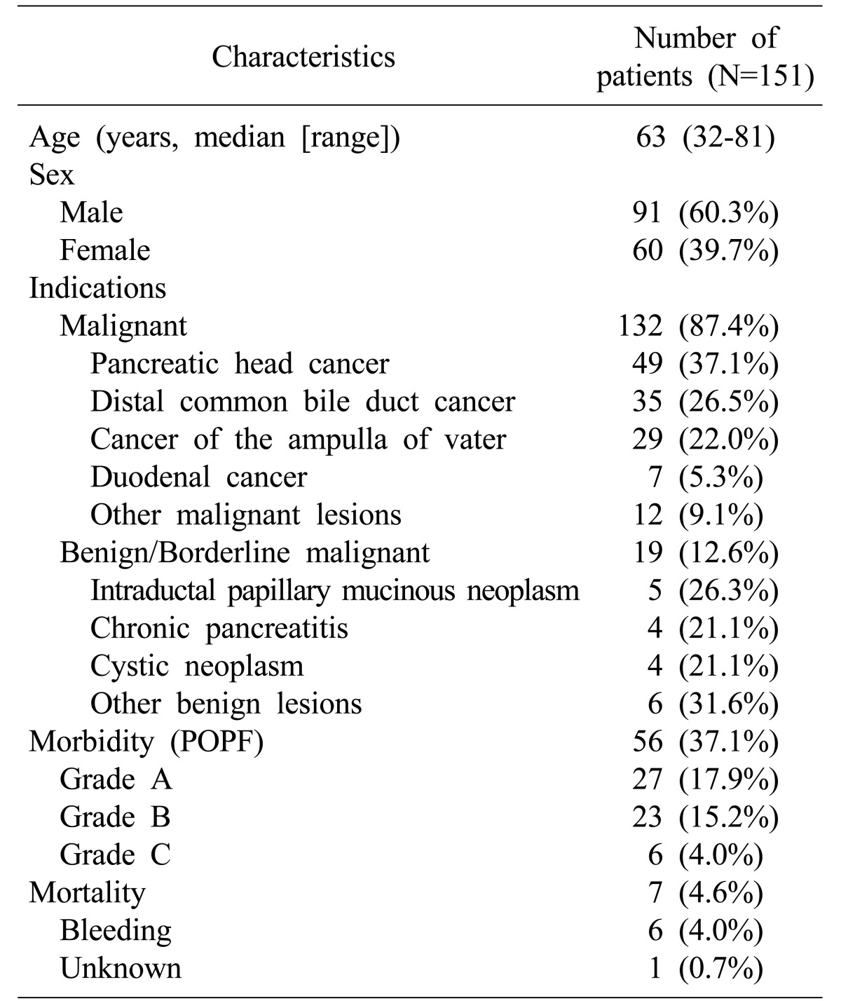
From June 2003 to May 2016, a total of 151 patients underwent PD with the modified PJ method. The patient characteristics are presented in Table 1. Seven deaths occurred within 30 days of PD; a mortality rate of 4.6%. Six out of the seven patients died of postoperative bleeding and one patient died due to an unknown cause, presumably pulmonary embolism.
Overall, the cumulative postoperative pancreatic fistula rate (POPF) was 37.1%, according to the definition by the International Study Group of Pancreatic Surgery (ISGPS).15 There were 27 (17.9%) grade A POPFs, 23 (15.2%) grade B POPFs, and 6 (4.0%) grade C POPFs.
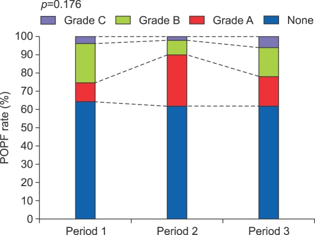
When the patient group was equally divided into three consecutive periods, POPF and mortality rates were not different among the 3 groups (POPF, period 1 vs. period 2 vs. period 3=18/51 (35.3%) vs. 19/50 (38.0%) vs. 19/50 (38.0%), p=0.176, Pearson's chi-square test; Mortality, period 1 vs. periods 2 and 3=3/51 (5.9%) vs. 4/100 (4.0%), p=0.689, Fisher's exact test) (Fig. 7).
To date, randomized controlled trials conducted to identify a better method for performing PJ have failed to discover a technique superior to the others in terms of operation-related morbidity and mortality.816 As a result, the surgeons can choose the preferred PJ method after achieving a sufficient level of experience of the procedure.
Duct-to-mucosa PJ may be a reasonable choice because it has a lower rate of clinically relevant POPF,17 although some studies have concluded otherwise.818 Since Warren and Cattell described a detailed method for duct-to-mucosa PJ in 1956, many technical modifications have been reported, and today it appears that every large volume center has its own method.16171819 Regardless of the technique adopted, all techniques have two common features. Firstly, the anastomosis consists of two layers; an inner true duct-to-mucosa layer and an outer seromuscular (Lembert) layer. Secondly, the suture sequence is posterior outer, posterior inner, anterior inner, followed by anterior outer.161819
We agree that the outer Lembert suture layer is required to protect the inner duct-to-mucosa layer by reducing the possible tension.17 However, in our experience, once the posterior outer layer has been sutured and tied, it is difficult to identify the pancreatic duct lumen and stent insertion and suturing are challenging. Furthermore, this technical difficulty is exacerbated when the pancreatic duct is small. Some authors have tried to avoid this problem by performing ductal20 or stump invagination PJ.21 Although the outer layer can be sutured after the inner layer has been completed,17 suturing and cannulation of a small pancreatic duct are still difficult, if the posterior inner layer is tied first.
The unique feature of our technique is that the inner layer is completed first, and the jejunum and the pancreatic stump are separated spatially throughout the process which provides sufficient working space and a clear visual field. This maneuver also allows free needle movement even for operators not accustomed to pancreatic surgery. Also, by anchoring and retracting the anterior inner row sutures, the whole circumference of duct-to-mucosa sutures can be performed in a single step, without worrying about making knots in the posterior row in the face of intervening anterior row sutures. Once the inner layer has been completed, there should be no problem in suture-tying for the seromuscular layer, as described by the other authors.1722
Some details of the described technique need to be clarified further. First, during duct-to-mucosa PJ, minor pancreatic ducts are excluded and they can be the origin of POPF.23 For patients with a soft, swollen pancreatic stump, simple horizontal mattress sutures can be applied to close minor pancreatic ducts,1224 whose fates are not a point of concern. A closer look at our reoperation case (Fig. 6B) revealed gradual tapering of the pancreatic stump with passage of time in a herringbone fashion. This implies that the minor pancreatic ducts close to the duct-to-mucosa PJ are waisted because we had not performed mattress sutures in that particular patient. Additionally, there is a debate regarding pancreatic duct stenting and its drainage methods. Although some authors have reported similar complication rates between stented and non-stented patients,2526 we believe that it is necessary to stent the anastomosis to promote duct-mucosa healing, especially in patients with a small pancreatic duct.27 With respect to the stent drainage methods, external drainage showed somewhat better results in terms of morbidity.28 However, internal drainage was reported to be simpler and to involve a shorter hospital stay.29 Finally, 8 stitches around the inner layer are sufficient and possible to perform in most cases. In cases with an extremely small pancreatic duct, even 4 stitches may adequately secure the duct-to-mucosa anastomosis, provided the stent can be inserted. When the pancreatic duct is unidentifiable, gentle milking of the remnant pancreas usually reveals the pancreatic duct stump. However, if the location of the pancreatic duct orifice is still doubtful, duct-to-mucosa PJ should be abandoned and end-to-side PJ or PG should be used instead, in order to avoid missing the main pancreatic duct. Although we have encountered few such cases, it was embarrassing because a peripherally located main pancreatic duct could be suture occluded, regardless of the anastomotic method used.
The operative results achieved using the modified PJ method are comparable to those described in a recent multicenter study, in terms of POPF and mortality rates.30 Although our method was not better than the other PJ methods, it achieved fairly constant POPF and mortality rates throughout the study period, indicating that the method was standardized31 and it was independent from the surgeons' experience. In fact, the described method is a slight modification of the widely practiced PJ technique, whose basic principles have also been applied to other anastomoses such as hepaticojejunostomy. We believe that our method is intuitive, simple, and can be readily adopted by beginner surgeons confronted with this complex operation.
In conclusion, here we propose a modified PJ method that is straightforward. The essentials of our technique are two-fold. First, we start PJ anastomosis with the jejunal Roux-limb and the pancreatic stump located apart to create space to enable free needle work even by surgeons not accustomed to the procedure. Second, the inner layer of duct-to-mucosa anastomosis is completed in one step with the anterior row of sutures anchored away, which allows for knot tying of the posterior row after suturing the anterior row. This technique is especially useful in cases with a small pancreatic duct. The operative results of our PJ method have remained fairly stable, and we hope that both hepato-biliary-pancreatic surgery specialists and beginner surgeons, who encounter unavoidable PD as in cases of pancreatic head trauma, will find this technique useful.
ACKNOWLEDGEMENTS
This work was supported by Inha University Research Grant. Authors would like to thank to Ms. Seung-Min Lee, for the enlightening drawings included in this article.
References
1. Barens SA, Lillemoe KD, Kaufman HS, Sauter PK, Yeo CJ, Talamini MA, et al. Pancreaticoduodenectomy for benign disease. Am J Surg. 1996; 171:131–134. PMID: 8554127.
2. Diener MK, Knaebel HP, Heukaufer C, Antes G, Büchler MW, Seiler CM. A systematic review and meta-analysis of pylorus-preserving versus classical pancreaticoduodenectomy for surgical treatment of periampullary and pancreatic carcinoma. Ann Surg. 2007; 245:187–200. PMID: 17245171.
3. Machado NO. Pancreatic fistula after pancreatectomy: definitions, risk factors, preventive measures, and management-review. Int J Surg Oncol. 2012; 2012:602478. PMID: 22611494.
4. Tewari M, Hazrah P, Kumar V, Shukla HS. Options of restorative pancreaticoenteric anastomosis following pancreaticoduodenectomy: a review. Surg Oncol. 2010; 19:17–26. PMID: 19231161.
5. Fernández-Cruz L, Belli A, Acosta M, Chavarria EJ, Adelsdorfer W, López-Boado MA, et al. Which is the best technique for pancreaticoenteric reconstruction after pancreaticoduodenectomy? A critical analysis. Surg Today. 2011; 41:761–766. PMID: 21626319.
6. Crippa S, Cirocchi R, Randolph J, Partelli S, Belfiori G, Piccioli A, et al. Pancreaticojejunostomy is comparable to pancreaticogastrostomy after pancreaticoduodenectomy: an updated meta-analysis of randomized controlled trials. Langenbecks Arch Surg. 2016; 401:427–437. PMID: 27102322.
7. Chen YJ, Lai EC, Lau WY, Chen XP. Enteric reconstruction of pancreatic stump following pancreaticoduodenectomy: a review of the literature. Int J Surg. 2014; 12:706–711. PMID: 24851718.
8. Hua J, He Z, Qian D, Meng H, Zhou B, Song Z. Duct-to-Mucosa versus invagination pancreaticojejunostomy following pancreaticoduodenectomy: a systematic review and meta-analysis. J Gastrointest Surg. 2015; 19:1900–1909. PMID: 26264363.
9. Zhang JL, Xiao ZY, Lai DM, Sun J, He CC, Zhang YF, et al. Comparison of duct-to-mucosa and end-to-side pancreaticojejunostomy reconstruction following pancreaticoduodenectomy. Hepatogastroenterology. 2013; 60:176–179. PMID: 22773303.
10. Osada S, Imai H, Sasaki Y, Tanaka Y, Nonaka K, Yoshida K. Reconstruction method after pancreaticoduodenectomy. Idea to prevent serious complications. JOP. 2012; 13:1–6. PMID: 22233940.
11. Ball CG, Howard TJ. Does the type of pancreaticojejunostomy after Whipple alter the leak rate? Adv Surg. 2010; 44:131–148. PMID: 20919519.
12. Warren KW, Cattell RB. Basic techniques in pancreatic surgery. Surg Clin North Am. 1956; 36:707–724. PMID: 13324659.
13. Akamatsu N, Sugawara Y, Komagome M, Shin N, Cho N, Ishida T, et al. Risk factors for postoperative pancreatic fistula after pancreaticoduodenectomy: the significance of the ratio of the main pancreatic duct to the pancreas body as a predictor of leakage. J Hepatobiliary Pancreat Sci. 2010; 17:322–328. PMID: 20464562.
14. Kennedy EP, Yeo CJ. Dunking pancreaticojejunostomy versus duct-to-mucosa anastomosis. J Hepatobiliary Pancreat Sci. 2011; 18:769–774. PMID: 21845376.
15. Bassi C, Dervenis C, Butturini G, Fingerhut A, Yeo C, Izbicki J, et al. International Study Group on Pancreatic Fistula Definition. Postoperative pancreatic fistula: an international study group (ISGPF) definition. Surgery. 2005; 138:8–13. PMID: 16003309.
16. Bassi C, Falconi M, Molinari E, Mantovani W, Butturini G, Gumbs AA, et al. Duct-to-mucosa versus end-to-side pancreaticojejunostomy reconstruction after pancreaticoduodenectomy: results of a prospective randomized trial. Surgery. 2003; 134:766–771. PMID: 14639354.
17. Bai X, Zhang Q, Gao S, Lou J, Li G, Zhang Y, et al. Duct-to-Mucosa vs Invagination for Pancreaticojejunostomy after pancreaticoduodenectomy: a prospective, randomized controlled trial from a single surgeon. J Am Coll Surg. 2016; 222:10–18. PMID: 26577499.
18. Berger AC, Howard TJ, Kennedy EP, Sauter PK, Bower-Cherry M, Dutkevitch S, et al. Does type of pancreaticojejunostomy after pancreaticoduodenectomy decrease rate of pancreatic fistula? A randomized, prospective, dual-institution trial. J Am Coll Surg. 2009; 208:738–747. PMID: 19476827.
19. Batignani G, Fratini G, Zuckermann M, Bianchini E, Tonelli F. Comparison of Wirsung-jejunal duct-to-mucosa and dunking technique for pancreatojejunostomy after pancreatoduodenectomy. Hepatobiliary Pancreat Dis Int. 2005; 4:450–455. PMID: 16109535.
20. Xu J, Zhang B, Shi S, Qin Y, Ji S, Xu W, et al. Papillary-like main pancreatic duct invaginated pancreaticojejunostomy versus duct-to-mucosa pancreaticojejunostomy after pancreaticoduodenectomy: a prospective randomized trial. Surgery. 2015; 158:1211–1218. PMID: 26036878.
21. El Nakeeb A, El Hemaly M, Askr W, Abd Ellatif M, Hamed H, Elghawalby A, et al. Comparative study between duct to mucosa and invagination pancreaticojejunostomy after pancreaticoduodenectomy: a prospective randomized study. Int J Surg. 2015; 16:1–6. PMID: 25682724.
22. Ohwada S, Ogawa T, Kawate S, Tanahashi Y, Iwazaki S, Tomizawa N, et al. Results of duct-to-mucosa pancreaticojejunostomy for pancreaticoduodenectomy Billroth I type reconstruction in 100 consecutive patients. J Am Coll Surg. 2001; 193:29–35. PMID: 11442251.
23. Maharaj R, Naraynsingh V, Shukla PJ. Concept of a duct-to-mucosa pancreaticojejunostomy. J Am Coll Surg. 2010; 211:143. PMID: 20610265.
24. Liu C, Long J, Liu L, Xu J, Zhang B, Yu X, et al. Pancreatic stump-closed pancreaticojejunostomy can be performed safely in normal soft pancreas cases. J Surg Res. 2012; 172:e11–e17. PMID: 22079848.
25. Imaizumi T, Harada N, Hatori T, Fukuda A, Takasaki K. Stenting is unnecessary in duct-to-mucosa pancreaticojejunostomy even in the normal pancreas. Pancreatology. 2002; 2:116–121. PMID: 12123091.
26. Smyrniotis V, Arkadopoulos N, Kyriazi MA, Derpapas M, Theodosopoulos T, Gennatas C, et al. Does internal stenting of the pancreaticojejunostomy improve outcomes after pancreatoduodenectomy? A prospective study. Langenbecks Arch Surg. 2010; 395:195–200. PMID: 20082094.
27. Ohwada S, Tanahashi Y, Ogawa T, Kawate S, Hamada K, Tago KI, et al. In situ vs ex situ pancreatic duct stents of duct-to-mucosa pancreaticojejunostomy after pancreaticoduodenectomy with billroth I-type reconstruction. Arch Surg. 2002; 137:1289–1293. PMID: 12413321.
28. Zhou Y, Zhou Q, Li Z, Lin Q, Gong Y, Chen R. The impact of internal or external transanastomotic pancreatic duct stents following pancreaticojejunostomy. Which one is better? A meta-analysis. J Gastrointest Surg. 2012; 16:2322–2335. PMID: 23011201.
29. Tani M, Kawai M, Hirono S, Ina S, Miyazawa M, Shimizu A, et al. A prospective randomized controlled trial of internal versus external drainage with pancreaticojejunostomy for pancreaticoduodenectomy. Am J Surg. 2010; 199:759–764. PMID: 20074698.
30. Keck T, Wellner UF, Bahra M, Klein F, Sick O, Niedergethmann M, et al. Pancreatogastrostomy versus pancreatojejunostomy for RECOnstruction after PANCreatoduodenectomy (RECOPANC, DRKS 00000767): perioperative and long-term results of a multicenter randomized controlled trial. Ann Surg. 2016; 263:440–449. PMID: 26135690.
31. Shrikhande SV, Barreto G, Shukla PJ. Pancreatic fistula after pancreaticoduodenectomy: the impact of a standardized technique of pancreaticojejunostomy. Langenbecks Arch Surg. 2008; 393:87–91. PMID: 17703319.