

 PDF
PDF ePub
ePub Citation
 Print
Print


INTRODUCTION
During the last decade, technical improvements in liver surgery have resulted in an expansion of indications for major liver resections, especially in high-risk patients with various underlying liver conditions (fibrosis, steatosis, or chemotherapy-induced injury). However, the risks of postoperative liver failure (PLF) and fatal outcome have remained important concerns.123 The reported incidence of PLF ranges between 0.7% and 9.1%.12456 Over the years, mortality after liver resection ranged from 0 to 5%, and although multifactorial, PLF seems to be the main cause (18-75%).789
Preoperative risk assessment ideally consists of clinical, biochemical, volumetric and functional factors. When planning a major liver resection, evaluation of the liver remnant volume by computed tomogram (CT) volumetry is important. The most common test for functional evaluation is the indocyanine green retention test at 15 minutes (ICG-R15); it is widely used in eastern countries for cirrhotic patients with hepatocellular carcinoma (HCC) requiring liver surgery.10 It accurately assesses the liver function reserve, and an ICG-R15 of 14% is considered as the safety limit for major liver resections.1112 Various laboratory data, imaging techniques and complex functional assessment methods are used to complement the Child-Pugh score to predict PLF and to assess functional hepatic reserve, especially in the high risk group of cirrhotic patients; however, these have not gained popularity.
We proposed to study the oral glucose tolerance test (OGTT), a simple and easily performed test, for the preoperative assessment of liver function in liver resection. Ozawa et al.13 determined glucose tolerance in 14 patients with liver malignancy. They observed that patients with parabolic GTT patterns fared relatively better than those with linear GTT patterns. They had earlier suggested that the parabolic glucose tolerance pattern is indicative of compensated damage to the liver, while the linear glucose tolerance pattern shows critically decreased hepatic functional reserve.14 The 3-hour OGTT is preferred over the 2-hour OGTT, as the derivative of glucose concentration is better defined in the third hour of the OGTT, and the rate of glucose appearance is least variable at 120 minutes.15 The present study proposes to evaluate the utility of OGTT in addition to CT volumetry, for improving the outcomes of liver resection.
MATERIALS AND METHODS
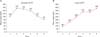
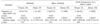
At our tertiary care hospital, adults (both cirrhotic and non-cirrhotic) who had undergone hepatic resections over a period of 2 years, were studied using a prospective longitudinal cohort study design. The management of liver lesions included in the study was as per the standard protocols of the hepatopancreaticobiliary unit of the hospital. A standard 3-hour 75-g OGTT was performed as per the 1999 World Health Organization (WHO) specifications. Blood samples were collected at 0, 30, 60, 90, 120, and 180 minutes for the measurement of plasma glucose.16 The blood sugar curve was classified as having either a parabolic or linear pattern. A parabolic pattern referred to the blood sugar curve in which the blood sugar value at 2 hours decreased with respect to the preloading value; otherwise, it was classified as linear (Fig. 1).13 Linearity index (LI) was calculated from the ratio of the 60-minutes blood glucose to the 120-minutes blood glucose level in the peripheral blood after the 75-g OGTT.17 In diabetics, LI was calculated after glycemic control, and the decrement between 180-minutes and 120-minutes blood sugar in percentage was measured. In patients showing a linear pattern with no proven diabetes, glycosylated hemoglobin (HbA1C) was measured to exclude undetected diabetes causing glucose intolerance and the resultant linear curve.
The total liver, tumor and future remnant liver were delineated on every image, using the portal and hepatic veins as markers for segmental division on CT. Integrated software calculated the volume of the total liver, total tumors, and remnant liver volume (RLV) anticipated after surgery. All delineations were made by an experienced radiologist. Tumor volume (TV) was subtracted from total hepatic volume. Total hepatic volume (TLV) was corrected for weight and length differences and expressed as ml/BSA. The liver volume to be resected (including tumor) was designated as RV. The volume of the future remnant was expressed as a percentage of the total hepatic volume. Parenchymal hepatic resection rate (PHRR) was defined as the proportion of resected liver to the entire liver volume, after excluding the volume of tumor in both. It was calculated as PHRR=(RV–TV)/(TLV–TV).18
Following were the exclusion criteria for the study: patients who did not undergo liver resection due to lack of medical fitness, inoperability because of vascular encasement, or advanced disease; patients who did not provide informed consent for the study (with no consequences on their clinical care); patients who could not be preoperatively evaluated by CT volumetry or OGTT; when a linear curve of OGTT coexisted with deranged HbA1C in a previously undetected diabetic, and the linearity of the curve could not be attributed to liver function alone; any patient who could not be followed up for a minimum period of 90 days after surgery.
The Brisbane 2000 terminology of liver anatomy and resections was used to define the extent and type of liver resection.19 Resections were considered extended when five or more segments were involved (extended right lobectomy and extended left hepatectomy), major when three or four segments were involved (right and left hepatectomy), and minor when one or two segments were resected, or a non-anatomical resection was performed.20
Postoperative surveillance included clinical examination during hospitalization and laboratory tests during the first week, including prothrombin time (PT), international normalized ratio (INR), total and direct bilirubin, aspartate transaminase (AST), alanine transaminase (ALT) on postoperative day (POD) 1, 3, 5 and 7. Blood analysis after the first week was as per the clinical indications. Abdominal ultrasound and/or computed tomography were performed in cases where postoperative intra-abdominal fluid collection was suspected.
The primary end points were mortality and postoperative complications. Postoperative mortality was defined as any death within 90 days of operation, or during the concurrent hospital stay.
Any deviation from the standard postoperative course other than normal sequelae (inherent to a specific surgical procedure), failure of therapy (goal of treatment not attained), or death, were considered as complications. They were classified as major when they resulted in organ failure, required another surgery or radiologic intervention, or required a red blood cell transfusion for postoperative bleeding. These included major postoperative bleeding, any organic failure, intra-abdominal abscess, sepsis and portal vein thrombosis. Other complications with no fatal potential were considered minor, and included pleural effusion, wound infection, urinary tract infection and atelectasis.21
As per the 50-50 criteria, postoperative liver failure is defined at the POD 5, with persistence of either PT <50% or serum bilirubin (SB) >50 mmol/L on POD 5. The value 50% of normal for PT corresponds to an INR of 1.7, and SB 50 mmol/L corresponds to 3 mg/dl.3 Secondary end points considered were intraoperative variables such as operative time, blood loss and transfusion requirement. In addition, intensive care unit (ICU) and hospital stay was also recorded.
Liver resection specimens were evaluated for the presence of steatosis using hematoxylin and eosin-stained sections. The degree of total steatosis was graded as mild (10%-30%), moderate (31%-60%), or severe (>60%), based on the percentage of hepatocytes with fat droplets.22 Liver fibrosis was quantified according to the Ishak score as follows - 0: no fibrosis; 1: fibrous expansion of some portal tract areas; 2: fibrous expansion of most portal tract areas; 3: fibrous expansion of portal tract areas with occasional portal-portal bridging; 4: fibrosis with portal-portal and portal-central bridging; 5: pronounced bridging with occasional nodules; 6: probable or definite cirrhosis.23
Statistics and ethics
Statistical analysis was performed using the SPSS version 16.0 (SPSS INC). The mean±standard deviation, median and range of continuous variables, and frequency distributions of categorical variables are presented. Categorical variables were compared using Fisher's exact test or the chi-square test, and continuous variables were compared using Student's t-test or Wilcoxon test, as appropriate. Significant variables (using a cutoff p<0.05) in a univariate analysis were considered for a multivariate analysis.
Analysis of variance test (ANOVA) and/or receiver operating characteristic (ROC) curve analysis was performed to identify variables affecting the outcomes of liver resection. Statistical significance was considered at p <0.05. The study adhered to the tenets of the Declaration of Helsinki, and informed consent was sought from each subject prior to enrollment.
RESULTS
Patient characteristics
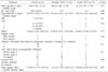
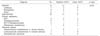
Demography: Mean age of the total 33 patients undergoing liver resection was 48±15 years, ranging from 21 to 73 years (median: 49 years). OGTT was parabolic in 22 (66.7%) patients and linear in 11 (33.3%) patients. The mean LI in those with parabolic curve was 1.31, while in the linear group it was 1.05. Known diabetic cases (5 patients), had good glycemic control prior to their undergoing OGTT. HbA1c was normal in all patients with a linear curve. The distribution of age, gender, body mass index, diabetes status, ASA grade, PHRR, viral hepatitis serology status and modified Child-Pugh class stratification between the OGTT parabolic and linear groups was comparable (Table 1).
Background liver status: The non-tumor-bearing liver was histologically unremarkable in 21 patients (63.64%). Of the 5 patients with evidence of cirrhosis on preoperative workup, subsequent histopathology confirmed grade 6 fibrosis in 2 cases, and grade 3 fibrosis in 3 patients. Preoperative workup or subsequent histology did not reveal any evidence of cirrhosis in 1 patient with hepatitis B.
Steatosis was evident in the histopathology of 9 patients. Both steatosis and fibrosis were positive in 2 patients. Stratification based on fibrosis grade and steatosis was comparable between the OGTT parabolic and linear groups.
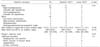
Indication: HCC was the leading indication for liver resection in our study, attributing to 30.3% of all cases. Nineteen (57.6%) patients underwent resection for malignant pathology. Liver resection as a part of metastatectomy were all metachronous. No statistically significant difference was noted in the distribution of indications between the two OGTT groups (Table 2).
Major liver resections were carried out in 23 (69.7%) patients. The average duration of surgery was 6.3±2.1 hours. The types of resections and additional associated procedures, as well as the operative parameters (including duration of surgery and blood loss) did not differ between the two OGTT groups (Table 3).
Postoperative outcomes
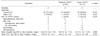
There was no mortality either during the hospital stay or within 90 days after surgery. Overall morbidity was observed in 24 (72.7%) patients; however, major complications occurred in only 3 (9.1%) patients.
Two patients had bile leak, of which one was self-limiting, while the other needed percutaneous drainage. One patient was re-explored for primary hemorrhage. Overall morbidity and major morbidity was higher in the linear OGTT group (90.9% and 18.2%, respectively) compared to the parabolic group (63.6% and 4.6%, respectively), but it did not reach statistical significance (Table 4).
OGTT curve correlated with the overall hospital stay; this association was statistically significance (p=0.04). Only 3 (9.1%) patients met the 50-50 criteria used to define PLF. The patients with a linear OGTT curve met 50-50 criteria more often (18.2% vs 4.5% in parabolic), but did not reach statistical significance (p=0.25).
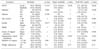
To determine factors affecting outcomes of liver resection (overall morbidity, major morbidity and PLF by 50-50 criteria), none of the demographic, perioperative (including steatosis or fibrosis in background liver) or volumetric parameters were found to significantly affect outcomes on univariate analysis (Table 5); hence, a multivariate analysis was not necessitated.
The OGTT was more often linear in presence of morbidity (41.7% vs 11.1%), major morbidity (66.7% vs 30%) and PLF by 50-50 criteria (66.7% vs 30%), but did not reach statistical significance. While the linearity index was comparable in the presence or absence of overall morbidity, it was insignificantly lower in presence of major morbidity (0.9 vs 1.2) and PLF by 50-50 criteria (0.9 vs 1.2). These linearity indices did not differ statistically (Table 6).
DISCUSSION
Recent advances in hepatic surgery and perioperative care, and refinements in patient-selection criteria and indications, have enabled an increasing number of patients to undergo liver resections. The past decade has seen reduced mortality after hepatic resection to less than 5%, and although the cause of death after hepatic resection is multifactorial, PLF seems to be the main cause.8 A great number of hepatic resection have been mainly reported from the Western countries and the Far East. Besides, a North Indian single-center study analyzed 241 consecutive cases of hepatic resections to define the risk factors associated with postoperative morbidity and mortality. Very few studies have been reported from South Asia.24 Further, the studies on objective preoperative quantitative and qualitative functional assessment prior to liver resection, have rarely been reported from the Indian subcontinent.
Among the various tests for functional evaluation, ICG-R15 is widely used in the Eastern countries for cirrhotic patients with HCC requiring liver surgery.10 It is considered to be accurate for assessment of liver function reserve in cirrhotic patients undergoing hepatectomy.12 However, there is a known risk of complications reported due to anaphylactoid reactions, including death.252627 The results of ICG-R15 depend on the hepatic blood flow, and regional variations can alter the retention value.28 Also, acute cholestasis directly influences the ICG test and should be taken into account while interpreting the ICG test in jaundiced patients.29
A close association exists between glucose intolerance and the decrease in hepatic energy charge and the derangement of mitochondrial function.3031323334 This formed the basis for utilizing OGTT in preoperative assessment for liver resection. The linear index of OGTT is related to the mitochondrial activity of the hepatocytes. Especially in cirrhosis, shunting occurs in portal vein blood, reducing the blood supply to the liver parenchyma, leading to insulin resistance and a lowered glucose tolerance.163536 In clinical application, the assessment of hepatic functional reserve by oral glucose tolerance test was shown to provide a predictive postoperative prognosis in post hepatectomy patients.13 In the study by Yamanaka et al.,37 the oral glucose tolerance test pattern was linear in 80% of the non-survivors compared with 20% of the survivors, in patients who underwent liver resection for HCC and metastatic tumor (p<0.05). Linear OGTT was found to be one of the 4 variables that significantly affect mortality, following resection for biliary malignancies. These variables, along with liver resection rate, were used to validate a discriminant formula to predict postoperative liver failure.38 More recently, OGTT LI was incorporated with 5 other parameters to construct a complex score for preoperative functional evaluation for resection in HCC.16
In our study, we observed a statistically significant correlation of the type of OGTT curve with the overall hospital stay. In patients who experienced overall morbidity and major morbidity, the OGTT was linear in 42% (10/24) and 67% (2/3) respectively, compared to 13% (1/8) in the absence of morbidity. Patients with linear GTT met the 50-50 criteria of POLF more often (18%) than those with a parabolic curve (4.5%), but this difference was statistically not significant. The linearity index was marginally lower in patients with major morbidity and POLF by 50-50 criteria. A larger sample size powered adequately in a prospective cohort study, can highlight the difference between linear and parabolic OGTT groups, if any.
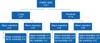
The main preoperative factors affecting outcomes include the presence of impaired preoperative liver function, extent of liver resection, and the presence of a small remnant liver.3 To analyze OGTT along with volumetric evaluation by PHRR, we subcategorized the study group into four subgroups: 1) PHRR >50% with a. Linear OGTT and b. Parabolic OGTT; 2) PHRR <50% with a. Linear OGTT and b. Parabolic OGTT (Figs. 2 and 3). None of the patients with a parabolic OGTT and PHRR less than 0.5 experienced any major morbidity. Minor morbidity was observed in all patients with a linear OGTT and a PHRR greater than 0.5, who underwent resections. Caution may need to be exercised when considering a major resection in a patient with linear OGTT and PHRR >50%.
OGTT was found to be an efficient tool for the prognosis of cirrhotic patients.3940 It was found to be effective in our study group which was predominantly non-cirrhotic, and its predictive value may improve in a cohort of predominant cirrhotic resections, wherein the concern of PLF is greater and the resections are more challenging. However, in view of the small sample size, our study stands underpowered to detect factors affecting mortality, morbidity and severe postoperative liver insufficiency. We did not perform a direct comparison of OGTT and ICGR 15. While it may be argued that OGTT did not fare well in predicting the 50-50 criteria for liver failure, this may not be the failure of OGTT, but the fallacy of the 50-50 criteria. The 50-50 criteria were developed in a rather heterogeneous cohort rather than a predominantly non-cirrhotic group like ours. It was defined based on fatal liver failure, while none of our patients experienced mortality. However, on applying the International Study Group of Liver Surgery (ISGLS) definition of liver failure, the grades did not differ significantly between parabolic and linear OGTT (data not shown). In addition to its efficacy in predicting outcomes, OGTT is advantageous due to its simplicity, standard technique, easy interpretation and low cost.15
In conclusion, our study paves the way for designing a prospective study to assess OGTT, compared to a validated preoperative method like ICGR 15, in future. OGTT is a relatively cheap, safe, simple and effective test, having the potential to substitute ICGR 15 and predict outcomes reliably. In a larger cohort with utilization of appropriate statistical tools, it should be possible to incorporate OGTT LI and PHRR into a simplified formula with a relevant cutoff, to help guide the extent of resection and anticipate postoperative liver insufficiency and morbidity, and to consider preoperative augmentation or for early postoperative prophylactic and therapeutic measures.
 XML Download