Abstract
Objective
Treatment of spontaneous intracerebral hemorrhage (ICH) remains controversial. However, an extensive hemorrhage with a poor mental status is suitable for surgical evacuation. Our experience with the transsylvian-transinsular (TS-TI) microsurgical approach for deep-seated basal ganglia (BG) ICH was investigated.
Material and Methods
A retrospective review was conducted on 86 patients with BG ICH who underwent an operation at the Department of Neurosurgery of our Hospital from September 2011 to October 2014. Thirteen patients underwent craniotomy and the TS-TI microsurgical approach for hematoma evacuation. Twenty-seven patients underwent conventional craniotomy with the trans-cortical transtemporal (TC-TT) approach, and 46 patients underwent a burrhole operation and hematoma drainage using a frameless stereotaxic device (ST).
Results
The average age distribution was similar. The preoperative Glasgow coma scale (GCS) was similar for the TC-TT and TS-TI groups. The pre-operative hematoma levels were higher in the TC-TT (109.4 ± 48.6 mL) and TS-TI (96.0 ± 39.0 mL) groups than in the ST group (46.5 ± 23.5 mL). The hematoma removal rate was 77% in the TC-TT group, 88% in the TS-TI group, and 34% in the ST group. The mean maintenance period of a hematoma catheter was 3.6 days in the ST group. The clinical outcome showed correlation with the preoperative neurological symptoms.
Spontaneous intracerebral hemorrhage (ICH) is a well-known pathologic condition that commonly results in severe morbidity and a high mortality rate. Most patients can be managed conservatively; however, in patients with an extensive ICH, surgical evacuation of the hematoma is required for hematoma volume reduction and immediate reduction of the intracranial pressure (ICP).4)5) Several conventional surgical methods are available for treatment of ICH. Stereotactic-guided aspiration of hematomas is a preferred treatment modality; however, due to its significant mass effect for effectively controlling ICP, open craniotomy remains indispensable for many ICH patients. Conventional craniotomy for evacuating a hematoma can be performed directly through the frontal or temporal cortex closest to the hematoma. The purpose of the current study is to present the transsylvian-transinsular (TS-TI) approach for evacuating a basal ganglia (BG) hematoma, and the possible reasons for achieving good results are evaluated.
A total of 86 consecutive patients with deep-seated BG hematomas who underwent surgical management from September 2011 to October 2014 in our department were included in the current retrospective study. The operations were performed by the same group of neurosurgeons.
The criteria for patient selection were the presence of a hematoma located in the BG with a volume >30 mL and an interval <24 hours between the onset of the hemorrhage and operation.
The level of bleeding was calculated using the following formula: height × width × length × 0.5.4) Midline shifting was the distance from midline to the fornix or foramen of Monro. Preoperative clinical grading of the disease was performed using the Glasgow coma scale (GCS). Patients with a mainly thalamic or subcortical hemorrhage were excluded.
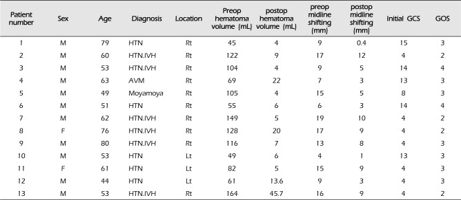
The total number of patients was 13, 11 men and 2 women, ranging in age from 49 to 80 years (mean age, 56.8 ± 11.6 years). There was one case of arteriovenous malformation (AVM) rupture, one case of moyamoya disease, and 11 cases of hypertensive ICHs (Table 1). The level of hemorrhage measured on preoperative axial computed tomography (CT) was between 45 and 149 mL (mean, 96.0 ± 39.0 mL). Midline shifting measured on preoperative CT was between 4 and 19 mm (mean, 12.3 ± 4.8). The preoperative GCS score was 8.0 ± 4.8 (Table 2).
The total number of patients was 27 including 19 men and 8 women, ranging in age from 44 to 72 years (mean age, 56.8 ± 8.1 years). The level of hemorrhage measured on preoperative axial CT was between 36.8 and 217.5 mL(mean, 109.4 ± 48.6 mL). The midline shifting measured on preoperative CT was between 4 and 23 mm (mean, 12.4 ± 4.9). The preoperative GCS score was 8.7 ± 4.0 (Table 2).
The total number of patients was 46, including 27 men and 19 women, ranging in age from 37 to 83 years (mean age, 58.6 ± 12.8 years). The level of hemorrhage measured on preoperative axial CT was between 30.6 and 118.3 mL(mean, 45.5 ± 23.5 mL). The midline shifting measured on preoperative CT was between 2 and 12 mm (mean, 5.7 ± 2.5). The preoperative GCS score was 12 ± 3.3.
The patients were placed in the supine position with their heads turned toward the healthy side 30 to 45 degrees and tilted back 15 degrees. Standard pterional craniotomy was performed as follows: the lower margin of the skin incision began from 1 cm anterior to the tragus and 1.5 cm superior to the zygomatic arch. The size of the bone flaps was similar to that in a ruptured middle cerebral artery aneurysm surgery. The sylvian fissure was sharply separated under a microscope from the lateral to medial direction near the frontal side, where no vessel is evident. Identification of the M3 branches within the fissure provided a reference for further proximal dissection to M2 divisions and the insular cortex.6) A 3-cm entry site on the lateral sylvian fissure is sufficient for entry to the cortical surface of the insula. A small 1.5-cm cortisectomy site was created in the insula cortex, and the hematoma cavity was entered.3) Hematoma evacuation was performed using suction and bipolar cautery. The lenticulostriate artery (LA) can be exposed well, and its bleeding can be stopped. A minimal amount of hematoma remained untouched near the AVM margin to avoid rebleeding. The hematoma cavity was covered with Surgicell and wet Gelfoam. Watertight closure of the dura was performed. In three cases, ventricular extension of the hematoma was observed and ventricular drainage was added. Replacement of the skull bone was determined based on the condition of the brain after ICH evacuation.
A 63-year-old male patient presented with sudden left-side weakness. His initial GCS score was 13. CT showed 69 mL of right BG ICH with 7-mm midline shifting. CT angiography showed an AVM (1.7 cm) in the right inferior BG that was fed by multiple LAs from the right M1. Emergent craniotomy and hematoma evacuation were performed using the TS-TI approach 3 hours after admission. Postoperative CT showed partial removal of ICH and a shrunken brain (Fig. 1A, B). He underwent gamma knife radiosurgery after 15 days after the craniotomy. No injuries to the frontal and temporal cortexes were observed on the CT performed at 3 months after the operation (Fig. 1C). The patient survived with left hemiparesis.
The patients were placed in the supine position with their heads turned toward the nearest hematoma with the cortical distance upside. If necessary, a navigation system was used. The craniotomy included a small craniotomy to large dandy craniotomy depending on the preoperative neurological condition. A small cortisectomy site was created, and the hematoma cavity was entered. Hematoma evacuation was performed using suction and bipolar cautery. The hematoma cavity was covered with Surgicell and wet Gelfoam. A hematoma catheter was added on the hematoma cavity in cases of remnant hematoma. Replacement of the skull bone was determined based on the condition of the brain after ICH evacuation.
A 61-year-old male patient presented with sudden stupor. His initial GCS score was 12. A high level of right BG ICH with 9-mm midline shifting was observed on CT. Emergent craniotomy and hematoma evacuation were performed using the craniotomy and transcortical approach 1 hour after admission. Postoperative CT showed total removal of ICH and a shrunken brain (Fig. 2A, B). Encephalomalatic changes in the temporal cortex were observed on the postoperative 2-month CT scan (Fig. 2C). The patient survived with mild disability.
The patients were placed in the supine position with their heads slightly flexed. A Medtronic navigation StealthStation® AxiEM7™ system was used. The entry point was an ipsilateral Kocher's area. A 12 French drainage catheter was usually inserted in the center of the hematoma cavity. Hematoma evacuation was performed by manual suction using a 5 mL syringe. The total level of the evacuation hematoma was approximately 5-15 mL. Irrigation with urokinase (6,000 µ) and saline (2.6 mL) was performed according to the immediate post-operative CT scan. The mean maintenance duration of the drainage catheter was between 1 and 11 days (mean, 3.6 ± 2.1 days).
After hematoma evacuation, the patients were managed in the neurosurgical intensive care unit. Once the patient's mental status had recovered to obeying commands, extubation of the endotracheal tube was performed. If the patient's mental status was not recovered to obeying commands, tracheostomy was performed approximately 1 week after surgery. Thereafter, the patients were moved to the general ward and active rehabilitation treatment was initiated. The remaining hematoma volume was measured using CT immediately following the operation, 1 week after surgery, and at discharge. The postoperative GOS scores and activities of daily living were assessed at discharge.
No significant difference in the preoperative hematoma volume was observed between the TC-TT and TS-IS groups (p = 0.318). The volume of the remaining hematoma measured by CT immediately following the operation was 11.6 ± 11.8 mL in the TS-IS group, 25.3 ± 24.4 mL in the TC-TT group, and 31.2 ± 22.2 mL in the ST group. The hematoma removal rate was 77% in the TC-TT group, 88% in the TS-IS group, and 34% in the ST group. The hematoma evacuation rate was higher in the TS-IS group than in the TC-TT group (p = 0.067). Midline shifting improved from 12.4 mm to 8.5 mm in the TC-TT group, from 12.3 mm to 6.2 mm in the TS-IS group, and from 5.7 mm to 4.2 mm in the ST group. There were 4 cases of mortality in the TC-TT group; all were comatose before the operation.7)11) The number of rebleeding cases, which showed increased hematoma on the postoperative CT scan, was 5 (10.8%), and the number of inadequate decompression cases, which showed 90% or more remaining hematoma on the postoperative CT scan, was 7 (15.2%) in the ST group. The mean maintenance period of hematoma catheter was 3.6 days in the ST group.
The mean GOS value at the time of discharge was 3.2 ± 1.0 in the TC-TT group, 2.8 ± 0.6 in the TS-IS group, and 2.0 ± 0.9 in the TC group.
The short-term outcome of ICH was determined by the severity of bleeding, as measured by clinical conditions, hematoma size, and presence of an intraventricular hematoma. Surgical intervention increases the chance of survival or prolongs the survival time, despite the presence of severe neurological deficits. Although a few methods are available for treatment of spontaneous cerebral hemorrhage, the treatment is generally classified according to 2 major groups: hematoma evacuation by craniotomy or drainage by burrhole.1)
Drainage of the hematoma by burrhole requires a small incision in the skin and brain. This method has the advantage of causing minimal brain tissue injury; however, the hematoma cannot be removed all at once and effective hemostasis is not possible, resulting in a recurrent hemorrhage after surgery. However, these disadvantages can be corrected by neuroendoscopy and use of a navigation system.2)14) According to our results, the ST group was more reliable than the TS-IS and TC-TT groups for treating a small BG hemorrhage. However, the number of rebleeding cases, which showed increased hematoma on the postoperative CT scan, was 5 (10.8%) and the number of inadequate decompression cases, which showed 90% or more remaining hematoma on the postoperative CT scan, was 7 (15.2%). The average hematoma drain retention period was 3.6 days.
For patients with extensive ICH, hematoma evacuation by craniotomy can be required for hematoma volume reduction and rapid reduction in intracranial pressure (ICP). This approach required incising the cerebral cortex to access the hematoma, resulting in effective hematoma evacuation, while causing considerable trauma to the brain tissue. The two standard craniotomy approaches are the TS-TI and TC-TT approaches.13)
Comparing the level of pre-operative hematoma in the two groups, a higher level was observed in the TC-TT group, which was not statistically significant.
Out study demonstrates the superiority of the TS-TI approach compared with the TC-TT approach in terms of the hematoma evacuation rate and outcome status. The hematoma removal rate was 77% in the TC-TT group and 88% in the TS-IS group. Midline shifting improved from 12.4 mm to 8.5 mm in the TC-TT group and from 12.3 mm to 6.2 mm in the TS-IS group. According to our results, the hematoma evacuation rate of the TS-IS group was superior to that of the TC-TT group (p = 0.067).
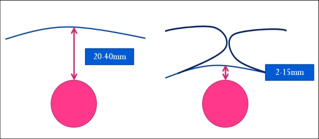
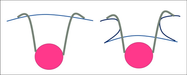
The TS-TI approach decreased the injury to normal cerebral tissue in the temporal lobe and shortened the distance from the cortex incision to the hematoma. The depth of the sylvian fissure ranges from 20 to 40 mm. The distance from the insular cortex to the hematoma is shorter when using the TS-TI approach compared with that using the direct TC-TT approach. Using the TS-TI approach, the distance is approximately 2 to 15 mm from the hematoma to the insular cortex (Fig. 3).7)11) In addition, the cortical transsectional area is reduced for the same surgical corridor (Fig. 4).
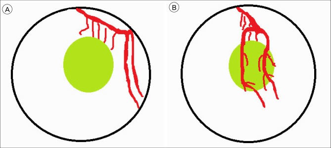
In addition to reducing brain tissue injury, the TS-TI approach has several advantages. First, the M2 branches are initially dissected during the TS-TI approach, which facilitates the localization and treatment of bleeding during surgery. The LA, as a responsible vessel, is located at the anterior route through the transinsular approach. Unlike with the TC-TT approach, the hematoma is located between the LA and M2. The bleeding LA is difficult to control (Fig. 5).12) Second, the incised insular cortex is located at a position lower than that of the hematoma, which results in a more satisfactory hematoma evacuation around deep neurostructures, such as the midbrain, internal capsule, and thalamus. Third, there is no need for navigation systems.
The disadvantages of the TS-TI approach include a risk of major vessel injury, sacrifice of the cortical and sylvian veins, manipulative transgression of the thin arachnoids around the sylvian area, and vasospasm due to mechanical damage to the vessel walls.8)9) In addition, a longer surgery time is required for novice neurosurgeons.10)
In our experience, the advantages of the TS-TI approach in treatment of deep-seated BG compared with the transcortical approach are that the TS-TI approach (1) minimizes normal brain tissue injury; (2) provides good access to the responsible vessels; (3) provides suitable decompression of important neurostructures, including the brainstem; and (4) does not require a navigation system.
References
1. Bae HG, Lee KS, Yun IG, Bae WK, Choi SK, Byun BJ, et al. Rapid expansion of hypertensive intracerebral hemorrhage. Neurosurgery. 1992; 7. 31(1):35–41. PMID: 1641108.

2. Dye JA, Dusick JR, Lee DJ, Gonzalez NR, Martin NA. Frontal bur hole through an eyebrow incision for image-guided endoscopic evacuation of spontaneous intracerebral hemorrhage. J Neurosurg. 2012; 10. 117(4):767–773. PMID: 22900841.

3. Jianwei G, Weiqiao Z, Xiaohua Z, Qizhong L, Jiyao J, Yongming Q. Our experience of transsylvian-transinsular microsurgical approach to hypertensive putaminal hematomas. J Craniofac Surg. 2009; 7. 20(4):1097–1099. PMID: 19634217.

4. Kaya RA, Türkmenoğlu O, Ziyal IM, Dalkiliç T, Sahin Y, Aydin Y. The effects on prognosis of surgical treatment of hypertensive putaminal hematomas through transsylvian transinsular approach. Surg Neurol. 2003; 3. 59(3):176–183. PMID: 12681546.

5. Li Q, Yang CH, Xu JG, Li H, You C. Surgical treatment for large spontaneous basal ganglia hemorrhage: retrospective analysis of 253 cases. Br J Neurosurg. 2013; 10. 27(5):617–621. PMID: 23406426.

6. Potts MB, Chang EF, Young WL, Lawton MT. UCSF Brain AVM Study Project. Transsylvian-transinsular approaches to the insula and basal ganglia: operative techniques and results with vascular lesions. Neurosurgery. 2012; 4. 70(4):824–834. PMID: 21937930.
7. Rhoton AL Jr. The supratentorial arteries. Neurosurgery. 2002; 10. 51(4 Suppl):S53–S120. PMID: 12234447.

8. Schaller C, Klemm E, Haun D, Schramm J, Meyer B. The transsylvian approach is "minimally invasive" but not "atraumatic". Neurosurgery. 2002; 10. 51(4):971–976. PMID: 12234405.

9. Schaller C, Zentner J. Vasospastic reactions in response to the transsylvian approach. Surg Neurol. 1998; 2. 49(2):170–175. PMID: 9457267.

10. Shin DS, Yoon SM, Kim SH, Shim JJ, Bae HG, Yun IG. Open surgical evacuation of spontaneous putaminal hematomas: prognostic factors and comparison of outcomes between transsylvian and transcortical approaches. J Korean Neurosurg Soc. 2008; 7. 44(1):1–7. PMID: 19096649.

11. Tanriover N, Rhoton AL Jr, Kawashima M, Ulm AJ, Yasuda A. Microsurgical anatomy of the insula and the sylvian fissure. J Neurosurg. 2004; 5. 100(5):891–922. PMID: 15137609.

12. Wang F, Sun T, Li X, Xia H, Li Z. Microsurgical and tractographic anatomical study of insular and transsylvian transinsular approach. Neurol Sci. 2011; 10. 32(5):865–874. PMID: 21863272.

13. Wang X, Liang H, Xu M, Shen G, Xu L. Comparison between transsylvian-transinsular and transcortical-transtemporal approach for evacuation of intracerebral hematoma. Acta Cir Bras. 2013; 2. 28(2):112–118. PMID: 23370924.

14. Zhu H, Wang Z, Shi W. Keyhole endoscopic hematoma evacuation in patients. Turk Neurosurg. 2012; 3. 22(3):294–299. PMID: 22664995.

Fig. 1
(A) A 63-year-old male patient presented with sudden left-side weakness. Computed tomography (CT) shows 69 mL of right basal ganglia hemorrhage with 7-mm midline shifting. (B) After craniotomy and hematoma evacuation were performed, postoperative computed tomography (CT) shows partial removal of the hemorrhage and a shrunken brain. The trajectory site through the sylvian fissure is noted. (C) No injuries of frontal and temporal cortex are seen on the CT performed 3 months after the operation.

Fig. 2
(A) A 61-year-old male patient presented with sudden stupor. His initial Glascow coma scale (GCS) score was 12. Computed tomography (CT) shows a high level of right BG intracerebral hemorrhage (ICH) with 9-mm midline shifting. (B) After emergent craniotomy and hematoma evacuation were performed, postoperative CT shows total removal of ICH and a shrunken brain. The hematoma is almost completely removed, resulting in a shrunken brain. (C) Encephalomalatic changes in the temporal cortex and hematoma evacuation trajectory site are observed on the CT scan performed 2 months after the operation.

Fig. 3
Schematic diagram: In the transtemporal approach, the distance from the temporal cortex to the hematoma (circle) is approximately 20 to 40 mm (right). In the transsylvian-transinsular approach, the distance from the insular cortex to the hematoma is shorter (2 to 15 mm [left]) than that in the direct transcortical approach.

Fig. 4
Schematic diagram: To maintain the same surgical conditions, the cortical transsectional area of the transtemporal approach (right) is larger than that of the transsylvian-transinsular approach (left).

Fig. 5
(A) Schematic diagram: In the transtemporal approach, the hematoma (circle) is located between the lenticulostriate artery (LA) and M2. (B) Schematic diagram: In the transsylvian-transinsular approach, the M2 branches are initially dissected and retracted. The LA, as a responsible vessel, is located at the anterior route (left).

Table 1
Summary of 13 patients who underwent craniotomy with the trans-sylvian trans-insular approach for deep-seated basal ganglia hemorrhage (Jan 2013 to Oct 2014)

Table 2
Comparison between the surgical modalities for treating a basal ganglia hemorrhage (Sep 2011 to Oct 2014)

*Values are reported as the mean ± standard deviation (range).
TS-IS = transsylvain-transinsular group; TC-TT = transcortical-transtemporal group; ST = stereotactic-guided aspiration group; M = male; F = female; Rt = right; Lt = left; ICH = intracerebral hemorrhage; GCS = Glasgow coma scale; GOS = Glasgow outcome scale


 PDF
PDF ePub
ePub Citation
 Print
Print


 XML Download