

 PDF
PDF ePub
ePub Citation
 Print
Print


INTRODUCTION
Coil embolization is a safe and effective treatment for unruptured and ruptured intracranial aneurysms. Some studies comparing coil embolization to clip ligation for ruptured aneurysms have reported more favorable outcomes for patients treated with coil embolization compared with those treated with surgical clipping, with low rates of procedure-related complications, ischemic infarction, vasospasm, and shunt-dependent hydrocephalus.12)13) Despite these good clinical results, rebleeding and recanalization requiring further treatment remain major concerns of coil embolization. Although the rebleeding rate after coil embolization of ruptured intracranial aneurysms may seem low at 1.0-3.6%,6)8)16) poor and fatal outcomes of rebleeding patients have been reported. We reviewed the clinical data of patients with rebleeding after coil embolization and evaluated the risk factors and possible mechanisms of rebleeding in these patients.
MATERIALS AND METHODS
Patient population
Between July 2006 and October 2014, 391 consecutive patients with spontaneously ruptured intracranial aneurysms were treated using the endovascular method at our institution. Patients with dissecting aneurysms, blood blister-like aneurysms, and pseudoaneurysms and those with absolute evidence of intraprocedural rupture were excluded. Finally, 330 consecutive patients with saccular aneurysms treated using conventional coil embolization were included in this study. Presence of subarachnoid hemorrhage (SAH) was confirmed by brain computed tomography (CT), brain magnetic resonance imaging, or cerebrospinal fluid tapping. Ruptured aneurysms were identified using digital subtraction angiography (DSA) and three-dimensional rotational angiography (3 DRA), and coil embolization was performed within 24 h from hospital admission. Postprocedural brain CT was performed immediately after coil embolization. Patients underwent follow-up brain CT immediately after occurrence of at least one of the following symptoms or signs: neurological deterioration, sudden severe headache, and sudden fluctuation of vital signs. Rebleeding of intracranial aneurysms was defined as a significant increase in amount of SAH on follow-up brain CT compared with immediate postprocedural brain CT. In patients with combined intracerebral hemorrhage (ICH), cases of increased or newly developed ICH without increased SAH were not considered as rebleeding cases. The reason we did not consider this situation as rebleeding of a coiled aneurysm was due to a possible mechanism reported by Cho et al., who hypothesized that delayed hemorrhage or propagation of initial ICH may be caused by vulnerability of parenchyma adjacent to the ICH rather than rebleeding of a coiled aneurysm.4)
Radiological changes observed between brain CT scans performed immediately after embolization and those performed during follow-up were analyzed by an experienced neuroradiologist. Each patient's clinical condition at admission was evaluated according to the Hunt-Hess grading system, and SAH conditions were examined using the Fisher grading system. The radiological characteristics of the aneurysms and the coiling procedures were reviewed carefully on the basis of DSA and 3 DRA findings. The outcomes were classified using the Glasgow Outcome Scale (GOS).
Endovascular treatment
Coil embolization was performed immediately after diagnostic DSA, which was typically on the day of ictus. This procedure was performed under general anesthesia, except in patients with poor neurologic condition. A bolus of heparin was administered intravenously according to patient body weight after femoral artery sheath placement, and additional boluses of 1,000 IU of heparin were administered every hour for maintenance of an activated clotting time of 250-300 s during the procedure. In the majority of cases, coil embolization using detachable coils was performed using the classic single-catheter method. Various methods including stent-assisted, balloon-assisted, double-catheter, and catheter protection were used in cases of necessitating neck protection. A loading dose of dual antiplatelet medication (100 mg aspirin and 300 mg clopidogrel) was administered orally before or just after intervention, and a continued maintenance dose (100 mg aspirin and 75 mg clopidogrel, daily) was administered thereafter. If thromboembolic complications occurred, various rescue therapy regimens such as intraarterial or intravenous infusions of urokinase, abciximab, tirofiban, or low-molecular-weight heparin (LMWH) were used during or after the procedure. Aneurysm occlusion was classified using the Raymond-Roy scale, as follows: grade I, complete occlusion; grade II, neck remnant; grade III, residual aneurysm flow.14)
RESULTS
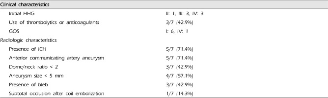
Rebleeding after coil embolization occurred in 7 (2.1%) of the 330 consecutive patients, whose clinical and radiological data are summarized in Tables 1 and 2. All cases of rebleeding occurred in the immediate postoperative period within 72 h (5-65 h) after coil embolization. The mean patient age was 60.0 ± 20.0 years (mean ± SD, range: 43-79 years), and there were 4 male patients. Three patients (42.9%) had poor clinical grades (Hunt-Hess grade [HHG] IV), and 5 patients (71.4%) had ICH. Of the 7 patients, 5 (71.4%) had anterior communicating artery (ACoA) aneurysms, 1 had a middle cerebral artery aneurysm, and 1 had a distal anterior cerebral artery aneurysm. The angiographic characteristics were as follows: aneurysm size < 4 mm (57.1%, 4/7); dome-to-neck ratio < 2 (42.9%, 3/7); presence of bleb (42.9%, 3/7); good treatment results for total or near-total occlusion of the aneurysm (85.7%, 6/7).
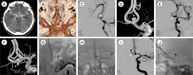
In one interesting case a size and morphological discrepancy of the aneurysm was observed between computed tomography angiography (CTA) and DSA in patient No. 6. Brain CT showed a large amount of SAH (Fig. 1A), and CTA showed an elongated aneurysm (Fig. 1B) with a dome-to-neck ratio of 2.47 (neck diameter, 1.44 mm; neck-to-dome diameter, 3.57 mm). However, 3 DRA performed after CTA showed a small saccular aneurysm (Fig. 1C, D) with a dome-to-neck ratio of 1.83 (neck diameter, 1.22 mm; neck-to-dome diameter, 2.24 mm). The aneurysm was treated successfully using 3 detachable coils with no neck and lumen remnants (Fig 1E). Unfortunately, sudden deterioration of mental status occurred after 8 h from coil embolization, and brain CT showed an increased amount of SAH and newly developed intraventricular hemorrhage.
Thrombolytics or anticoagulants were administered to 3 patients (42.9%). Urokinase was administered in 1 patient because of thrombus formation on the distal artery of the aneurysm, and continued anticoagulation using LMWH was performed in 2 patients to prevent thrombus formation caused by coil protrusion into the parent artery. Of the rebleeding patients, 2 underwent additional coil embolization and decompressive surgery, 1 underwent surgical clipping, 2 underwent only decompressive surgery, and 2 patients could not be treated. In all patients, mental status after rebleeding was worse than that before rebleeding. Six patients were in poor condition and presented with stuporous mentality or worse symptoms, and 3 of these patients were in a coma. Although we tried to secure the aneurysms and stabilize the brain condition, rebleeding patients showed a very poor outcome (GOS 1, 85.7%, 6/7).
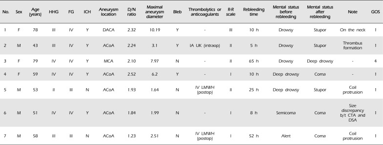
Illustrative case (patient no. 7)
A 58-year-old male presented with decreased consciousness after a sudden headache (HHG III). Brain CT showed a dense SAH (Fig. 2A), and CTA showed a saccular aneurysm of the ACoA (Fig. 2B). Emergency cerebral DSA was performed, and 3DRA showed 2 ACoA aneurysms located in the superior (Fig. 2C , D) and anterior (Fig. 2E, F) directions. Under general anesthesia, the 2 ACoA aneurysms were embolized using detachable coils, with no evidence of neck remnants (Fig. 2G, H), and the patient recovered well with a nearly alert mental status. However, there was a risk of thrombus formation because a coil loop protruded into the ACoA (Fig. 2H), and therefore, LMWH was administered intravenously in the postoperative period. Eight hours after coil embolization, the patient presented with a comatose mentality, and brain CT showed increased SAH in the basal cistern. Follow-up DSA performed immediately showed contrast leakage from the ACoA aneurysm in the anterior direction (Fig. 2I). The aneurysm was secured by performing additional coil embolization (Fig. 2J) and decompressive hemicraniectomy. Unfortunately, the patient died 6 days after rebleeding.
DISCUSSION
In this study, the incidence of rebleeding after coil embolization for ruptured intracranial aneurysms was 2.1% of 7 patients among 330 patients. This incidence rate was similar to those reported in previous studies. The Cerebral Aneurysm Rerupture After Treatment study reported a 1.9% incidence rate of rebleeding after aneurysm treatment using coiling and clipping.7) In that study, the risk of rebleeding after coil embolization was greater than that after clipping (3.4% after coiling vs. 1.3% after clipping), and the median time to rebleeding was 3 days.7) Recently, 2 large retrospective studies evaluated the incidence of rebleeding after coiling of ruptured intracerebral aneurysms.4)8) Kang et al.8) reported the incidence of rebleeding as 1.0% (6 of 591 patients) and reported that rebleeding occurred within 24 h in all cases and no further episodes of rebleeding occurred after the first 24 h. In another study the rebleeding rate was 1.1% (13 patients), and of the 13 patients, 10 experienced rebleeding within 3 days after coil embolization.4) Similarly, in the current study, all rebleeding cases occurred within 3 days after coiling. Our study confirmed that recurrent hemorrhage after coiling occurred most often within 3 days after coil embolization, and careful management and neurological workup should be performed during this period.
The risk factors known to influence rebleeding are incomplete embolization, anticoagulant or thrombolytic agent use, coil compaction, and coil migration.1)2)7)10)15)16) Coil compaction and coil migration were reported to show association with delayed rebleeding,4) whereas incomplete embolization of ruptured aneurysms is a strong risk factor associated with early rebleeding.6)7)13) However, complete occlusion of aneurysms does not always guarantee prevention of rerupture. Rebleeding may occur after successful embolization with satisfactory occlusion on angiography because uneven distribution of the coil masses may cause a channel of blood stream to the ruptured point of the aneurysm, and early recanalization may occur with spontaneous resolution of the thrombi through the inserted coil loops.8) In our study, subtotal occlusion of the aneurysm was observed in only 1 patient; however, other risk factors including the use of thrombolytics or anticoagulants, presence of ICH, and presence of bleb were also identified. Use of anticoagulants and thrombolytics was a risk factor for rebleeding, because spontaneous resolution of thrombi or disturbances in thrombus formation inside the coiled aneurysm could occur. Coexistence of ICH was another risk factor for rebleeding. Some studies have reported that ICH is an important risk factor for rebleeding and that thrombosed pseudoaneurysms inside the ICH may cause rebleeding.6)16)
The most common location of reruptured aneurysms was the ACoA (71.4%, 5/7). An ACoA aneurysm was considered a risk factor for rebleeding in some previous studies.4) Sluzewski and van Rooij16) suggested that ruptured ACoA aneurysms were more often associated with ICH. An interesting finding of the current study was related to ACoA aneurysms. Four of the 5 reruptured ACoA aneurysms had unilateral hypoplasia of the A1 segment of the anterior cerebral artery. Unilateral hypoplasia of the A1 segment is not a rare vascular variation, and frequency of this variation up to 13% was reported in an autopsy series.11) Chen and Li3) reported that the prevalence of ACoA aneurysms with A1 segment hypoplasia patients was 14%, and 43-57% of patients with ACoA aneurysms had unilateral hypoplasia of the A1 segment. Strong association of hemodynamic stress caused by unilateral hypoplasia of the A1 segment with ACoA aneurysms has been reported.3)9) In this series, the incidence of A1 hypoplasia in rebleeding patients was relatively high (80.0%) compared with previous reports, and we hypothesized a possible association of rebleeding with early minor compaction of the coils owing to hemodynamic stress caused by unilateral hypoplasia of the A1 segment. In addition, these ACoA aneurysms in our series with unilateral A1 hypoplasia were small-sized aneurysms with a maximal diameter of < 4 mm (patient nos. 2, 5, 6, and 7 in Table 1). Small aneurysm size has been reported as a risk factor for rebleeding.16) Overestimated coil packing density and masking of contrast filling into the aneurysm after coil embolization could explain the reason why small size was a risk factor. Hemodynamic stress due to A1 segment hypoplasia and small aneurysm could lead to some synergic effect on rebleeding after coil embolization.
In this study, rebleeding patients showed a very poor outcome. Only 1 patient showed a good outcome (GOS 4), and the remaining 6 patients showed poor outcomes (GOS 1). Most previous studies have reported very poor outcomes in rebleeding patients.1)2)4)5)6)8)16)17)18) In particular, the outcomes of early rebleeding were worse than those of delayed rebleeding.4) Our study has some limitations. This was a retrospective study and was subject to selection bias and deviations. In addition, the sample size was small, and statistical analysis was not possible.
CONCLUSION
Rebleeding of ruptured intracerebral aneurysms after coil embolization is rare, but the outcomes of rebleeding are serious or fatal. In this study, we suggested the risk factors of rebleeding, including the presence of ICH, presence of bleb, use of anticoagulants or thrombolytics, and occurrence of ACoA aneurysms. In cases of small ACoA aneurysms, unilateral hypoplasia of the A1 segment may influence rebleeding owing to hemodynamic stress.

 XML Download