Abstract
Owing to the rapid development of intervention techniques and devices, endovascular coil embolization of cerebral arteries has become standardized. It is particularly preferred when a patient presents with an unruptured intracranial aneurysm of the posterior communicating artery (PcomA). However, the risk of thrombogenic complications of the coil migration may also result in a large cerebral infarction.
When coil migration occurs during embolization, a procedure for removal of the embolic coil should be performed immediately. We experienced a clinically rare case of migration of a framing coil to the distal middle cerebral artery aneurysm during endovascular embolization of an unruptured PcomA aneurysm. The migrated coil was barely retrieved using snare techniques.
Along with microsurgical clipping, endovascular coil embolization has become a widely selected option for treatment of intracranial aneurysms. With the growing use of endovascular therapeutic procedures, more unexpected procedure-related complications have been reported, and coil migration is reported to occur in 2-6% of intracranial aneurysms.5) Only a few clinical cases have been reported in relation to coil migration, and no proven treatment guidelines have yet been established; however, the results of coil migration complications are still devastating, and affected patients require immediate salvage therapy.10)11)
In general, it is the final helical coils that tend to migrate as a part of a procedural complication. Except in situations where the migration occurs with the helical type or due to a mismatch with the size of the aneurismal sac, migration of the framing coils towards the end of the procedure is very rare. We experienced the migration of a framing coil to the distal middle cerebral artery after filling the aneurysmal sac with the framing and helical coil. We report on a case of framing coil migration where its retrieval was managed using a gooseneck snare (Amplatz Goose Neck® snare, SK200, eV3, Plymouth, MN, United States) leaving no neurologic sequelae in the patient.
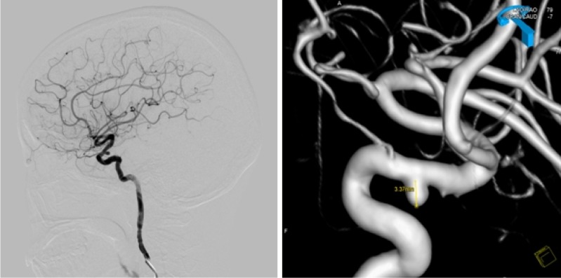
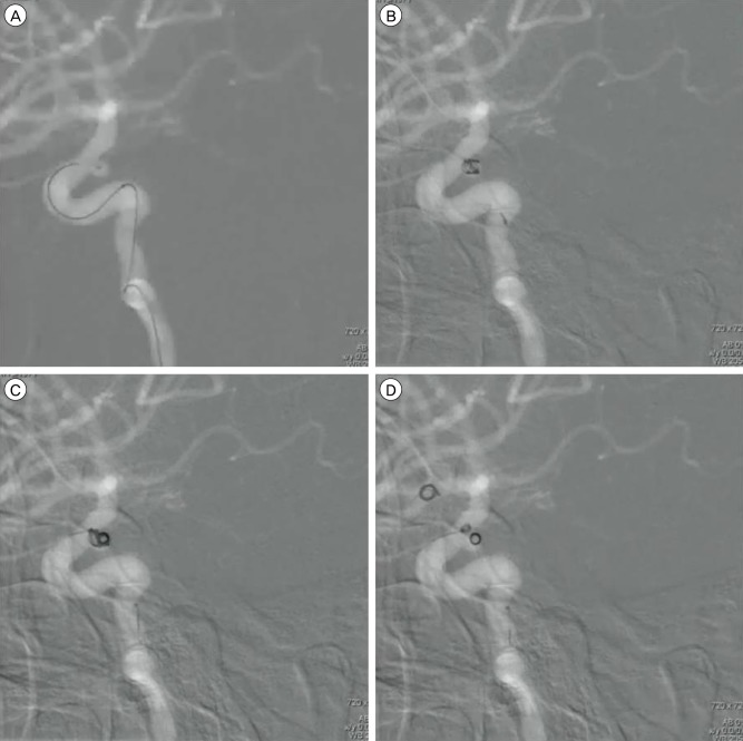
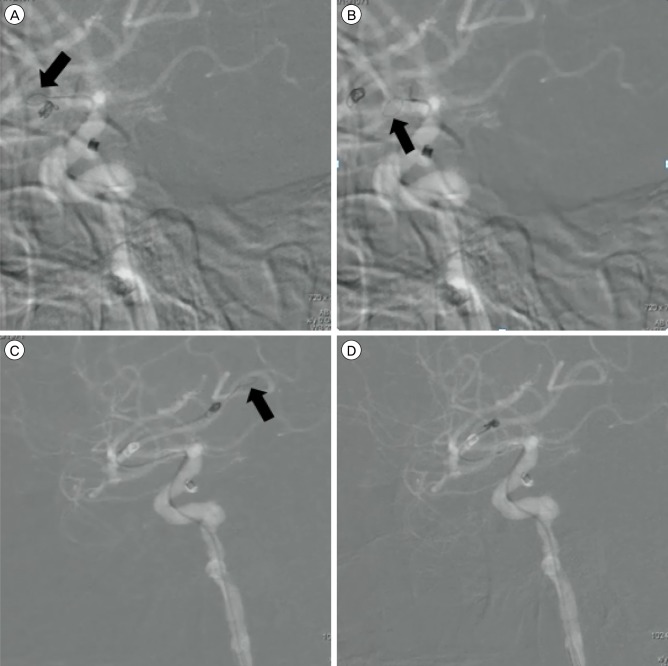
A 40-year-old female presenting with chronic headaches for several years was admitted to our hospital. Through imaging studies of computed tomography angiography (CTA), a right unruptured posterior communicating artery (PcomA) aneurysm was discovered. She had no known medical history or past neurological disability. Results of transfemoral cerebral angiography (TFCA) confirmed an unruptured aneurysm measuring 3.37 mm × 3.01 mm in size with a 2.75 mm neck arising from the right PcomA (Fig. 1). We inserted an Excelsior® SL-10® micro catheter (45° shape) into the aneurysm using a Synchro®-14 microwire. Without stent placement, two coils (eV3 3D® 3 mm × 6 cm, eV3 Helix® 2 mm × 4 cm, Plymouth, MN, United States) were successfully introduced and detached. In the process of complete obliteration by introducing the third coil (eV3 Helix® 2 mm × 3 cm, Plymouth, MN, United States), we detected a coil loop being abnormally released out from the aneurysmal sac when the detachment point was 2 mm away from the distal marker of a microcatheter. To avoid the risk of coil migration, an attempt was made to withdraw the last coil; nonetheless, the initially detached coil (eV3 3D® 3 mm × 6 cm, Plymouth, MN, United States) was forced out from the aneurysm and migrated into the proximal middle cerebral artery (MCA) (M1) (Fig. 2). The other coil (eV3 Helix® 2 mm × 4 cm, Plymouth, MN, United States) remained within the aneurysmal sac. We attempted to capture the migrated framing coil using a gooseneck snare (Amplatz Goose Neck® Microsnare Kit, SK200, eV3, Plymouth, MN, United States), but the initial retrieval attempt failed as the coil had moved further towards the distal MCA (M3). After this failure, a 0.014 inch microcatheter (PROWLER® SELECTTM PLUS, Codman®, Raynham, MA, United States) was placed in the MCA, distal to the migrated coil, and the coil was then secured using a gooseneck snare (Fig. 3). Immediately after the successful retrieval of the migrated framing coil, a stent (ENTERPRISETM VRD 4.5 mm × 28 mm, Codman®, Raynham, MA, United States) was deployed in order to prevent further coil migration. Consequently, an intra-stent aneurysmal selection and one more coil (eV3 3D® 3 mm × 6 cm, Plymouth, MN, United States) insertion was carried out, and then the entire procedure was completed. The patient experienced no clinical symptoms, and was discharged two days after the procedure.
This clinical case reports on the process of securing a coil migration and the subsequent stent-assisted coil embolization of an unruptured PcomA aneurysm. In 1995, Watanabe et al. reported the first case of retrieval of a migrated coil during the coil embolization of a ruptured aneurysm both in the superior cerebellar artery and basilar bifurcation using the "twist" technique.10) Prestigiacomo et al., Koseoglu et al., and Fiorella et al. reported cases of securing a partial or total coil migration using a goosenecksnare.1)3)7)
This case is authentic compared to the other cases of coil migration, as it involves the migration of a framing coil alone after embolization of an aneurismal sac using a framing coil and a helical coil. There are two different points to consider that distinguish this case from other cases of helical coil migration. First, there is a difference in the degree of difficulty in retrieving the framing coil compared to the helical coil. This is a rare case, thus not much comparable study has been done. However, when using the snare retrieval technique, one must remember that the retrieval catheter must be able to encircle the migrated coil. In this situation, it may be difficult for the retrieval catheter to pass through the framing coil, as it already has its own shape. On the other hand, the helical coil is usually soft and untangled, hence it is easier for the retrieval catheter to pass through the coil and capture it. The framing coil that had moved out from the aneurismal sac was migrating farther away distally during its retrieval, and there was the possibility of migration of the remnant helical coil within the sac. Theoretically, retrieval of the migrated coil after deployment of a stent within the PcomA is ideal. However, this delays the retrieval start time, and stent insertion may burden the coil retrieval. In this case, we first captured the migrated coil, and then deployed a stent in order to perform an intra-stent selection.
There are two methods for retrieval of a migrated coil: Surgical or endovascular removal. Usually, surgical removal is only performed when the endovascular retrieval procedure fails. Because there may be a delay in the flow opening of the vessel, and the difficulty itself of the surgical approach, the prognosis of the surgical method of removing a coil may not always be good.
In reviewing the published reports, three popular techniques and methods are used for endovascular coil retrieval: wire techniques, snare devices, and retriever devices. When using wire techniques for coil retrieval, which were used in the reports by Standard et al. and Lee et al., a microwire is shaped into a pigtail or J-shape Xpedeion balloon guidewire (MTI, Richmond, CA, United States); 0.010-in.microwires (Agility, Cordis, Bridgewater, NJ, United States) were used.4)8) Snare techniques use a distally looped wire. In 2004, Koseoglu et al. reported several cases of effective foreign body retrieval in various parts of vascular structure, such as the MCA and superior vena cava.3) Koseoglu et al. used a nitinol gooseneck snare (Microvena, White Bear Lake, MN, United States), which has five sizes (5, 10, 15, 25, and 35 mm). The 5 mm and 10 mm snare loops are applied using a 4 Fr guiding catheter, while the larger snares are applied using a 6 Fr guiding catheter. In cases similar to ours, Koseoglu et al. decided on the size of the snare loop based on the vessel size first, and then the snare loop was advanced to encircle and pull out the migrated coil. The Amplatz Goose Neck® Microsnare Kit, which was used in this article, is a commonly used device. The Amplatz Goose Neck® Microsnare is a one-loop device; hence, the rescue of migrated coils or stents may fail in certain situations. To overcome such limitations, multi-loop devices such as En Snare® (Merit Medical, South Jordan, UT, United States) have recently been developed, and are popular in practical fields. Unlike the Goose Neck Snare®, En Snare® is a three-loop device system, which is adaptable to a wide range of vessel types. The third type of medical device is a retriever device. There are usually two types of retriever devices: alligator and stent. Zoarski et al. reported on a case of retrieval of a migrated coil using an alligator retrieval device® (Chestnut Medical Technologies, Menlo Park, CA, United States), while Vora et al. used an L5 Merci® retriever (Concentric Medical, Mountain View, CA, United States) and O'Hare et al. used a X6 Merci® retriever (Concentrie Medical, Mountain View, CA, United States), Solitare® stent (ev3, Plymouth, MN, United States) in an effort to remove foreign bodies in the vascular structure.2)6)9)
The snare device is designed with loops where the shapes are smooth and flexible for intravascular safety; however, there are no sharp margins in the device in which we might encounter difficulty in capturing and securing the intravascular fragments. The alligator retrieval device has sharp ends, thus, when using it, the operator is required to use caution and to have abundant experience and skill in order to minimize damage to the inner vessel walls. The stent-assisted retrieval technique is derived from the idea of the stent-assisted removal of a thrombus. In 2014, Liu et al. reported a case of endovascular embolization of a PcomA aneurysm where the authors used Trevo devices (Concentric Medical, Mountain View, CA, United States) in an attempt to retrieve the migrated coil after experiencing failure in their attempt to retrieve the coil using the Alligator device (Chestnut Medical Technologies, Menlo Park, CA, United States). The stent-assisted method has some limitations compared to the snare or the alligator retrieval techniques. The stent-assisted technique is not efficient at capturing foreign fragments in tortuous and small vessels.
To the best of our knowledge, few studies comparing and contrasting the advantages and disadvantages of the various retrieval methods have been reported. In addition, the rather low incidence of coil migration may not allow the operators to conduct large-scale prospective studies. Thus, it is natural for the operators to have a preference when choosing the appropriate device for uncommon but potentially catastrophic cases of coil migration.
Loss of a device is an unwanted experience for the operator. In practice, the fracture and migration of endovascular system devices can occur at any time, resulting in various clinical complications for patients. Coil migration is a disastrous situation where the operator is required to decide promptly to troubleshoot the problem by mobilizing expert help, if necessary. Even if the coil migration is detected in situ during the endovascular procedure, the distal occlusion of the affected vessel may not occur for hours, as long as a heparin infusion is maintained. When this happens, the operator must decide whether to use a salvage method to retrieve the migrated coil or to shift the migrated coil to the most distal part of the affected vessel in order to avoid a major cerebral infarction. Then, the optimal method of retrieval must be planned, based on the availability of the devices. We report on a case where an intra-procedural framing coil migration was retrieved using a snare technique, in which the patient was treated without neurological deficit. However, in our retrospective review of the situation, the stent insertion should be considered initially. Despite successful introduction of the first and second coil embolizations, when an abnormal coil action is noted during introduction of the third coil, the operator must stop and consider a stent insertion using the jailing technique. The coil migration could have been prevented if stent placement had been considered earlier in the process of reviewing the CTA. Possible causes of coil migration in this case are as follows: a wide aneurysmal neck, instability of the coil in the aneurysmal sac, excessive coil embolization, and removal of the last coil. All of these complex factors could have contributed to the resultant coil migration during the procedure.
Coil migration during endovascular treatment of an unruptured aneurysm is an emergent situation in which the operator is required to decide on the most appropriate salvage management technique in order to avoid devastating consequences. According to our experience, when there is a migration of a framing coil after embolization with the framing coil and the subsequent helical coil, there is a higher risk of further migration of an embolic coil within the main cerebral vessel. In addition, there is also a higher risk of migration of the remnant coil from the aneurismal sac after migration of the framing coil. The lower rate of such incidents leaves operators who have less experience with no standard protocol on hand. Nonetheless, operators must be prepared for unexpected situations during the procedure. The authors intend to share their unusual experience of framing coil migration to distal MCA during endovascular embolization of an unruptured PcomA aneurysm.
References
1. Fiorella D, Albuquerque FC, Deshmukh VR, McDougall CG. Monorail snare technique for the recovery of stretched platinum coils: technical case report. Neurosurgery. 2005; 7. 57(1 Suppl):E210. discussion E210. PMID: 15987594.

2. Hopf-Jensen S, Hensler HM, Preiss M, Muller-Hulsbeck S. Solitaire (R) stent for endovascular coil retrieval. J Clin Neurosci. 2013; 6. 20(6):884–886. PMID: 23623613.
3. Koseoglu K, Parildar M, Oran I, Memis A. Retrieval of intravascular foreign bodies with goose neck snare. Eur J Radiol. 2004; 3. 49(3):281–285. PMID: 14962660.

4. Lee CY. Use of wire as a snare for endovascular retrieval of displaced or stretched coils: rescue from a technical complication. Neuroradiology. 2011; 1. 53(1):31–35. PMID: 20352417.

5. Liu KC, Ding D, Starke RM, Geraghty SR, Jensen ME. Intraprocedural retrieval of migrated coils during endovascular aneurysm treatment with the Trevo Stentriever device. J Clin Neurosci. 2014; 3. 21(3):503–506. PMID: 24332812.

6. O'Hare A, Brennan P, Thornton J. Retrieval of a migrated coil using an X6 MERCI device. Interv Neuroradiol. 2009; 3. 15(1):99–102. PMID: 20465937.
7. Prestigiacomo CJ, Fidlow K, Pile-Spellman J. Retrieval of a fractured Guglielmi detachable coil with use of the goose neck snare "twist" technique. J Vasc Interv Radiol. 1999; 10. 10(9):1243–1247. PMID: 10527203.

8. Standard SC, Chavis TD, Wakhloo AK, Ahuja A, Guterman LR, Hopkins LN. Retrieval of a Guglielmi detachable coil after unraveling and fracture: case report and experimental results. Neurosurgery. 1994; 11. 35(5):994–998. PMID: 7838356.
9. Vora N, Thomas A, Germanwala A, Jovin T, Horowitz M. Retrieval of a displaced detachable coil and intracranial stent with an L5 Merci Retriever during endovascular embolization of an intracranial aneurysm. J Neuroimaging. 2008; 1. 18(1):81–84. PMID: 18190501.

10. Watanabe A, Hirano K, Mizukawa K, Kamada M, Okamura H, Suzuki Y, et al. Retrieval of a migrated detachable coil-case report. Neurol Med Chir (Tokyo). 1995; 4. 35(4):247–250. PMID: 7596469.
11. White PM, Lewis SC, Nahser H, Sellar RJ, Goddard T, Gholkar A, et al. HydroCoil endovascular aneurysm occlusion and packing study (HELPS trial): procedural safety and operator-assessed efficacy results. AJNR Am J Neuroradiol. 2008; 2. 29(2):217–223. PMID: 18184832.

Fig. 1
Pre-operative transfemoral cerebral angiography (TFCA). TFCA shows a right posterior communicating artery aneurysm.

Fig. 2
Roadmap images of the intra-operative angiography. (A, B) A microcatheter is located in the posterior communicating artery aneurysm, and two coils are inserted in the aneurysmal sac. (C) At the time of the third coil insertion, a coil loop protrudes towards the parent vessel. (D) The initially detached coil has migrated to the middle cerebral artery.

Fig. 3
The process of retrieving the migrated coil. (A, B, C) A Goose Neck Snare® (black arrow) is introduced for retrieval of the migrated coil, but the coil has further migrated to the distal middle cerebral artery from the proximal middle cerebral artery. (D) At the end, the coil is captured by the snare retriever and removed.


 PDF
PDF ePub
ePub Citation
 Print
Print


 XML Download