Abstract
Objective
To clarify the prognosis of the patients with intra-sylvian hematoma (ISH) and intracerebral hematoma (ICH) in ruptured middle cerebral artery (MCA) aneurysms.
Methods
We categorized hematoma into ISH and ICH by the presence of intra-hematomal contrast enhancing vessel (IHCEV) on computed tomography angiography (CTA). Forty-four ruptured MCA aneurysm patients with ICH or ISH were grouped by the grading system proposed by the authors in our previous study. We investigated the relevance of the following factors: patient's age, gender, Hunt-Hess grade, Glasgow outcome scale (GOS) and changes in Glasgow coma scale (GCS) between pre-operation and 7 days after operation.
Results
There were no significant differences statistically in age, gender, Hunt-Hess grade, and GOS between the ISH and ICH groups. In their peri-operative GCS change, the ICH group showed greater improvement compared to the ISH group (p = 0.0391). The hematoma grade had a significant relevance with the patients' GOS.
Conclusion
Although there were no significant statistic differences in the GOS of the 2 hematoma groups, there were prominent improvements of post-operative GCS in the ICH group. Unlike in the ISH group, effective removal of hematoma was possible in most patients of the ICH group. Thus although there is no difference in the prognosis of the 2 groups, early surgical evacuation of hematoma seems to be effective in improving the short-term GCS score in peri-operative period.
Intracerebral hematoma (ICH) and intra-sylvian hematoma (ISH) occur in 35-55% of patients with subarachnoid hemorrhage (SAH) from the ruptured middle cerebral artery (MCA) aneurysms and that hematomas are associated with poor outcome.8)10)15) However, their presence is known to have little effect on the prognosis of the patients following an early operation.1)13)14) In our last study, we applied the hematoma grading system to predict the prognosis of ruptured MCA patients. Hematoma volume, midline shifting and diffuse SAH were used as variables.11) The result showed that the grading system had a significant relationship with the patients' Glasgow outcome scale (GOS). Van der Zande et al.15) used computed tomography angiography (CTA) to categorize the hematoma into pure ICH and ISH depending on the presence of intra-hematomal contrast enhancing vessel (IHCEV). The difference in prognosis of ICH and ISH patients was not significant. However, from the surgical point of view, there seemed to be a definite difference between ICH and ISH. ICH is easily removable in contrast to ISH. This led us to evaluate the differences in the peri-operative Glasgow coma scale (GCS) in the 2 hematoma groups, and we wished to clarify the effect on the patients' prognoses. We evaluated 44 ruptured MCA aneurysm patients with hematoma who were admitted at our hospital between August 2005 and December 2012.
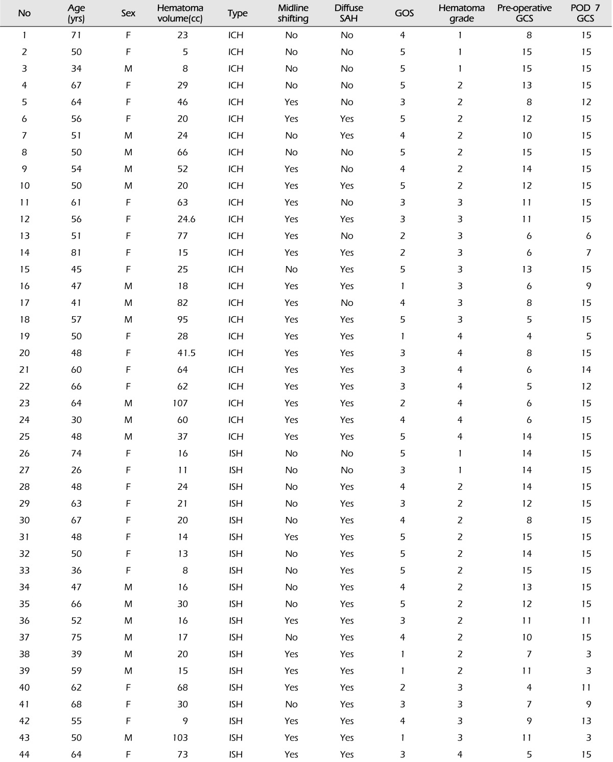
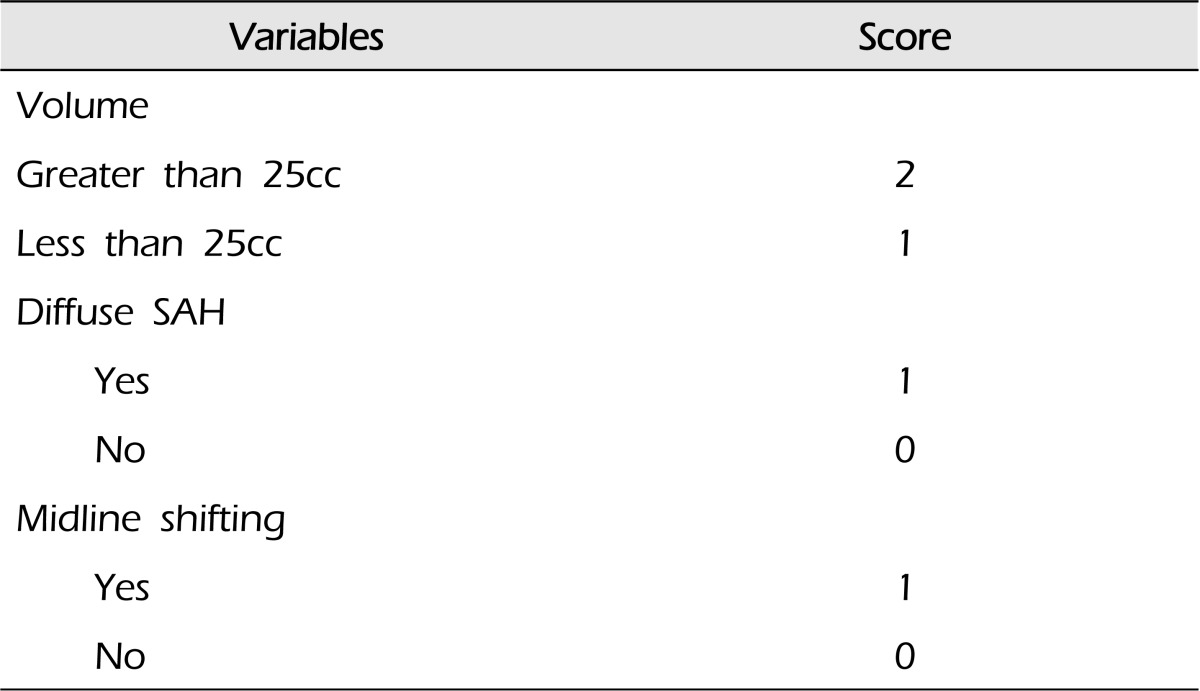
One hundred five patients with SAH from ruptured MCA aneurysms were admitted from August, 2005 to December, 2012. Forty-four patients (41.9%) among them had ICH or ISH of more than 5 cc in CT scan (Table 1). Their age ranged from 26 to 81 years old and the male to female ratio was 1:1.44 (M:18, F:26). Among the 44 cases, 25 patients (56.8%) had ICH while 19 (43.2%) had ISH. The presence of enhancing vessels were noted in these patients by comparison of their non-enhanced CT and CTA.15) We suggested the hematoma grading system, 1 or 2 points were assigned depending on the volume being less or greater than 25 cc. The presence of diffuse SAH was assigned 1 point in the hematoma scale and 0 point for focal SAH, and 1 point was assigned to midline shifting of more than 5 mm versus 0 point for those less than 5 mm (Table 2).11)
We investigated each patient's hematoma grade, type in CT and CTA, and we compared their age, gender, Hunt-Hess grade and GOS. The change in GCS from admission to 7 days after surgery was also compared.
Multiple logistic regression analysis was used to identify the independent factors predicting the patients' prognoses.
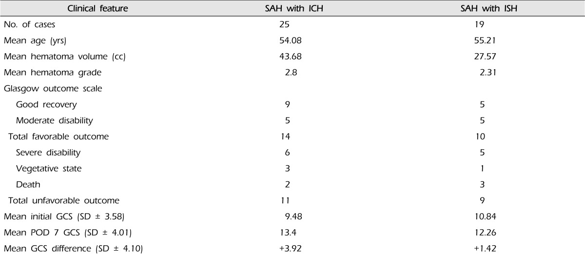
The Hunt-Hess Grades at admission were 2 in 7 cases (15.9%), 3 in 11 cases (25%), and 4 in 24 cases (54.5%). Thirty two cases (72.7%) showed diffuse SAH in the initial brain CT, while 12 cases (27.3%) did not. Twenty six had midline shifting of average 7.5 mm (range 1-18 mm) and 16 among these patients had midline shifting of more than 5 mm. The average hematoma volume was 36.73 cc (range 5-107 cc). By hematoma grade, 5 cases (11.4%) were grade 1, 19 cases (43.2%) grade 2, 12 case (27.2%) grade 3, and 8 cases (18.2%) grade 4. The GOS were evaluated in these patients; 5 patients were categorized as grade 1, 4 as grade 2, 11 as grade 3, 10 as grade 4, and 14 as grade 5. Most of these patients needed rehabilitation and therefore their final GOS was checked at the point of discharge after rehabilitation (about 1-1.5 months after admission). The average GOS of the ICH patients were 3.64, where 2 patients were categorized as grade 1, 3 as grade 2, 6 as grade 3, 5 as grade 4, and 9 as grade 5. The average GOS of the ISH patients was 3.42, where 3 patients were categorized as grade 1, 1 as grade 2, 5 as grade 3, 5 as grade 4, and 5 as grade 5. Assuming the patients with GOS 4 and above to have good prognoses and those below 4 to have poor prognoses, 24 patients had GOS equal to or more than 4 and 20 had less than 4 (Table 3). Comparing the GOS of ICH and ISH patients, the group of hematoma type did not hold any statistical significance (p = 0.824). However, in the change of GCS, the result was different when comparing the pre-operative GCS and the post-operative GCS at day 7 (Table 4). The ICH group had average pre-operative GCS of 9.48 and average post-operative GCS at day 7 of 13.4, an average improvement of 3.92 with no patient having lower GCS after operation. The ISH group had average pre-operative GCS of 10.84 and average post-operative GCS of 12.26, an average improvement of 1.42. Subsequently, in ruptured MCA aneurysm patients, those with ICH improved more after surgery compared to those with ISH statistically (p = 0.0391).
The ICH patients had a larger average hematoma volume of 43.68 cc (range 5-107 cc) compared to that of the ISH patients, which was 27.57 cc (range 8-103 cc). Consequently, the 14 of 25 (56%) ICH patients had midline shifting of more than 5 mm, while 2 of 19 (10.5%) ISH patients had midline shifting of more than 5 mm.
The higher hematoma grade resulted in lower GOS (p = 0.041).
Intracerebral hemorrhage is associated with an increased mortality rate in patients with ruptured aneurismal SAH,2)5) and emergent craniotomy with evacuation of the hematoma and aneurysm clipping should be considered.3)4)6)16) Many studies have categorized hematoma into types and analyzed the difference in prognoses.
Shimoda et al.12) subdivided hematomas into temporal ICH, ISH, and ICH with diffuse SAH, and noted that the patients with ISH/ICH with diffuse SAH fared worse compared with those with only ISH/ICH. Prat et al.9) categorized hematomas into temporal ICH and ISH, and observed better outcomes in patients with temporal. Oh et al.7) grouped hematomas into frontal ICH, temporal ICH, and ISH, and found that temporal ICH had more favorable clinical outcome when compared to the other hematoma groups.
van der Zande et al.15) stated that when categorizing the hematoma types into ICH and ISH depending on the existence of IHCEV, admission status and outcome were similar for both groups.
In our previous study, we proposed a grading system for hematoma accompanied by ruptured MCA aneurysm.11) This grading system is currently being used in our hospital as a useful tool in evaluating the patient prognosis.
We attempted to confirm van der Zande's statement on prognosis by different hematoma types. We also investigated the relationship between our hematoma grading system and the types categorized by van der Zande. The average hematoma grade was 2.3 (SD ± 0.89) in the ISH group and 2.8 (SD ± 1.04) in the ICH group.
The group with ICH generally had a larger hematoma volume and more frequent midline shifting compared to the ISH group. Generally, the ICH group had higher hematoma grades, and this had a direct association with their GOS.
In our study of the ruptured MCA aneurysm patients, the group with accompanying ICH had a lower average GCS at admission, but improved more after surgery compared to the group with accompanying ISH.
This was because in ICH, the hematoma is located within the brain parenchyma and thus is easily removable. ICH has a larger volume in general, and therefore greater improvement in consciousness can be expected by removing this lesion. Although there is no significant difference in the outcome of the 2 hematoma groups, the degree of improvement might be higher in ICH group compared to ISH group.
Ruptured MCA aneurysm with hematoma, as in our previous study, proved to have a clear relationship between the hematoma grade and prognosis and this was confirmed to be statistically significant in this study. There were no notable differences in prognoses when simply subdividing the hematoma into ICH and ISH. However, when comparing their peri-operative GCS, a greater improvement in consciousness level after surgery was noted in ICH patients.
References
1. Abbed KM, Ogilvy CS. Intracerebral hematoma from aneurysm rupture. Neurosurg Focus. 2003; 10. 15(4):E4. PMID: 15344897.

2. Adams HP Jr, Kassell NF, Torner JC. Usefulness of computed tomography in predicting outcome after aneurismal subarachnoid hemorrhage: A preliminary report of the Cooperative Aneurysm Study. Neurology. 1985; 35(9):1263–1267. PMID: 4022373.

3. Bailes JE, Spetzler RF, Hadley MN, Baldwin HZ. Management morbidity and mortality of poor-grade aneurysm patients. J Neurosurg. 1990; 4. 72(4):559–566. PMID: 2319314.

4. Batjer HH, Samson DS. Emergent aneurysm surgery without cerebral angiography for the comatose patient. Neurosurgery. 1991; 2. 28(2):283–287. PMID: 1997899.

5. Kawamura S, Suzuki A, Sayama I, Yasui N. [Clinical significance of intracerebral hematomas following aneurysmal rupture]. Neurol Med Chir (Tokyo). 1987; 12. 27(12):1158–1166. Japanese. PMID: 2452362.

6. Le Roux PD, Dailey AT, Newell DW, Grady MS, Winn HR. Emergent aneurysm clipping without angiography in the moribund patient with intracerebral hemorrhage: The use of infusion computed tomography scans. Neurosurgery. 1993; 8. 33(2):189–197. discussion 197. PMID: 8367040.
7. Oh JW, Lee JY, Lee MS, Jung HH, Whang K. The meaning of the prognostic factors in ruptured middle cerebral artery aneurysm with intracerebral hemorrhage. J Korean Neurosurg Soc. 2012; 8. 52(2):80–84. PMID: 23091663.

8. Pasqualin A, Bazzan A, Cavazzani P, Scienza R, Licata C, Da Pian R. Intracranial hematomas following aneurysmal rupture: Experience with 309 cases. Surg Neurol. 1986; 1. 25(1):6–17. PMID: 3484561.

9. Prat R, Galeano I. Early surgical treatment of middle cerebral artery aneurysms associated with intracerebral haematoma. Clin Neurol Neurosurg. 2007; 6. 109(5):431–435. PMID: 17449171.

10. Rinne J, Hernesniemi J, Niskanen M, Vapalahti M. Analysis of 561 patients with 690 middle cerebral artery aneurysms: Anatomic and clinical features as correlated to management outcome. Neurosurgery. 1996; 1. 38(1):2–11. PMID: 8747945.

11. Shim YS, Moon CT, Chun YI, Koh YC. Grading of intracerebral hemorrhage in ruptured middle cerebral artery aneurysms. J Korean Neurosurg Soc. 2012; 5. 51(5):268–271. PMID: 22792422.

12. Shimoda M, Oda S, Mamata Y, Tsugane R, Sato O. Surgical indications in patients with an intracerebral hemorrhage due to ruptured middle cerebral artery aneurysm. J Neurosurg. 1997; 8. 87(2):170–175. PMID: 9254078.

13. Su CC, Saito K, Nakagawa A, Endo T, Suzuki Y, Shirane R. Clinical outcome following ultra-early operation for patients with intracerebral hematoma from aneurysm rupture-focussing on the massive intra-sylvian type of subarachnoid hemorrhage. Acta Neurochir Suppl. 2002; 82:65–69. PMID: 12378994.
14. Tokuda Y, Inagawa T, Katoh Y, Kumano K, Ohbayashi N, Yoshioka H. Intracerebral hematoma in patients with ruptured cerebral aneurysms. Surg Neurol. 1995; 3. 43(3):272–277. PMID: 7792692.

15. van der Zande JJ, Hendrikse J, Rinkel GJE. CT angiography for differentiation between intracerebral and intra-sylvian hematoma in patients with ruptured middle cerebral artery aneurysms. AJNR Am J Neuroradiol. 2011; 2. 32(2):271–275. PMID: 21071532.

16. Wheelock B, Weir B, Watts R, Mohr G, Khan M, Hunter M, et al. Timing of surgery for intracerebral hematomas due to aneurysm rupture. J Neurosurg. 1983; 4. 58(4):476–481. PMID: 6827342.


 PDF
PDF ePub
ePub Citation
 Print
Print


 XML Download