

 PDF
PDF ePub
ePub Citation
 Print
Print


INTRODUCTION
The morbidity and mortality rates of ischemic stroke are high all over the world. Research on stroke mechanisms reveals secondary injury develops through the inflammatory reaction induced by the leukocytes that adhere to the micro vessels during cerebral ischemia-reperfusion.9) This mechanism was verified in animal experiments using middle cerebral artery occlusion and reperfusion (MCAO-R) models.5)12) Experiments using the MCAO-R model, found reduced leukocyte infiltration and enhanced neurologic functions in mice that were injected with adhesion-molecule-specific monoclonal antibodies (mAbs), or in mice whose adhesion molecules were removed through genetic manipulation, compared with wild-type mice.13)21)25)
Studies on adiponectin are active, and many favorable effects of adiponectin on the vascular system reported. Adiponectin activates angiogenesis in ischemic damages,19) and inhibits atherosclerosis.15) In addition, a negative association between the initial infarction volume and the adiponectin level in ischemic stroke was confirmed.6) In serial measurement of adiponectin level in ischemic cerebrovascular disease patients, a gradual decrease was observed.24)
In this study, the effects of adiponectin on the interaction between leukocytes and endothelia were investigated through experiments based on the hypothesis that adiponectin might inhibit the secondary inflammatory reaction in cerebral ischemia.
MATERIALS AND METHODS
Animal preparation
All experiments were conducted according to the guidelines issued by the Institutional Animal Care and Use Committee and complied with the National Institutes of Health (NIH) guide for the care and use of laboratory animals. For the experiments of this study, 10 to 12-week-old adiponectin-deficient mice (APN-KO) (n = 8) and wild-type mice (WT) (n = 8) were prepared. Each group was sub-divided into 2 groups by reperfusion time; 6h APN-KO, 12h APN-KO, 6h WT and 12h WT. General anesthesia was given to mice using 2% isoflurane and 70% nitrous oxide, and 30% oxygen was supplied during the experiments via a face mask. The mice's body temperatures were maintained at 36.5-37.5℃ during the experiments, as measured by a rectal thermometer.
Model of middle cerebral artery occlusion and reperfusion
Focal cerebral ischemia was induced using the intraluminal filament technique.11) After the left common carotid artery was dissected and completely exposed, the external carotid artery was tied by 4-0 silk. Thereafter, silicon-coated 8-0 monofilament was headed to the middle cerebral artery through the common carotid artery. The inserted monofilament was tightened with a 6-0 silk suture to prevent bleeding while advancing the monofilament and removing the monofilament during reperfusion. One hour after the occlusion, the filaments were removed for reperfusion (Fig. 1).
Cranial-window preparation
After preparation of the focal cerebral ischemia and reperfusion model, the mice were placed on a stereotactic frame to perform 4×6 mm-sized craniotomy in the left parietal area using a dental drill. Extreme caution was required to avoid damage to the dura mater and the tissues beneath it. After the craniotomy, the dura mater was opened using 26-gauge needles to expose the brain cortex of the MCA territory and the artificial cerebrospinal fluid was arranged to flow over the surface of the brain (Fig. 2).
Intravital microscopy
As a target vessel, a straight pial venule (100 µm long and 30-40 µm in diameter) was selected. To observe the microcirculation, a fluorescence intravital microscope was used at 780× specimen-to-monitor magnification. Using 1 ml fluorescein isothiocyanate-albumin that was intravenously injected shortly before data collection, a bright contrast was obtained, and the length and diameter of the microvessel were measured. Using 1 ml of the 0.1% rhodamine that was intravenously injected 10 minutes before data collection, the leukocytes and platelets were selectively stained.
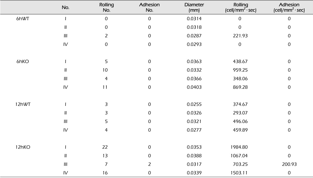
Data were collected at 6 and 12 hours after 1-hour occlusion. At each data selection, the target venule was recorded for 30 seconds through a microscope, and the number of rolling leukocytes and leukocytes that adhered to endothelium were counted and documented (Fig. 3).
RESULTS
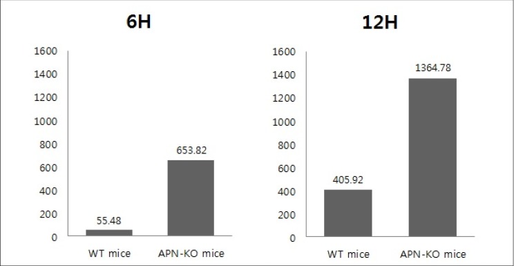
The behaviors of the leukocytes in the target vessel of APN-KO and WT mice was observed and recorded for 30 seconds at 6 and 12 hours after the occlusion, using an intravital fluorescence microscope. The number of rolling and adhesion leukocytes of each experimental group was counted and recorded and converted to per mm2 · sec (Table 1). In 6hWT mice, a mean of 55.48 per mm2 · sec of rolling leukocytes were observed while a mean of 653.82 per mm2 · sec of rolling leukocytes were observed in 6hAPN-KO mice. In 12hWT mice, a mean of 405.92 per mm2 · sec of rolling leukocytes was observed in all 4 groups. In the 12hAPN-KO mice, rolling leukocytes were observed in all 4 groups, and in particular, leukocyte adhesion was observed in experimental group III, with a mean of 1364.78 per mm2 · sec of total leukocytes.
Mann-Whitney U-test was used to test for statistical significance of the outcomes. Leukocyte numbers between the APN-KO and WT mice 6 hours after the occlusion differed significantly with p = 0.029. The difference at 12 hours after the occlusion was also statistically significant with p = 0.029 (Fig. 4).
DISCUSSION
Adiponectin is an adipocyte-derived bioactive protein that initially attracted attention in the treatment of metabolic syndrome.14) Clinically, the plasma adiponectin level is negatively associated with dyslipidemia, hypertension, and the C-reactive protein level,17) and hypoadiponectinemia (defined as plasma adiponectin level < 4 µg/mL) increases the risk of type 2 DM, hypertension, and coronary heart disease.16)17) Studies of the role of adiponectin in the cardiovascular system have been conducted using APN-KO mice. These studies found that APN-KO mice showed exaggerated myocardial remodeling under pressure-overload conditions, and severe cardiac injuries were reported during ischemia-reperfusion.20)22) By contrast, when adiponectin was injected into the APN-KO mice, pathological cardiac hypertrophy and ischemia-induced myocardial damages were inhibited.18) These protective effects have driven research on the effects of adiponectin on the cerebrovascular system.
The most recent studies find that plasma adiponectin level is closely related with the cerebrovascular system. Chen at al. described hypoadiponectinemia as an independent and significant risk factor for cerebrovascular disease while Efstathiou et al. reported an increased risk of mortality for hypoadiponectinemia patients who suffered ischemic insults.4)6) In addition, advanced intracranial atherosclerosis patients were observed to have had significantly low plasma adiponectin levels.1)
In this study, the effects of adiponectin on the early stage of inflammation reaction in cerebral ischemia-reperfusion were investigated. At both 6 and 12 hours after the occlusion, the APN-KO mice group showed significantly more rolling and adhesion leukocytes than the WT mice group did. Although firm adhesion of the leukocytes to the endothelium was observed only in experimental group III of the 12hAPN-KO mice, the rolling leukocytes would form a firm adhesion on the endothelium through the media of leukocyte adhesion molecule CD11b/CD18 (integrin) and its endothelial ligand ICAM-1. The activated leukocytes that adhered to the endothelium release toxic mediators to damage the surrounding vasculatures or parenchymal cells, or induce a change in blood rheology and accelerated thrombosis by inducing platelet aggregation.7)8)10)
Inhibition of the endothelial adhesion-accumulation of the leukocytes after cerebral ischemia-reperfusion improves electrophysiological and neurological function, and reduces cerebral edema and cerebral infarction size.3)5)23) Therefore, adiponectin that restrains the leukocyte-endothelium interaction is considered to inhibit the secondary inflammatory reaction and is neuroprotective in cerebral ischemia-reperfusion.
The limitation of this study is the limited number of its experimental groups. In addition, the patterns beyond 12 hours after ischemia were not observed. Although the leukocyte accumulation in cerebral ischemia-reperfusion steadily increased to reach a peak after 24-48 in other studies,2)10) more advanced studies with a larger sample size may be needed in the future.
CONCLUSION
In this study, the effects of adiponectin on the early inflammatory reactions in cerebral ischemia-reperfusion were investigated. As a result, statistically more rolling and adhesion leukocytes were observed in the APN-KO mice than in the WT mice in the experiments with the MCAO-R model. In conclusion, adiponectin was observed to inhibit the interaction between the endothelium and leukocytes in cerebral ischemia-reperfusion, and accordingly, adiponectin might prevent the secondary insult caused by the inflammation reaction.


 XML Download