

 PDF
PDF ePub
ePub Citation
 Print
Print


INTRODUCTION
Aneurysms at the origin of the posterior inferior cerebellar artery (PICA) remain a challenge for neurosurgeons because the close proximity to the brainstem and lower cranial nerves poses a high risk for surgical clipping.1)14)15) Aside from such anatomic unchangeability of adjacent structures, complex anatomic features of PICA origin aneurysms themselves also add high risks for both surgical and endovascular treatments.16)19) However, in the past decade, there have been substantial advances in device technologies and endovascular techniques. As a result, the number of patients who are not candidates for endovascular treatment is decreasing. In particular, the "self expanding stent technology" and "supercompliant balloon technology" have revolutionized the methods and results of endovascular treatment.14)22) Combining a self expanding stent with a "cross-over technique", which uses an inter-arterial communication as an access route to the other vascular part,9)12) the authors herein report on a bilateral approach for stent-assisted coiling of two complex PICA origin aneurysms.
CASE REPORT
Case 1
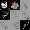
A 71-year-old woman who had no past medical history presented to our emergency room with a sudden thunderclap headache, nausea, and vomiting. On neurological examination, the score on the Glasgow Coma Scale was 11 (E3V3M5) and isocoric pupil response was observed. Initial computed tomography (CT) and CT angiography showed Fischer grade 4 subarachnoid hemorrhage (SAH) and a ruptured left PICA aneurysm (Fig. 1A and 1B). Cerebral angiography and 3D image reconstruction showed a left PICA origin aneurysm measuring 7 mm in length with a dome to neck ratio of 0.7 and PICA incorporation into the sac (Fig. 1C and 1D). As a treatment option, in view of anticipated surgical difficulty and patient's preference, our neurovascular team decided on endovascular treatment. The use of a multi-catheter or balloon remodeling technique is likely to have a high risk of PICA occlusion during coil embolization of this complex aneurysm; therefore, coiling with placement of a stent in the vertebral artery (VA)-aneurysm-PICA path was planned. However, an antegrade approach to the PICA appeared to be very difficult and too risky, so a retrograde approach via the contralateral VA was decided. For selection of the left PICA arising from the aneurysm, a 6F guiding catheter was placed in the right VA, an Excelsior SL-10 microcatheter (Striker, Fremont, CA) was introduced into the right VA via the guiding catheter to cross over the vertebrovertebral junction to the left VA, and the microcatheter over a Synchro-14 guidewire (Striker, Fremont, CA) was able to select the left PICA smoothly (Fig. 1E). After being placed in the left PICA, the Excelsior microcatheter was exchanged for a Rebar-18 microcatheter (ev3 Inc, Irvine, CA) that was compatible with a stent delivery system. Another 6F guiding catheter was introduced into the left VA and an Excelsior SL-10 microcatheter was placed in that aneurysm through the guiding catheter for coil delivery. The right vertebral artery was non-dominant and smaller than the left vertebral artery. However, the size of the right vertebral artery was sufficient to accept the Rebar-18 microcatheter. After deployment of a Solitaire™AB stent (ev3 Inc, Irvine, CA) for protection of the PICA (Fig. 1F), a total of eight detachable coils were packed in the aneurysm. As only a 20-mm-long stent (4 × 20) was available at that time, it covered the left PICA, aneurysm neck, left VA, and a short segment of the right VA, in order. Completion angiography showed complete occlusion of the aneurysm (Fig. 1G) and no procedural complication was identified. The patient received dual anti-platelet therapy for 30 days, and continued life-long aspirin. At the four-month follow-up evaluation, the aneurysm was well excluded (Fig. 1H and 1I). The patient continues to live independently at home and is doing well.
Case 2
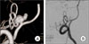
A previously healthy 43-year-old man visited our emergency room complaining of a severe headache for six hours. Except for nuchal rigidity, neurological examination was unremarkable. Brain CT showed right premedullary SAH and minimal intraventricular hemorrhage mainly in the fourth ventricle, three-dimensional CT angiography showed a ruptured right PICA origin aneurysm, and cerebral angiography was successively performed for visualization of anatomic details of the aneurysm. It had a maximal size of 11 mm and a low dome-to-neck ratio of 0.9, and gave rise to the PICA, which was incorporated into the sac (Fig. 2A). Aiming at both aneurysm occlusion and PICA preservation, coiling with placement of a stent in the VA-aneurysm-PICA path was considered the best method. All procedural steps were similar to those of Case 1, except for the use of two micro-catheters for coil delivery and use of a 4 × 22 mm Enterprise stent. A total of eight detachable coils were placed in the aneurysm. After placement of the first two coils, eliminating contrast filling of the aneurysm dome, systemic heparinization (3,000 IU) was administered intravenously. However, thrombosis occurred in the aneurysm neck during placement of the last coil, which resulted in near occlusion of the PICA flow. Tirofiban was given as an intraarterial bolus of 0.5 mg over five minutes, resulting in recanalization of the occluded PICA. On delayed angiograms (Fig. 2B), no progressive thrombosis was observed and the PICA flow was intact, so that the patient was awakened. On neurological examination, no neurological deficit was identified immediately or at one month after the procedure. The patient received dual anti-platelet therapy for 30 days, but was lost to follow-up afterwards.
DISCUSSION
PICA origin aneurysms frequently have unfavorable anatomic findings, including a wide neck and PICA incorporation into the sac.1)10)14) Endovascular treatment of such aneurysms is associated with risk for PICA occlusion or procedural failure. Although special techniques have recently been addressed,6)8) two goals of aneurysm occlusion and PICA preservation appear to be mostly secured by stent-assisted coiling, where a self expanding stent successively covers the PICA, aneurysm neck, and VA. However, in the case of PICA incorporation, selecting the PICA antegrade through the ipsilateral VA is extremely difficult and risky. Anatomy-wise, if the contralateral VA were non-interrupted and large enough to accept a catheter, a contralateral and retrograde approach to the PICA would be easier than the ipsilateral antegrade one. A few case studies regarding the "cross-over technique" using the communicating artery or vertebrovertebral junction as an access route have been reported in the literature.5)9) Moret et al.14) suggested that the anatomic benefits afforded by this technique outweigh the potential risks associated with catheterization of another major cerebral artery. We believe that, in two cases in this study, without stent-assisted coiling using the "cross-over technique", PICA occlusion would have been inevitable. However, long-term follow-up results of this procedure are not available. In particular, the size of the PICA is so variable that the issue of long-term stent patency remains unanswered.
After far-lateral suboccipital exposure, PICA origin aneurysms can be approached between the 11th cranial nerve inferiorly and the ninth and tenth cranial nerves superiorly. Therefore, the 10th and 11th cranial nerves are at risk of injury during the operation, which can result in lower cranial nerve dysfunction.3)7)10)15) In addition, complex aneurysmal geometry may make it difficult for neurosurgeons to complete aneurysm clipping without occlusion of the PICA. Occlusion of the PICA close to its origin may lead to serious morbidity and mortality, depending on the distribution of the medullary perforators and the presence of collateral supply from other cerebellar arteries. Aneurysm clipping following occipital artery-PICA bypass surgery may be a solution to this serious complication,2)19)20) however, it is too early to generalize the staged treatments.
CONCLUSION
Due to recent instrumental and technical developments leading to good results, endovascular treatment is considered the primary treatment for posterior circulation aneurysms. However, complex PICA aneurysms, having a wide neck and PICA incorporation, remain a challenge for both surgical and endovascular treatments. In such cases, a bilateral approach for stent-assisted coiling may be a novel method toward achievement of good results.
 XML Download