

 PDF
PDF ePub
ePub Citation
 Print
Print


Organized hematoma is a rare pseudo-tumorous lesion mostly occurs in confined and vascularized region such as sinonasal cavity.12 Even though it is not a true tumor, it continually makes expansion and aggressively destructs adjacent structures, thus frequently misdiagnosed as malignant tumor. The pathogenesis of the lesion is not clearly elucidated, and it is suspected to occur when hematoma is not absorbed and became chronic under the condition of drainage absence.
While, various kinds of tumors and pseudo-tumors can be shown in temporomandibular joint (TMJ) region, including synovial chondromatosis, osteochondroma, pigmented villonodular synovitis, metastatic tumors and plasmacystoma, the organized hematoma is uninformed on this area. Additionally, the clinical detection of neoplasms involving TMJ is often delayed due to the similar manifestations of it to ordinary TMJ internal derangement.34 In fact, approximately 20% of patient, actually having mass on TMJ, initially diagnosed and treated as joint derangement. The mean delayed in time in diagnosis of tumor or tumor-like lesion of TMJ was 2.5 years. Thus, diagnosis of organized hematoma might be challenging for both clinician and radiologist.34
Therefore, this report introduced the useful imaging features of organized hematoma occurred on TMJ for precise diagnosis. Also, diagnostic point for early detection was described for the TMJ tumors and pseudo-tumors to avoid delayed diagnosis.
Case Report
A 68-year-old male patient presented with a swelling and pain on the left pre-auricular region initiated about two to three years ago. He also had pain increases during opening his mouth. He had botulinum toxin injected on the affected area three month ago, and the symptom was not relieved at all. There was no particular underlined systemic disease in his medical record.
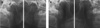
A TMJ panoramic radiography, an imaging technique especially for assessing TMJ structure, was performed to evaluate if there is degenerative change of condyle and articulating eminence as well as relative position of condyle to articular fossa. The image revealed that joint space was slightly widened on left TMJ compared with the right side (Fig. 1A). While, there was no restriction of both condyles during mouth opening (Fig. 1B). No significant destruction or remodeling of bony structure was seen on joint components.
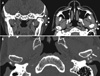
A computed tomography (CT) and magnetic resonance (MR) imaging, both without contrast-media, were also obtained to evaluate any minor degenerative change of condyle and disc problem of TMJ. On the CT images, round soft tissue attenuation was localized on the posterolateral aspects of left condyle head (Fig. 2A). The lesion showed soft-tissue equivalent attenuation compared with the adjacent muscles. As it was shown in the TMJ panoramic radiography, there was no significant bony destruction; however, slight thinning and focal discontinuity of cortical bone was detected on the condyle head contacting with the lesion. Also, mild subchondral sclerosis was found on this site (Fig. 2B).
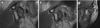
On MR image of proton density and T1-weighted sequence, the lesion was well-encapsulated and showed heterogeneous signal indicating solid mass rather than cyst (Fig. 3). The left mandibular condyle showed anterior dislocation due to a mass on the posterior aspect. However, TMJ discs were in normal position during the mouth open and close conditions. Local T1 hyper-intensity was recognized as it indicated that there was hematogenous component included in this lesion (Fig. 3C).
With preliminary diagnosis of organized hematoma or giant cell tumor based on the images, the ovoid mass located on the postero-lateral aspect of left condylar head was surgically removed (Fig. 4A). On the histopathologic exam, the lesion was composed of blood cells intermixed with granulation tissue. It was encapsulated with fibrous band and there was slight neovascularization was observed on the border of the capsule (Fig. 4B). With the above findings, organized hematoma has been made as final diagnosis. No significant complications were found during a month of follow-up period.
Discussion
Histopathologically, this lesion is composed of multinucleated foreign-body-type giant cells, thick fibrotic capsule with neovascularization.2 Its pathogenesis is not clear, however, blood accumulation without proper drainage is suggested as the initial stage. The known cause of this initial stage is traumatic hemorrhage, while, the time interval between the trauma and the symptom occurrence may vary from a month to years.5 The hematoma usually dissolves in several days, however it is not absorbed and became chronic hematoma, it starts organizing with neovascularization.125
Even though very rare, there have been reports of organized hematoma on other body part including retroperitonium, thigh and lower leg rather than sinonasal cavity.678 In addition, there was one case with similar pathology, involving TMJ, and authors introduced this lesion as a hematic cyst.9 However, Bergin et al.10 stated that hematic cyst was basically describing the same pathologic entity as organized hematoma, thus, the current case was the second report of this unusual disease, as far as the author knows. Considering TMJ region is confined space with capsule and rich in vascular supply close to maxillary artery, the possibility of organized hematoma occurrence cannot be disregarded in this area.
Currently, MR imaging feature of organized hematoma involving sinonasal cavity has been well described. The heterogeneous internal signal indicating various stages of hemorrhage is characteristic in MR imaging.1 This feature was also useful for diagnosis of TMJ case. Both previous case9 and our case showed the heterogeneous signal including T1-hyperintesity, which could be correlated with hemosiderin component of the mass. Still, this feature might also be suitable for describing tumors showing internal hemorrhage signal such as vascular malformation or giant cell tumors.1112 Compared with vascular malformation and giant cell tumors, organized hematoma showed hypointense peripheral rim indicating fibrous capsule around the mass.9 In addition, giant cell tumors occurred in this region showed more aggressive expansion involving cranial base or condyle head destruction compared to the organized hematoma.12
Early diagnosis is challenging for TMJ tumors and pseudotumors. The patient in our case also had been diagnosed with joint disorder, initially and underwent botolinium toxin injection. Considering trauma is suspected as the etiology of organized hematoma, thus, the injection might also be a causative factor. Thus, clinician may give a careful diagnosis and close check-up after such process including the arthrocentesis on TMJ, which is one of a known treatment option for TMJ disorder. Also, for the TMJ showing intact bony contour of condyle and articular eminence while joint space increased in TMJ panoramic radiography, as it was shown in our case, the advanced imaging modality might be useful for differential diagnosis between the mass and the joint problems. In the internal derangement of TMJ, according to previous research, decreased joint space or retropositioned condyle was shown in the radiography.13
Organized hematoma in sinonasal cavity shows aggressive expansion of the mass occupying the whole cavity, which may not distinguishable from malignant tumor. For the TMJ lesion, both cases did not show aggressive feature. This might be due to the small size of the lesion. Even with the small size, the previous organized hematoma occurred in TMJ region caused deformity and erosion of lateral condyle head. In our case, very subtle but reactive subchondral sclerosis was appeared on the contact surface of the condyle. Overall, this lesion seemed to possess potential to show mass effect on bone structure, surgical removal would be the primary choice as stated in previous studies.129 Since the surgical intervention of TMJ region is complex and may accompany complications including nerve damage, infection and malocclusion, early diagnosis and removal would be important.14
In conclusion, organized hematoma of TMJ is rare but possibly occurring disease and MR imaging is helpful for diagnosis. This lesion may involve bony structure of this lesion, thus careful and early diagnosis is essential to prevent challenging surgical procedure and post-operative complications.

 XML Download