

 PDF
PDF ePub
ePub Citation
 Print
Print


Introduction
Appropriate diagnosis and treatment planning depend on data derived from diagnostic aids such as clinical examinations, study models, and the relevant radiographs. Lateral cephalography and panoramic radiography are important tools for treatment planning and are often recommended to orthodontic patients. A lateral cephalogram can be used for evaluating the skeletal relationship, growth pattern, dentition, and alveolar process. In these evaluations, certain landmarks or points on the skull are used for the quantitative analyses and measurements.
The gonial angle is an important parameter for determining the growth pattern of an individual, assessing the rotation of the mandible1 and the extraction pattern in class II patients,2 making decisions regarding whether to perform surgery in class III skeletal base patients,3 and estimating age in forensic medicine.4 It is also an important parameter for evaluating the symmetry of the facial skeleton. Usually, this angle is measured using a lateral cephalogram. However, according to Larheim and Svanaes,5 the accuracy of measurements of the gonial angle using lateral cephalograms is questionable because of the superimposition of the right and the left angles. In other words, the presence of the superimposed images on a lateral cephalogram makes it difficult to reliably measure the gonial angle in an individual, particularly when planning any type of jaw surgery.
Panoramic radiography, which is considered the standard of care for dental diagnosis and treatment planning, is used by dentists and orthodontists alike. It provides a significant amount of information about the teeth and the supporting bone, and is used for screening for cysts, cancer, extra teeth, the congenital absence or premature loss of teeth, teeth fused to the bone or abnormally retained teeth, tooth eruption path, bone pathology, and mandibular asymmetry.67 An appropriate diagnosis of asymmetries before treatment is prudent for addressing the treatment limitations and therapeutic options. Although most practitioners do not use panoramic images for the diagnosis of a mandibular asymmetry,89 some researchers do support their use. According to Mattila et al.,7 panoramic radiography can be used for determining the gonial angle more accurately than lateral cephalography, as the right and left gonial angles can be measured individually without any superimposition.
A panoramic radiograph provides information about both the left and right sides; hence, it would be prudent to check whether the visualization of both sides is equally reliable. Therefore, the aim of this study was to evaluate the accuracy of panoramic imaging with respect to the measurement of the right and left gonial angles by comparing the measured angles with the gonial angles determined using a lateral cephalogram of adult patients with class I malocclusion.
Materials and Methods
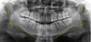
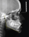
Lateral cephalograms and panoramic radiographs of 50 patients (25 females and 25 males; mean age, 23±3 years) were obtained from the Department of Orthodontics, Amrita School of Dentistry. Panoramic and cephalometric images were acquired with a Cranex D digital X-ray unit, version 3 (Soredex Co., Tuusula, Finland). An X-ray energy of 85 kVp for the panoramic radiography and an energy of 73 kVp for the lateral cephalometric radiography were used. An exposure time of 14.1 seconds was selected for panoramic radiography and 9.4 seconds was selected for lateral cephalography. The inclusion criteria for the radiographs were as follows: the radiographs had to be of high quality and sharpness; all radiographs had to be taken using the same apparatus; and all the selected patients had to have skeletal class I malocclusion. The exclusion criteria were a history of trauma, previous facial/mandibular surgery, syndromes affecting the face/jaw, and facial asymmetry. Cephalometric landmarks were located, identified, and marked on the selected radiograph using a 2H pencil. In the panoramic radiographs, the gonial angle was measured by drawing a line tangent to the lower border of the mandible and another line tangent to the distal border of the ascending ramus and the condyle on both sides (Fig. 1). In the lateral cephalograms, the gonial angle was measured at the point of intersection of the plane tangential to the lower border of the mandible and that tangential to the distal border of the ascending ramus and the condyle (Fig. 2). The gonial angle at the intersection of these planes was traced on a piece of tracing paper and measured using a protractor. Gonial angle measurement was done twice, with an interval of 1 month, to determine any errors in measurement. To confirm that all the considered patients had class I malocclusion, the sella-nasion-A point, sella-nasion-B point, and A point-nasion-B point angles were measured and a Wits analysis was performed. The paired t-test was used for evaluating the difference between the gonial angle in the lateral cephalograms and that in the panoramic radiographs, and the Student t-test was used for evaluating the difference in the gonial angle with respect to gender. The analyses were performed using SPSS version 20.0 (IBM Corp., Armonk, NY); a P-value of <.05 was considered to indicate statistical significance.
Results
The study sample consisted of lateral cephalograms and panoramic radiographs of 50 patients (25 females and 25 males; mean age, 23±3.5 years) with skeletal class I malocclusion. The mean value of the gonial angle measured using the lateral cephalograms was 122.74° with a standard deviation of 0.90°, and that of the gonial angle measured using the panoramic radiographs was 122.79° with a standard deviation of 1.00°. No statistically significant difference was found between the gonial angle measured using lateral cephalograms and that using panoramic radiographs (P=.53, Table 1). The mean value of the gonial angle measured using the lateral cephalograms in females was 122.72°, and in males, it was 122.76°; the P-value was .89, which was statistically nonsignificant. Further, the mean value of the gonial angle measured using the panoramic radiographs in females was 122.74°, and in males, it was 122.84°; the P-value was .71, which was also not statistically significant (Table 2). Thus, no statistically significant difference was observed in the measured gonial angle with respect to both the patient's gender and the diagnostic tool used.
Discussion
The gonial angle describes the shape and the form of the mandible and plays an important role in forecasting the growth pattern of an individual and the extraction pattern in class II patients,2 deciding whether to perform surgery in class III skeletal base patients,3 and estimating age in forensic medicine. It is also an important parameter for evaluating the symmetry of the facial skeleton.12 The aim of this study was to evaluate the accuracy of panoramic imaging in measuring the right and the left gonial angles by comparing the measured angles with the gonial angles determined using lateral cephalograms in patients with class I malocclusion, in order to enhance the application of panoramic radiography in clinical practice for the determination of the gonial angle.
According to Mattila et al.10 and Dahan et al.,11 the size of the gonial angle depends on the method of measurement used. The measurement could either involve the horizontal side of the gonial angle formed by the tangent to the lower border of the mandible or be based on a line passing through the gnathion. On a lateral cephalogram, both planes can be easily determined, but on a panoramic radiograph, the determination of the gnathion could be difficult and might result in an inaccurate measurement of the gonial angle. Thus, in this study, to avoid any inaccuracies in measurement, the horizontal plane of the gonial angle in both the panoramic radiograph and the lateral cephalogram was formed by a line drawn tangentially to the lower border of the mandible.
The mean values of the external gonial angle measured using the panoramic radiographs and the lateral cephalograms were 122.79° and 122.74°, respectively. Further, no statistically significant difference was observed in the gonial angle measured using these 2 diagnostic tools (P=.53). The mean values of the right and the left gonial angles measured using the panoramic radiographs also did not show a statistically significant difference. The results of this study were in accordance with the findings of Larheim et al.5 and Bhullar et al.,12 who reported no statistically significant differences in the gonial angle measured using lateral cephalograms and panoramic radiographs. Mattila et al.7 measured the gonial angle using panoramic radiographs and lateral cephalograms, compared the values with those found using dry skulls, and concluded that the measurements made using the panoramic radiographs were more accurate, which is contrary to the findings of the present study. Shahabi et al.13 compared the external gonial angle determined using lateral cephalograms and panoramic radiographs of class I patients and concluded that panoramic radiography could be used for determining the gonial angle as accurately as a lateral cephalogram. Araki et al.,14 in 2015, compared the gonial angles measured using 49 panoramic radiographs with the gonial angle estimated using lateral cephalometric radiographs taken from 2 dry mandibles and found that the gonial angle measurements were slightly smaller on the panoramic radiographs than on the lateral cephalometric radiographs. Alhaija et al.15 evaluated the potential of panoramic radiographs to measure mandibular inclination and steepness; they observed a high correlation between the measurements taken using both types of radiographs. Thus, according to their study, panoramic radiographs are a useful tool for the measurement of the gonial angle, which is an indicator of mandibular steepness and, subsequently, the mandibular growth direction; this is in agreement with the results of this study. In a previous study, the reliability of cephalometric measurements made using panoramic radiographs was compared with that of the actual measurements obtained from dry skulls. The results revealed the highest correlation between panoramic and cephalometric radiographs with respect to the measurement of the gonial angle, whereas the least correlation was observed for the length of the mandibular body. Further, in the case of different growth patterns, the gonial angle and the ramus height showed the closest correlation between the 2 types of radiographs.12 Thus, the ability to determine the growth direction from a panoramic radiograph is useful because most dentists request a panoramic radiograph for patients during routine dental examinations.
In this study, no statistically significant difference was observed in the gonial angle with respect to gender. Similar results were obtained by Dutra et al.16; they did not observe any gender-based difference in the gonial angle, but Ghosh et al.,17 Bhardwaj et al.,18 Huumonen et al.,19 and Xie and Ainamo20 reported that females had a larger gonial angle, which might be due to the impact of masticatory forces.
According to Slagsvold and Pedersen,21 angles are not correctly reproduced in lateral cephalograms unless the angle plane is parallel to the film. The gonial angle measured in a lateral cephalogram is geometrically an intermediate angle between the right and the left gonial angle. Arithmetically, it is the mean. Any distortion of the right and the left gonial angles is reflected in this angle. According to Nohadani and Ruf,22 angular values from panoramic radiographs are more reliable, as the angular values in the posterior and the lateral aspects of the mandible are not influenced by the image distortion inherent to panoramic radiography, whereas Fischer-Brandies et al.23 preferred only lateral cephalograms for determining the gonial angle.
In the present study, as no statistically significant difference was observed between the gonial angles measured using lateral cephalograms and panoramic radiographs, panoramic radiography can be considered reliable for measuring the gonial angle, particularly in cases where the outlines of the 2 sides are not clearly visible on a lateral cephalogram and in patients presenting with asymmetry, as the right and the left gonial angles can be accurately visualized in a panoramic radiograph without any interference due to superimposed images.


 XML Download