

 PDF
PDF ePub
ePub Citation
 Print
Print


Introduction
The maxillary sinus, the largest paranasal sinus, is a pyramid-shaped structure. The base of this pyramid is in the lateral wall of the nasal cavity pointing towards the zygomatic process of the maxilla.1 After tooth loss, the alveolar process of the posterior maxilla is resorbed and, as a result, the vertical bone dimension is reduced. Many older individuals do not have enough bone volume to support an implant between the sinus floor and alveolar crest, and thus, implant surgery can be complicated.2
Today, the sinus lift procedure is a standard procedure for the treatment of atrophic posterior maxilla before the insertion of implants. It is one of the most reliable procedures in pre-prosthetic surgery.3 Perforation of the sinus membrane is one of the complications of this surgery. Some sinus anatomical variations increase the risk of sinus membrane perforation. One of these variations is the existence of a maxillary sinus septum.45
Maxillary sinus septa have been studied more than other structures inside the maxillary sinus. Several studies25678910111213 have evaluated different aspects of these septa, including their prevalence, location, height, and morphology.
Al-Faraje et al.1 classified the morphology of septa into 6 different patterns and described specific clinical considerations for each one. Wen et al.8 presented a classification system for septa based on the location, number, orientation, and size of the antral septa and treatment considerations for each type to avoid complications during sinus lift surgery. These studies demonstrated the importance of detailed knowledge of maxillary sinus septa morphology to arrange a precise treatment plan and to avoid complications during surgery.
In a retrospective cohort study, the presence of antral septa and their interference in Schneiderian membrane elevation in 45 patients undergoing sinus lifts was assessed. Interfering septa were present in 28.8% of the cases having antral septa. Membrane perforation was reported in 11.5% of cases.14 Thus, pre-operative radiographic examinations using panoramic radiographs or cone-beam computed tomography (CBCT) could be helpful in successful surgical planning and to overcome such complications.
Therefore, the aim of this study was to determine the frequency of the different patterns of the maxillary sinus septa in an Iranian population and to predict the risk of sinus membrane perforation based on different scores.
Materials and Methods
In this statistical cross-sectional study, we evaluated cone-beam computed tomography (CBCT) images of 222 patients who were more than 20 years old and were referred to a maxillofacial radiology clinic in northern Iran between September 2014 and March 2016 for radiographic examination of the maxilla or paranasal sinuses. An expert maxillofacial radiologist with more than 15 years of experience reported the presence of maxillary sinus septa in 152 CBCT images. Thus, 152 CBCT images were included in the second step of this study. This study was approved by the Ethics Committee of Guilan University of Medical Sciences: IR.GUMS.REC.1394.514. All procedures followed were in accordance with the ethics standards of the responsible committee on human experimentation (Guilan University of Medical Sciences) and with the Helsinki Declaration of 1975, as revised in 2008. Informed consent to be included in the study was obtained from all patients.
Motion artifacts, the presence of maxillary sinus lesions, the destruction of maxillary sinus walls, fractures of the maxillary sinus walls, and a past medical history of maxillary sinus surgery were exclusion criteria.
The dental status on each side of the posterior portion of the maxilla was divided into 2 groups: adentate (less than 2 teeth in the premolar and molar region) and dentate (2 or more teeth in the premolar and molar region). Demographic data, including age and sex, were extracted from the information registered in CBCT software before performing the procedure.
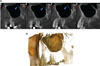
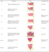
CBCT images were taken using a New Tom VG (QR Srl Company, Verona, Italy) by selecting the “zoom” mode. An expert maxillofacial radiologist and a final-year dental student evaluated the right and left maxillary sinuses in axial views with a 1-mm thickness and sequential slice interval for the presence of sinus septa. They reported the CBCT findings in agreement with each other. They then determined the classification and location of these septa based on mesiodistal cross-sectional and axial directions, and then totally reconstructed the panoramic views. To do so, they reconstructed mesiodistal cross-sectional images with a 1-mm thickness and distance of sequential slice with a 60-mm image width on premolar molar regions adjacent to the maxillary sinuses that had internal septa. To reconstruct cross-sectional images in the mesiodistal direction, the reconstruction line perpendicular to the alveolar bone in the premolar-molar area was drawn and then the software automatically reconstructed them from the buccal to the palatal side. They determined the location of the septa and then classified the sinus septa as well as the degree and score of the sinus membrane perforation risk according to a classification method modified from that of Al-Faraje et al.1 (Table 1).
Before starting this study, 2 maxillofacial surgeons with more than 10 years of experience in implant surgery provided input on the classification of Al-Faraje et al.1 (Fig. 1). Their comments were expertized on the CBCT findings by a maxillofacial radiologist as the observer of the CBCT images in this research. They established a modification of the Al-Faraje et al.1 classification method in agreement with each other. Finally, based on the risk descriptions in Al-Faraje et al., classifications, and the surgeons' experiences; the risk of the membrane perforation was scored from 0 (low) to 2 (high risk).
Cross-sectional images were further evaluated to measure the height of the maxillary sinus septa. The septa that had 3 mm or more height were considered to be ‘long’. Fifty images were reevaluated by the observers 2 weeks later. The intraobserver reliability was found to be 95%. Moreover, the locations of the septa were determined according to their presence in premolar or/and molar regions. Figures 1 and 2 reveal 2 different kinds of septal classifications.
All data were analyzed with SPSS version 22 (IBM Corporation, Armonk, NY, USA). A chi-squared test was used to compare the quantitative variables according to different patterns of maxillary sinus septa. The mean ages of the patients with different patterns of maxillary sinus septa were compared with a Kruskal-Wallis test. P<.05 was considered significant.
Results
In this study, CBCT images of 222 patients were evaluated. Among them, 152 of the patients (68.4%), 93 females (61.2%) and 59 males (38.8%), showed at least one pattern of septa in one of the maxillary sinuses. A total of 42.1% of 152 patients had internal septa only in one maxillary septum, and 57.9% had internal septa in both maxillary sinuses. Meanwhile, if all the patients (with and without septa) were considered, the prevalence of sinus septa in the female and male populations were 29% and 35.2%, respectively. In total, 240 maxillary sinuses had internal septa. Twenty-five maxillary sinuses showed more than one pattern of maxillary sinus septa in one sinus. Therefore, there were 265 instances of the varying pattern of maxillary sinus septa taken together. The ages of the patients ranged from 20 to 81 years old. The mean age was 44.1±14.7 years, and the median age was 43.5 years.
Class I and Class III patterns of maxillary sinus septa had the highest prevalence, which was 28.3% (75) and 25.3% (67), respectively. The lowest prevalence values belonged to Class VII-divI and Class VII-divII, which were 1.1% (3) and 0.8% (2), respectively (Table 2).
Table 2 also shows the mean age and distribution of different patterns of maxillary sinus septa based on sex, dental status, and location. The Class I pattern had the highest frequency in females (31%) and in the dentate group (27.7%). The Class III pattern was more frequent in males (30.8%) and was concurrent with the Class I pattern in the edentate group (30.8%). There was no significant difference in the mean age of the patients with different patterns of maxillary sinus septa.
In this study, there was no significant difference among different patterns of maxillary sinus septa according to sex (P value=.589) or dental status (P=.810) (Table 2).
The most common septal patterns were Class VI in the premolar region (27.3%) and Class I in the molar region (28.9%). Overall, the distribution of sinus septa varied significantly by location (P=.001).
The mean age of the patients with different score ranges for the risk of sinus membrane perforation are shown in Table 3. Age was not found to be significantly related to the degree of risk of sinus membrane perforation (P=.193).
The prevalence values of the scores for membrane perforation risk were 68% (low risk), 26% (moderate risk), and 6% (high risk). There was also no significant difference between genders (P=.216) or by dental status (P=.686) in the membrane perforation risk level. Overall, the risk of membrane perforation was higher in molar than premolar regions. However, the risk scores have significant differences between premolar and molar tooth regions. (P=.004) (Table 3).
Discussion
In this study, the prevalence of the presence of maxillary sinus septa was 68.4% in the patients' CBCT images studied and 54.05% of the maxillary sinuses evaluated. Van Zyl and van Heerden10 reported a prevalence of 69% in CT scans of 200 patients with one or more sinus septa and of 56% in the maxillary sinuses evaluated. Rancitelli et al.9 reported 38.1% as the prevalence of maxillary sinus septa in 228 maxillary sinuses in CBCT images.
Nevertheless, in several other studies, the prevalence of maxillary sinus septa in CT scans11121315 was reported to be 24%-37%. Gosau et al.16 examined 65 cadaver heads and reported an incidence of sinus septa of 27%. Ella et al.17 studied male cadavers and CT scans of male patients. In their study, 39% of the maxillary sinuses had sinus septa. In other cadaver studies, Rosano et al.18 and Naitoh et al.19 found the incidence of sinus septa was 37% and 33.3%, respectively. Their studies had limited sample sizes: 30 and 15 human skulls, respectively. This variability might be related to the differences in the populations examined, sample sizes, methods of evaluation and inclusion criteria, minimum septa height, and consideration of the septa as sinus septa. Malkinson et al.14 evaluated the sinus septa during sinus lift surgery. They reported the septa existed in 40% of the cases. Krennmair et al.5 reported a prevalence of 27.7% in 65 clinically examined maxilla and 16% in 200 CT-examined maxillary sinuses. Shen et al.20 evaluated a large sample size with CT scans obtained from 423 patients. Thirty percent of the patients had sinus septa. However, they used 2.5-mm thick CT images, while the present study used 1-mm thick CBCT images, which could explain the reason for the difference in reported results. Neugebauer et al.21 evaluated CBCT scans of 1,029 patients, and they found septa in 47% of the patients and 33.2% of the sinuses. Koymen et al.22 assessed 205 dental CT scans of maxilla. Septa were found in 35.4% of the maxillary sinuses.
In this study, the sinus pattern with the highest prevalence was the Class I classification. This finding means that this pattern was short enough to not interfere with sinus lift surgery and a lateral window design. Additionally, the Class VI pattern of septa (long and multiple perpendicular septa) had the highest membrane perforation risk and a low prevalence (6%). If this type of septum existed in the sinus, it would interfere with sinus lift surgery. Thus, the surgeons would have to design 2 separate windows.
On the other hand, the risk of sinus membrane perforation was low in 68%; however, in 32%, the estimated risk was medium to high.
We found there was a significant difference between the frequencies of septa presence in molar areas compared to premolar areas. Class III, IV, and VII-div I were considered medium- to high-risk groups relative to the others, and these patterns were in molar rather than premolar areas.
According to the results of this study, 26.7% of existing sinus septa were located in the premolar region, and 73.3% were in the molar region. This finding, that is, the higher prevalence of sinus septa in the molar region, was similar to the findings of Malec et al. (66%),11 Rancitelli (70.4%),9 Neugebauer et al. (79.6%),21 Shen et al. (81.44%),22 Park et al. (77.4%),12 Kim et al. (74.5%),13 Lee et al. (77.7%),2 and Velasquez-Plata et al. (76%).15 The molar area in these studies included the middle and posterior regions. However, the molar region must be investigated precisely to find maxillary sinus septa that interfere with sinus lift surgery.
There was no significant difference between dentate and edentate groups regarding the presence of different patterns of sinus septa. The prevalence of sinus septa in the dentate group (80.4%) was higher than in the edentate group (19.6%). This finding was similar to those of Shen et al.20 and Velásquez-Plata et al.15 The percentage of sinus septa in the atrophic alveolar ridge (21.28%) was less than the total non-atrophic and partially atrophic ridge (43.94).20 In contrast, the prevalence of sinus septa in edentulous (atrophic) patients was more than in the dentate group in Lee et al.,2 Pommer et al.,23 and Krennmair et al.5
In our study, the mean age of patients with maxillary sinus septa was 44.1±14.7 years old. This finding differed from those of Malec et al.11 (65±15 years in males and 65±15 years in females).
The findings of this study regarding the degree of membrane perforation risk and its relationship to the other variables of sex, dental status, and location of sinus septa were not comparable with other studies because other studies not consider similar variables.
This study suggests that maxillofacial surgeons must consider the type of maxillary sinus septal pattern with an accurate examination of radiographs before applying implants and performing sinus lift surgery. Additionally, considering the location of the surgery and the related membrane perforation risk can be helpful to design of a treatment plan with less complications during and after surgery.
In conclusion, according to this study, the molar region is the most frequent region for the presence of maxillary sinus septa. There was a significant difference between the location of sinus septa and the frequency of membrane perforation risk. We found a higher prevalence of low perforation risk in the molar region. Additionally, this study found that there was no relationship between dental status and the presence of sinus septa. Sex, age, and dental status were not factors related to sinus septal patterns.


 XML Download