

 PDF
PDF ePub
ePub Citation
 Print
Print


Introduction
Root surface area, or the area of the dental root in contact with the surrounding bone, plays an important role in orthodontic treatment, as well as in prosthodontic treatment and periodontal therapy. The root surface area of a tooth to be moved is vital in determining the proper force magnitude and in predicting the biological response during orthodontic tooth movement, especially during orthodontic intrusion. Moreover, the root surface area has also been associated with the anchorage value of teeth.
Previous root surface area measuring methods have been categorized as follows: 1) the membrane method, 2) the weighting conversion method, and 3) the division planimetry method. Those methods have some disadvantages, such as inaccuracy, imprecision, complicated measuring procedures, and the need for tooth extraction prior to root surface area measurements.1 In recent years, micro-computed tomography (micro-CT) has been applied to measure the root surface area of extracted permanent teeth because of its micrometer resolution and nondestructive nature.2 Nevertheless, the root surface area of non-extracted teeth, which is important for orthodontic treatment, cannot be measured using any of these methods. Hence, Tasanapanont et al.3 presented a novel cone-beam computed tomography (CBCT) approach for measuring root surface area, and suggested that this technique could be used to assess the root surface area of non-extracted teeth.
Normal occlusal loading and function are responsible for the normal development of alveolar bone and dental roots. Occlusal hypofunction decreases alveolar bone mass, accelerates bone resorption, leads to atrophic changes in the periodontal ligament, causes deficient root development, and affects root surface area.456 Some previous studies78 have reported that individuals with anterior open bite had a tendency to develop short dental roots from the incisors to the premolars. Occlusal hypofunction due to anterior open-bite malocclusion plays an important role in dental root morphology, leading to the risk of root resorption during tooth movement.910 Root morphology, such as dental root length, root size and root shape, might also be associated with root surface area.
The aim of our investigation was to compare the root surface areas of the maxillary permanent teeth in patients exhibiting anterior normal overbite and in those exhibiting anterior open bite, using CBCT.
Materials and Methods
Subjects and image acquisition
This study was approved by the Human Experimentation Committee of the Faculty of Dentistry, Chiang Mai University, Thailand (No. 53/2016). Informed consent was provided by all patients before CBCT images were obtained. The subjects were Thai orthodontic patients who required pretreatment CBCT images and met the following inclusion criteria: 1) age from 15 to 30 years; 2) class I sagittal skeletal relationship (A point, nasion, B point angle of 2°±2°); 3) complete root formation of the permanent teeth (except for the third molars); and 4) no history of previous orthodontic treatment. The exclusion criteria were 1) variations of tooth morphology (such as peg-shaped lateral incisors); 2) root resorption; 3) radiographic signs of severe periodontitis or periapical lesions; and 4) the presence of craniofacial anomalies.
The subjects were divided into 2 groups according to the following 6 cephalometric measurements: 1) the sella-nasion (SN) to gonion-gnathion (GoGn) angle; 2) the SN to palatal plane (PP) angle; 3) the PP-GoGn angle; 4) the gonial angle; 5) the facial index; and 6) the ratio of posterior to anterior face height. If 3 or more of the parameters indicated an open configuration, the subjects were categorized as having an open vertical skeletal configuration. The normal bite group (15 patients; 4 males and 11 females; mean age 20.4±3.7 years) had anterior normal overbite (overbite=0-2 mm) and a normal vertical skeletal configuration. The anterior open bite group (18 patients; 5 males and 13 females; mean age 19.0±3.1 years) had anterior open bite (overbite<0 mm) and an open vertical skeletal configuration.
CBCT images of maxillary permanent teeth, produced using a ProMax 3D (Planmeca OY, Helsinki, Finland) machine at 84 kVp, 10 mA, an 8-×8-cm field of view, and a voxel size of 0.16 mm, were categorized by tooth type as follows: central incisor, lateral incisor, canine, first premolar, second premolar, first molar, and second molar (maxillary third molars were excluded from the study).
Measurement of the root surface area
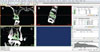
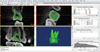
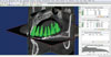
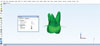
Following the method of Tasanapanont et al.,3 the patients' Digital Imaging and Communications in Medicine files, obtained by CBCT, was converted to the stereolithography (STL) format using Mimics Research version 17.0 (Materialise, Leuven, Belgium). To reconstruct the areas of interest, the threshold value for the tooth region was predefined. The outer boundaries of the tooth morphology in 2-dimensional (2-D) images were identified by each slice orientation and each slice in each orientation manually (Fig. 1). To easily identify the cementoenamel junction (CEJ), intentional extension spine markings were constructed from the 2-D images. The CEJ on each section was marked (Fig. 2). When the “Calculate 3-D” function was selected, the 3-D tooth models were constructed (Fig. 3). The CEJ was marked, and the root surface area of each tooth was calculated automatically by 3-Matic Research version 9.0 (Materialise) (Fig. 4).
To test the intraobserver reliability, all CBCT images were re-measured by the same examiner after a 4-week interval.
Statistical analysis
All data were tested for normality using the Shapiro- Wilk test. The root surface areas were compared between the normal bite group and the anterior open bite group using the independent t-test. The results were considered statistically significant at P<.05. The intraclass correlation coefficient was used to assess the intraobserver reliability.
Results
The intraobserver reliability test for measurement of the root surface area showed a high intraclass correlation (r=0.999), suggesting high reliability in the measurements. The root surface area measurements were normally distributed, and showed no statistically significant difference between the left and right sides. Therefore, the measurements from both sides were pooled for the statistical analysis.
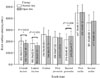
The means and standard deviations of the root surface areas of the teeth ranging from the maxillary central incisor to the maxillary second molar in the normal bite group were 202.08±33.31, 180.54±32.54, 259.59±58.86, 236.92±51.52, 214.02±49.45, 447.78±66.31, and 361.47±69.85 mm2, respectively. The means and standard deviations of the root surface areas of the teeth ranging from the maxillary central incisor to the maxillary second molar in the anterior open bite group were 182.70±27.48, 163.29±24.02, 244.80±53.19, 230.94±39.62, 245.52±44.37, 452.40±66.32 and 377.00±80.89 mm2, respectively (Fig. 5). In both groups, the maxillary first molar had the greatest root surface area and the maxillary lateral incisor had the least. The mean values of the root surface areas of the maxillary central and lateral incisors in the anterior open bite group were significantly less than those in the normal bite group. The mean value of the root surface areas of the maxillary second premolar in the anterior open bite group was significantly greater than in the normal bite group. No statistically significant difference between the 2 groups was found in the canines, first premolars, first molars, or second molars.
Discussion
Our CBCT-based investigation showed that the maxillary first molar had the greatest root surface area and the maxillary lateral incisor had the least root surface area in both groups. As in previous studies,11 the normal bite group showed the normal pattern of root surface area for the maxillary permanent teeth, arranged in descending order as follows: maxillary first molar, maxillary second molar, maxillary canine, maxillary first premolar, maxillary second premolar, maxillary central incisor, and maxillary lateral incisor. It should be noted that the central incisors to the first premolars, inclusively, in the anterior open bite group had less root surface areas than those in the normal bite group. In contrast, the second premolars to the second molars, inclusively, had greater root surface areas.
The decreasing root surface areas from the central incisors to the first premolars in the anterior open bite group might be associated with occlusal hypofunction due to the loss of occlusal contact. Normal occlusal loading and function are responsible for the normal development of alveolar bone, dental root, and associated supporting structures. Occlusal hypofunction decreases alveolar bone mass, accelerates bone resorption, causes deficient root development, and leads to atrophic changes in the periodontal ligament, such as narrowing of the periodontal space, vascular constriction, and deformation of the mechanoreceptors.456 Several previous studies78 have reported that individuals with anterior open bite had a tendency to develop short dental roots from the incisors to the premolars that could be associated with occlusal hypofunction. Experimental findings in rat molars have included external root resorption during tooth movement, in association with hypofunction.12 In addition, abnormal pressure from a tongue thrusting habit might cause root resorption of the anterior teeth.1314
In summary, the anterior open bite might affect root surface area. Orthodontic force magnitudes, especially in cases of vertical problems, should be carefully determined to avoid unwanted side effects during orthodontic treatment.
 XML Download