

 PDF
PDF ePub
ePub Citation
 Print
Print


Introduction
Three-dimensional (3D) imaging in dentistry offers many advantages with respect to diagnosis and treatment planning. Cone-beam computed tomography (CBCT) is a method of acquiring 3D radiographic images that is becoming increasingly popular in dentistry. The resulting images are user-friendly and provide much more information than conventional 2-dimensional radiographs.1
Cone-beam devices are capable of providing submillimeter resolution in images of high diagnostic quality, with a short scanning time and radiation dosages up to several times lower than those of conventional CT scans.2 The use of CBCT images has increased in many clinical applications, such as identifying and locating pathologic lesions, dental implants, temporomandibular joint imaging, orthodontic analysis, analyzing airway space, and the development of surgical guides. Such clinical applications require scans to have a sufficient geometric accuracy to achieve satisfactory results.
In terms of geometric accuracy, the currently available multidetector row computed tomography (MDCT) machines are commonly accepted as a reference standard against which other devices are evaluated. In phantom studies on stereotactic localization using MDCT imaging data, the mean localization error has been reported to be between 0.11 mm and 0.4 mm.34 The measurement accuracy of CBCT images has been studied on different machines with varied results. Some authors found no statistically significant differences between CBCT images and the underlying anatomic topography,58 whereas others reported differences that were statistically significant but not considered clinically relevant.678910 Those studies investigated linear relationships to determine the accuracy of known points in space as determined by the intersection of geometric lines. It could be suspected that volumetric accuracy depends proportionally on linear accuracy and does therefore not require a separate evaluation. Nevertheless, volume calculations for automatically, semi-automatically, or manually segmented objects play an important role in computer-assisted preoperative planning, follow-up, and image-guided surgical procedures.
Since specialized cone-beam devices for maxillofacial imaging represent a relatively new technology, few studies have focused on the volumetric accuracy of CBCT imaging. The purpose of this study was to investigate the influence of various parameters, including the object shape, distance from the center of the image, tube voltage, and tube current, on the volumetric accuracy of CBCT scans.
Materials and Methods
Phantom construction
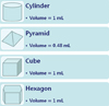
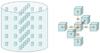
Four geometric objects (cylinder, pyramid, cube, and hexagon) with predefined dimensions were fabricated using a highly precise computer numerical-control milling machine. The volumes ranged between 0.48 mL and 1 mL (Fig. 1). The known volumes of the objects served as reference values for further statistical evaluation. The objects consisted of Teflon-perfluoroalkoxy (PFA) embedded in a hydrocolloid matrix (Dupli-Coe-Loid™, GC America Inc., Alsip, IL, USA), encased in a 150-mm diameter×150-mm height acrylic resin (polymethyl methacrylate) cylinder assembly (Fig. 2). The objects were positioned 0 mm, 30 mm, and 60 mm from the center and aligned in a strictly symmetrical set of 5 rows and 5 columns with a distance of 30 mm (Fig. 3).
Phantom computed CBCT scanning
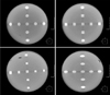
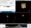
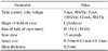
An Alphard Vega Dental CT system (Asahi Roentgen Ind. Co., Ltd, Kyoto, Japan) was used to acquire CBCT images (Fig. 4). In this device, the X-ray source revolves 360 degrees around the phantom in 17 seconds (Table 1). The field of view was cylindrical, with a diameter of 154 mm and a height of 154 mm. Each of the 512 captured projections was represented by a 512-×512-pixel matrix, with pixels defined on a 12-bit gray scale (4096 gray scale). The fixed field of view size was 15 cm, resulting in a scan volume of 15 cm×15 cm×15 cm. The reconstructed 3D volume consisted, therefore, of 512×512×512 isotropic voxels. The corresponding voxel sizes were 0.3 mm×0.3 mm×0.3 mm. The imaging data were collected using 3 different exposure settings (5 mA and 80 kVp, 5 mA and 100 kVp, and 12 mA and 80 kVp). The phantom was placed in the machine in a reproducible method, with the center of the phantom in the center of the scout image.
Using the ADR software system (Asahi Roentgen Ind. Co. Ltd., Kyoto, Japan), with which the standard Alphard-Vega model was equipped, the level and width of the gray scale values of the image were adjusted in the histogram to enable optimal interpretation.
Segmentation and volume measurement
OnDemand 3D (CyberMed Inc., Seoul, Korea) software was used for object segmentation (Fig. 5). Segmentation was semi-automated with manual intervention. The optimal grayscale threshold (−5 HU to −48 HU, determined by OnDemand 3D software) found in the preliminary analysis was then applied to all 540 image data sets for the subsequent analysis. The volume of each of the 4 objects scanned in 3 different image settings was calculated, acquiring a total of 180 measurements per image setting. All measurements were performed by the same trained examiner and repeated at 2 separate time intervals. Ultimately, with 3 different exposure conditions and 4 differently shaped objects, a total of 12 image sets were analyzed.
Data and statistical analysis
To evaluate the measurement accuracy, the measured volumes of the objects were compared to the true volumes. The accuracy was expressed by the volume error (VE). The VE was calculated as:
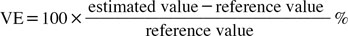
The VE was calculated for all objects under 3 different exposure settings.
To determine the effect of the object's position on the VE in the CBCT imaging data, the volumetric deviation of the 540 objects was correlated with the distance of the respective objects from the center of the imaging data set. The significance of the relationship between the measurement error and distance was assessed using the Pearson correlation coefficient. P values <.05 were accepted as significant.
The effect of the object's shape on volume estimation was tested using phantoms of a known volume. One-way analysis of variance (ANOVA) at P=.05 was used to determine whether there was a significant relationship between the object's shape and the volume measurements. To evaluate the effects of tube current and tube voltage, the independent 2-sample t test was used. P values <.05 were accepted as significant. Descriptive statistics were calculated with standard spreadsheet software (Microsoft Excel; Microsoft Co., Redmond, WA, USA), and statistical analysis was performed using SPSS 12.0K for Windows (SPSS Inc., Chicago, IL, USA).
Results
The measured volumes and the VE values for the objects are listed in Table 2. Overall, the average error in volume measurement ranged from −32.13% to 39.3%. The minimum error (−32.13%) was obtained for the pyramid objects imaged with 5 mA and 100 kVp. The maximum error (39.3%) was obtained for the hexagon objects imaged with 12 mA and 80 kVp. The average VE ranged from 4.47% to 2.35%.
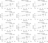
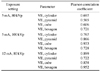
One-way ANOVA demonstrated no relationship between the object's shape and VE at any image setting (P>.05) (Table 2). After having run the imaging analysis, the positions of all 540 objects from CBCT imaging were listed. For all CBCT imaging data, the Pearson correlation analysis suggested that there was a significant (P<.05) correlation between the distance of objects to the center of the imaging data set and the volume measurements (Table 3, Fig. 6). As shown in Figure 6, all 12 combinations of volume measurements underestimated the volume in the center and overestimated it at the periphery.
Discussion
This study evaluated the effects of CBCT scanning parameters on the accuracy of volumetric measurements. Even though volumetric accuracy has been a major topic of discussion regarding conventional tomography and MDCT, few publications are available on this issue in CBCT.
Lascala et al.9 performed linear measurements on 13 distances between anatomical landmarks in dry human skulls scanned with the NewTom 9000 device (Quantitative Radiology, Verona, Italy). They concluded that the real distances measured on dry skulls were always larger than those obtained from the CBCT images. However, these differences were only significant for measurements taken between structures at the skull base, and not for other dentomaxillofacial structures. Marumulla et al.8 evaluated the geometric accuracy of the same CBCT scanner by using a grid phantom and a sophisticated mathematical test method. They determined that the mean error was 0.13 mm (±0.09 mm). Kobayashi et al.11 measured cross-sectional distances in cadaver mandibles and compared them to measurements obtained from CT scans. They reported a mean error of 0.22 mm (±0.15 mm) for CBCT scans and of 0.36 mm (±0.24 mm) for spiral CT scans. Although the errors were not large in either type of image, a statistically significant difference was found between the 2 methods, at P<.001. The mean absolute percentage errors were reported to be 1.4% for CBCT and 2.2% for spiral CT.
The effects of various CT parameters, such as window setting, slice thickness, segmentation threshold, field of view, peak voltage, and tube current, on volumetric accuracy have been previously documented.12131415 Different window width and level settings can also affect the measurement of the diameter. These factors affect the accuracy of the measured volume, resulting in inconsistency and uncertainty in detecting volume changes in serial CT scans. Goo et al.16 stated that for the accurate measurement of lung nodule volume, it was critical to select a section thickness and segmentation threshold that was appropriate for the size of a nodule. Various research groups have investigated the effects of image acquisition parameters using sphere objects with known volumes. Ko et al.17 reported computer-calculated volumes obtained from regions of interest marked by a radiologist. Using a threshold method for segmentation, they found that tube current-time, reconstruction algorithm, and object size significantly affected volume error. Way et al.14 reported that there was no statistically significant difference in the volume error for CT scans taken with a technique where only pitch, field of view, or tube current (mA) changed, whereas slice thickness significantly (P<.05) affected volume error. Several authors have reported an inverse correlation between measurement error and volume size.161819 However, all those studies were performed on relatively small spherical phantoms for the purpose of lung nodule evaluation. The effects of volume size variation on the measurement error of spherical objects cannot be automatically extrapolated to objects with a rectangular geometry.
The present study evaluated the volumetric accuracy of a CBCT scanner and analyzed the influence of different parameters on the measurement errors of CBCT images under various exposure conditions. The volumetric reference objects for measurements consisted of a homogeneous radiopaque material providing high contrast to the surrounding hydrocolloid matrix. The reference objects were made by a precision computer numerical-control drilling mill machine and had sharp and regular edges. Both properties allowed the 3-dimensional segmentation and volume calculation to be as accurate as possible. The choice of the material Teflon-PFA for the reference objects, and hydrocolloid gel for the phantom matrix, was based on prior experiments exploring the visualization of different materials. The radiopacity of Teflon-PFA resembles that of cortical bone on CBCT scans, so the contrast between Teflon-PFA objects and gels is similar to that between bone and soft tissue in vivo.
The segmentation process could affect the accuracy of volume measurements. Some authors reported that the volume errors associated with manual segmentation ranged from 150% to 350%. Image quality is the predominant factor affecting segmentation. CBCT imaging quality can be related to machine settings, patient positioning, management, volume reconstruction, and export to the Digital Imaging and Communications in Medicine format. There is no standard method of segmentation. Our segmentation procedures were largely based on image thresholding. The use of a global threshold value for the entire object has the advantage that only a single segmentation parameter is estimated. This is relatively simple and often used for bone segmentation, which commonly has a uniform density.2021
Since the segmentation process of the reference objects and the volume measurements were semi-automatically carried out in ideal conditions, the variability among the measurements may be attributed to other factors.
Blake et al.22 reported a mean absolute percentage error for MDCT-based volumetric measurements of Plexiglas phantoms between 3% and 5%. The scans were performed at a 1.3-mm slice thickness, with a reconstruction interval of 0.6 mm. Goo et al.16 estimated the mean absolute percentage error for MDCT-based volumetric measurements of acrylic spheres to be 5.4% for an object with a volume of 1.07 mL. The scans were performed at a 1-mm slice thickness, with a reconstruction interval between 0.5 and 2 mm. These results are certainly within the tolerance limits of 5% to 10% for volumetric quality assurance testing in the high-precision disciplines of stereotactic radiosurgery and radiotherapy postulated by Ramaseshan and Heydarain.24 Disler et al.24 stated that volume estimations are likely to be clinically useful even with errors of up to 10%.
In this study, the mean VE of −4.47 to 2.45% determined for the evaluated CBCT device closely matched the values reported by Blake et al.23 and Goo et al.16 Thus, the evaluated CBCT machine had clinically accurate and acceptable volume measurements.
Previous studies of volumetric accuracy were performed on phantoms containing spheres to simulate tumors or lesions, even though real anatomical forms are frequently non-spherical. In this study, 4 differently shaped objects were used to evaluate how the object's shape influenced the volume measurement. The results were independent of the object's shape. There was no statistically significant dependence of volume errors on the object's shape for any image setting. According to previous CT studies, errors may be dependent on the object size. Future studies must evaluate more sizes and geometries, and their effects on volume measurements.
In this study, the VE of CBCT imaging was not evenly distributed. Volumes were underestimated in the center of images and overestimated at the periphery. Attention should be paid to the distortion of objects located at the periphery of the scan volume. These consist of archshaped or curved defects and blurring of object boundaries. Katsumata et al.25 attribute such artifacts, which affect mostly solid, regularly shaped objects, to the halation of the image intensifier. Presumably, halation artifacts may be considered as a possibility in all CBCT systems equipped with an image intensifier/charge-coupled device detector unit. Object distortions at the periphery of the scan volume were observed in images generated by the device used in this study as well. Since this artifact appeared only when the area to be imaged was positioned near the facial surface, halation artifacts must be taken into consideration at the temporomandibular joint and the anterior dental arch.
Overestimation is also caused by partial volume averaging. The volume error for small objects is especially sensitive to uncertainties in the segmented boundary, as a slight deviation due to the partial volume effect and reconstruction artifacts such as a blurry or irregular edge would result in a substantial percentage error. When the slice thickness is large, the blurred boundary due to partial volume averaging contributes to extra slices for the object.
Furthermore, even in CBCT scans obtained under ideal imaging conditions, the acquired images contain statistical variations in the X-ray photons recorded at the detector and other uncertainties of the CBCT scanner. For example, the starting scan position of the CBCT scanner is not perfectly reproducible. The slice locations relative to the anatomical structures are therefore not identical in repeated scans even if the phantom is not repositioned. This has to be taken into consideration when using CBCT for volume measurements.
The volumetric software used in this study showed good overall performance, allowing the segmentation and volume measurements of all phantom objects. This study analyzed the effect of tube voltage and tube current on the accuracy of volumetric measurements. Our results indicated that there was no statistically significant dependence of volume errors on tube current, but we observed a significant dependence on tube voltage. We think that the reason for this result is that image noise may increase with decreased tube voltage. The increased noise and presence of more artifacts makes segmentation of the object from the surrounding structures more difficult, causing images to be more distorted. The area affected by artifacts was larger in images made using 100 kVp than with 80 kVp.
Therefore, volumetric errors were influenced by tube voltage, but were independent of tube current. For assessments of volume measurements, it may be sufficient to use serial scans with a high resolution but a low dose. Further studies are needed to clarify the optimal exposure conditions for observing objects in a clinical setting.
There were limitations in this study. First, this study used simple and similarly-sized geometries to evaluate general trends, but it would be useful to investigate more sizes and irregular geometries, since most anatomic structures vary in size and geometry. Second, it is not known whether the dependence of volumetric errors on imaging and reconstruction parameters is consistent for CBCT scans acquired with scanners from different manufacturers with different 3D analysis software. Third, it is also not known whether the trends observed in this phantom study would also be seen in real patients. Fourth, for a fuller evaluation of the effects of CBCT parameters on the VE, it would be necessary to make more measurements with a precisely machined phantom using a wider range of test materials. The custom phantom used in this study was not as dimensionally accurate as desired. These and other issues should be investigated in future studies.
In conclusion, we found that the evaluated CBCT device provided satisfactory volume measurements. Our goal was to evaluate the effects of CT scanning and reconstruction parameters on accuracy. Although the VE values estimated using phantom objects may be different from those of real anatomic structures, this study presented trends illustrating the dependence of VE on CBCT imaging conditions.
The results obtained from the experiment are as follows. The mean VE ranged from −4.47% to 2.35%. There was no relationship between objects' shape and the VE. There was a significant correlation between the distance of the object to the center and the VE. The tube voltage affected the volume measurements, but the tube current had no effect. This information may provide useful guidance for assessing volume measurements.


 XML Download