

 PDF
PDF ePub
ePub Citation
 Print
Print


Introduction
The extraction of third molars is one of the most frequently performed procedures in dental clinics, and is necessary when there is insufficient space for eruption of these teeth, when they are poorly positioned, or when there is a risk of formation of cysts or odontogenic tumors.1 The appropriate surgical procedure should be determined based on findings from preoperative examinations evaluating the morphology of the third molar and its relationships with adjacent structures.2
Conducting an assessment by a radiographic examination allows the evaluation of several factors that are related to the degree of difficulty of the surgical procedure, such as the depth of impaction and the position of the tooth. Identifying these factors can reduce the occurrence of surgical complications.34 Some systems classify impacted third molars according to their depth of impaction and their position on the mesial-distal axis. The most commonly used classification systems are those proposed by Winter5 and Pell and Gregory.6 Both classifications are based on 2-dimensional images, and do not allow for the evaluation of some positions, such as the buccolingual position.
A classification system of the depth of impaction and position of impacted third molars is expected to lead to improved interdisciplinary communication in treatment planning and to help estimate the degree of surgical difficulty of each case. Although panoramic radiography is very helpful in preoperative assessments, it may present limitations, such as distortion and blurring of the image.7
CBCT examinations allow a proper assessment of the 3-dimensional anatomic relationships between the third molar and surrounding tissues.2 At the time of writing, a search of the PubMed database using the descriptors “maxillary third molar,” “upper third molar,” “wisdom third molar,” “classification,” “position,” “cone beam computed tomography,” “CBCT,” “CT,” “CBVT,” “3D images,” and “three-dimensional reconstruction” revealed no studies that described the depth and angular position of impacted maxillary third molars (IMTMs) based on CBCT images.
Therefore, given the absence of studies classifying the position and the depth of impaction of IMTMs based on CBCT images, the present study proposes a 3-dimensional classification of the position and depth of impaction of IMTMs and an analysis of their prevalence with respect to gender and age.
Materials and Methods
This was a cross-sectional observational retrospective study analyzing the sagittal and coronal CBCT sections of 300 maxillary third molars from the database of the Radiology Clinic, School of Dentistry, Federal University of Juiz de Fora (UFJF). The exams of patients missing any upper molars were excluded. This study was approved by the Research Ethics Committee of the UFJF (opinion number 932.681).
The images analyzed were obtained using an i-CAT scanner (Imaging Sciences International, Hatfield, PA, USA) operating at 120 kVp and 3-8 mA with 0.25-mm voxels, a 26.9-s rotation time, and a 10-cm field of view. The images were stored in the Digital Imaging and Communications in Medicine format and later reconstructed with CS 3D Imaging software (Carestream Dental, Rochester, NY, USA). Linear and angular measurements were made in this program. The assessments were performed by a single examiner with experience in CBCT imaging.
The classification of third molars was developed using CBCT images and was based on 3 criteria: buccolingual position, mesial-distal position based on a modified version of the Winter classification,5 and depth of impaction according to a modified version of the Pell and Gregory classification.6
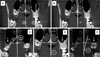
The position of the third molar on the buccolingual axis was assessed in a coronal section. The classification was made according to the angle formed between the reference line (line 1, Fig. 1A) drawn on the third molar long axis (longitudinal axis of the tooth) and the reference line (line 2, Fig. 1A) in the midsagittal plane. To measure the angle formed between the reference lines 1 and 2, line 1 was shifted until it intersected with line 2, and the angle was then measured (Fig. 1B). The maxillary third molars were then classified as buccal when the angle formed was greater than +10° (Fig. 1C), central when the angle formed was between +10° and −10° (Fig. 1D), and lingual when it was less than −10° (Fig. 1E).
The classification of the mesial-distal position was made in the sagittal section. The angles formed by the intersection of the reference line drawn on the long axis of the third molar (longitudinal axis) and another line passing through the occlusal plane of the maxillary first molar (line tangent to the cusps of the first molar) were analyzed. The alignment was considered distoangular when the angle formed was less than or equal to 75° (Fig. 2A), vertical when the angle formed was between 76° and 104° (Fig. 2B), and mesioangular when the angle was greater than or equal to 105° (Fig. 2C).
To assess the depth of impaction, the level of the occlusal plane of the maxillary third molar was analyzed. The tooth was classified according to the midpoint of its occlusal surface in relation to the occlusal plane of the maxillary second molar. The tooth was considered to be high when the midpoint of its occlusal plane was found coronally to the cementoenamel of the maxillary second molar (Fig. 3A), medium when the midpoint of the occlusal plane appeared between the cementoenamel junction and the occlusal plane of the maxillary second molar (Fig. 3B), and low when the midpoint of the occlusal plane was found at the same level or caudally to the occlusal plane of the maxillary second molar (Fig. 3C).
Three hundred CBCT images of maxillary third molars were classified, and associations with gender and age were examined using analysis of variance with the Scheffé post-hoc test. To determine the associations among the 3 classifications, the chi-square test was used. Statistical analyses were conducted using SPSS version 13.0 (SPSS Inc., Chicago, IL, USA), with a 5% significance level (P<.05).
Results
Among the 300 maxillary third molars, 151 (50.3%) teeth were on the right side, and 149 (49.7%) teeth were on the left. The age of patients ranged from 18 to 40 years, with a mean of 23.74±4.76 years. Regarding gender, 100 third molars belonged to male patients (33.3%), and 200 third molars belonged to female patients (66.7%).
The distribution of the 300 IMTMs according to the proposed classification is presented in Table 1. Table 2 shows the 3-dimensional classification data distributed by gender and age.
A significant association with age was found for the mesial-distal position (P=.005) because younger individuals exhibited an increased prevalence of the distoangular position, while there was a tendency toward verticalization with increasing age. In terms of the depth of impaction, the correlation analyses demonstrated that younger individuals exhibited a greater tendency toward medium and high depths of impaction, which tended to decrease with age (i.e., the frequency of a low depth of impaction increased; P=.0001).
The associations between gender and each of the classifications of maxillary third molars were also evaluated, and none were significant (buccolingual position: P=.417; mesial-distal position: P=.052; and depth of impaction: P=.738).
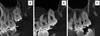
Significant associations were found between each of the classifications (buccolingual position with depth of impaction, mesial-distal position with buccolingual position, and mesial-distal position with depth of impaction). Figure 4 illustrates the spatial distribution of the associations between the classifications.
Discussion
To our knowledge, this was the first study to assess the depth and angular position of IMTMs based on CBCT images, and most importantly, this study was the first to determine the buccolingual position of these teeth. The scientific literature on IMTM depth and angular position that has been published in databases (PubMed) contains results based on 2-dimensional images.1348910111213 Because of the nature of radiographs (i.e., 2-dimensional images), it has been impossible to determine the buccolingual position. Indeed, none of the previous studies analyzed the buccolingual position of the IMTMs.1348910111213 Thus, the aims of this study were to present a new classification of the depth and angular position of IMTMs based on CBCT images and to determine the associations of gender, age, and depth with each angular position.
A preoperative CBCT examination is now considered an important assessment tool for planning third molar surgery and for predicting the risks related to surgical interventions.2 Some important findings that may affect the result of the removal of an IMTM can be obtained from CBCT images, but not from 2-dimensional images, such as the buccolingual position, the real proximity of the roots of the IMTM to the sinus floor,12 and the relationship of the IMTM with the second molar.14
In terms of the buccolingual position, 74.0% of IMTMs had a buccal position, 19.3% a central position, and 6.7% a lingual position. Because this is a novel classification system, it is not possible to compare these findings with other reports. Neither clinical examinations nor 2-dimensional imaging techniques can identify when third molars are impacted at such angulations. Panoramic radiography is considered quite imprecise for determining the amount of overlying bone. Thus, surgical difficulty may be expected when the tooth is in a deeper position inside the bone due to limited access to the surgical site and the need to remove a larger amount of bone. However, a CBCT examination can indicate the position of the tooth, the amount of overlying bone, and its relationships with the surrounding structures. Tomographic examination is thus an aid for diagnosis and therapy because it reveals the anatomical situation with greater precision. The procedures performed during extraction are more predictable because of the better visualization of the anatomical obstacles and the 3-dimensional position of the third molar.7 Further studies should investigate whether buccolingual angulation predisposes surgical procedures to complications.
The most frequently observed IMTM position was vertical (61.3%), and this finding is consistent with those reported by other authors.389101112 However, Topkara and Sari1 reported that the distoangular position was the most frequent position and that in many studies, the angulation of IMTMs was usually determined based on visual impressions according to the Winter classification. Although various authors have cited the Winter classification5 in their studies, the methodology used to evaluate the angles that determine the different positions is quite variable. Ventä et al.15 used the angle between the tooth's occlusal surface and the occlusal plane of the first and second molar for IMTM classification, and classified teeth as vertical (−10° to +10°), mesioangular (11° to 70°), distoangular (−11° to −70°) or horizontal (>70°). In contrast, Sandhu and Kaur13 used the angle between the axis of the second molar and that of the IMTM to classify IMTMs as vertical (0° to 25°), mesioangular (26° to 75°), horizontal (>75°), or distoangular (<0°). Hassan10 used the angle between the long axis of the third molar and the long axis of the second molar to classify IMTMs as vertical (−10° to +10°), mesioangular (11° to 79°), horizontal (80° to 100°), distoangular (−11° to −79°), or buccolingual (crown overlapping the root). De Carvalho et al.3 classified angles from 0° to 30° as horizontal, 31° to 60° as mesioangular, 61° to 90° as vertical, and >90° as distoangular. In the present study, the IMTMs were classified as follows: distoangular when the angle was ≤75°, vertical when the angle was 76° to 104°, and mesioangular when the angle was ≥105°. To establish these angle ranges, each third molar was classified visually using a modified version of the Winter classification.5 Next, the ranges of the angles were measured.
In our study, the most common IMTM impaction level was low (41.3%). In other studies, this classification has been performed using the nomenclature of class A, which corresponds to the low level; class B, which corresponds to the medium level; and class C, which corresponds to the high level. Our results are comparable to those reported by others because the same criteria were used.1112 Hassan,10 de Carvalho et al.,3 and Topkara and Sari1 reported that the most frequent impaction level was B, which corresponds to the medium level in our study. Evaluating the depth of impaction of an IMTM is important because depth is considered to be a predictor of surgical difficulty,3 and also serves as an indication of the potential for oroantral perforation.12 These authors stated that radiographs are not a reliable method for predicting the possibility of oroantral perforation. Therefore, surgeons should obtain reliable tomographic information regarding the depth of impaction when planning to remove such teeth.12
The associations of age with depth of impaction and classification of the mesial-distal position were significant. In older patients, maxillary third molars tended to be classified as low and in the mesioangular and vertical positions. These associations are important because the incidence of radiographic lesions increases with age, and partially impacted mesioangular third molars exhibit a high incidence of caries or periodontal bone loss in the adjacent second molar.8 Moreover, mesioangular and horizontal third molars are more strongly associated with the external root resorption of second molars, and one study reported that a significantly greater number of cases were diagnosed based on CBCT images than panoramic radiographs.14 According to Oenning et al.,14 CBCT images provided diagnostic information in the sagittal, axial, and coronal planes without an overlap of structures, whereas panoramic radiographs did not have the third dimension.
According to the results obtained from the tests of the associations between IMTM classifications, strong associations of the low position with the vertical and buccal positions were observed, and these positions represent the lowest degree of extraction difficulty. Regarding the association between the buccolingual and the mesial-distal categories, a cyclical distribution was observed, in which all positions were equally closely associated. Position and depth alone may not indicate an extremely difficult surgical procedure, but some combinations of these variables can elicit problems. Therefore, these variables should be evaluated in combination, not in isolation.
Until now, planning of third molar removal surgery was based on mesial-distal position and depth obtained from 2-dimensional images. With CBCT, in addition to these positions, it is possible to assess the buccolingual position. Two-dimensional radiographs provide information that can be useful for surgical planning; however, only CBCT examinations permit the evaluation of the real situation of the tooth.
This study underscores the role of CBCT in the diagnosis and treatment planning for the surgical removal of IMTMs. Although the expected surgical difficulty of IMTM removal is low, some cases may present unexpected difficulties. CBCT is the primary tool for identifying variables that predict surgical complications, because it enables a 3-dimensional evaluation, which led to a new proposal for the classification of the position and depth of impaction of IMTMs. A future study will investigate the relationships of the IMTM classifications with intraoperative complications and postoperative morbidity.


 XML Download