

 PDF
PDF ePub
ePub Citation
 Print
Print


Introduction
Three-dimensional (3D) image analysis of the mandible is possible due to the wide availability of computed tomography (CT) imaging technology. Accordingly, there have been several anatomical analyses of the mandibular foramen (MnF),1234 most involving distance-only measurement methods. Such measurement methods have limitations, even when 3D CT images are used. Moreover, the analytical data for the mandible are limited with respect to their relationship with skeletal malocclusion, the size of the mandible, and the measurement planes and reference points for locating the position of the MnF.
The position of the MnF has been studied for mandibular treatment (i.e. ramal surgery, including osteotomy; inferior alveolar nerve block).56 The present study used 3D images of the mandibles of patients diagnosed with skeletal class III malocclusion, and analyzed the position of the MnF (i.e. its proportional position in the mandible) using measurement planes and anatomical reference points. These data can be used in clinical cases, particularly for ramus surgery. The purpose of this study was to produce a useful guide of MnF positions in patients diagnosed with skeletal class III malocclusion.
Materials and Methods
This retrospective study was approved by our institutional review board. This study analyzed CT images of patients who underwent orthognathic surgery. Specifically, the study used CT images of mandibles that were taken during preoperative diagnosis work-ups. All patients were diagnosed preoperatively with skeletal class III malocclusion. The CT images were taken with a Siemens Sensation 64 system (1.0 mm slice thickness, Siemens Sensation 64 CT scanner; Siemens AG, Erlangen, Germany) using a pixel size of 0.4375 mm and a field-of-view of 22.40 cm. We excluded CT data from facial asymmetry patients with a mandibular shift greater than 5 mm (considered more than a moderate state of asymmetry).7 The CT data were collected for a total of 85 patients, and both right and left MnF images were analyzed.
Points, lines, and planes for measurements
Simplant version 14.0 (Materialise Dental, Leuven, Belgium) was used to reconstruct the CT images into 3D images. The anatomical reference points, lines, and planes were then set onto the image of the mandible. The 3D position of the MnF was measured based on the established reference points and planes. This process was intended to help the surgical team locate the MnF during mandibular ramus osteotomy. The anatomic points, lines, and planes that were used, as well as the measurements, are described below.
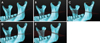
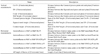
The MnF, coronoid process, sigmoid notch, condyle, gonion, antegonial notch, menton, mesiobuccal cusp of the mandibular first molar, and the most superior incisal contact point of the lower central incisor were identified in the images. In addition, the points of the anterior and posterior ramus were defined (Table 1). The anatomic lines used for defining the reference planes in this study are shown in Table 2. Three vertical lines were constructed: one connecting the sigmoid notch points and antegonial notch points, termed the SN-AGN line; another connecting a sigmoid notch point and gonion point, termed the SN-Gn line; and the other connecting a condyle point and a gonion point, termed the Cond-Gn line. In addition, five horizontal lines were constructed that connected each of the anterior ramus points and each of the posterior ramus points. The occlusal plane, mandibular plane, and five reference horizontal planes were defined (Table 3, Fig. 1A). Five vertical coronal planes were also defined, as shown in Table 3 and Figure 1B.
The vertical heights of anatomic points and horizontal length of the ramus
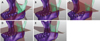
The vertical heights of the anatomic points were measured according to each of the five horizontal planes (Fig. 2). The vertical distances from the coronoid process, sigmoid notch, and condyle to each horizontal plane were added to the vertical distances from the gonion to each horizontal plane, and these distances were set as the vertical heights of the coronoid process, sigmoid notch, and condyle in the mandible for each horizontal plane (Table 4). The vertical distances from the gonion to the horizontal planes were set as the heights of the MnF in the mandible, according to each horizontal plane. Based on each of the five horizontal planes that pass through the MnF, the distance on each plane from the anterior ramus point to the posterior ramus point was designated the anteroposterior horizontal distance of the ramus (Table 4). The perpendicular distance from each anterior ramus point to each vertical plane was designated the horizontal distance from the anterior ramus to the MnF (Fig. 3).
Analysis of the measurement values
To investigate the positional relationship of the MnF in the mandible, regression analysis was used to examine the rational relationships between the vertical heights of the coronoid process, sigmoid notch, and condyle points and the vertical height of the MnF. Regression analysis was also used to determine the rational relationship between the anteroposterior horizontal distance of the ramus from each of the five horizontal planes passing through the MnF and the horizontal distance from the anterior ramus point to the MnF. The study data were analyzed with IBM SPSS Statistics version 21 (IBM Corp., Armonk, NY, USA), and statistical significance was established at p<0.05. The intra-observer error for anatomic point determination was calculated via repeated measurement (10 measurements) of the MnF. All measurement data are reported as mean±SD unless otherwise indicated.
Results
The sex ratio and age of the 85 patients with skeletal class III mandibular prognathism were 46 : 39 (men : women) and 22.2±5.5 years. The condyle height relative to the MnF-mandibular plane was the smallest (53.45±6.45 mm). The heights of the coronoid process and the MnF were the greatest when measured relative to the MnF-mandibular plane (60.09±5.96 mm and 21.31±3.83 mm, respectively). The height of the sigmoid notch was the greatest when based on the MnF-SN/Gn p plane (43.89±5.02 mm). The height of the condyle was the greatest when based on the MnF-Cond/Gn p plane (63.80±5.79 mm). The heights of the MnF, coronoid process, and sigmoid notch were the smallest when based on the MnF-Cond/Gn p plane (18.60±4.56 mm, 50.08±6.89 mm, and 40.50±5.45 mm, respectively). The anteroposterior length of the ramus and the distance from the anterior ramus point to the MnF were the longest when they were based on the MnF-mandibular plane (42.98±5.17 mm and 27.60±4.22 mm, respectively; Table 5). The intra-observer error for the MnF area was 0.60±0.24 mm.
Vertical position of the MnF
The heights of the coronoid process, sigmoid notch, and condylar head on the five horizontal planes were all significantly associated with the height of the MnF according to the regression analysis. The regression relationship was the strongest for the coronoid process, sigmoid notch, and condylar head when the measurements were based on the MnF-mandibular plane (coefficients of determination (R2) of 0.424, 0.597, and 0.604 respectively).
The heights of the coronoid process and the MnF were significantly associated according to the regression analysis (Table 6). The non-standardized regression coefficients were significant for the relationships between the heights of the coronoid process and the MnF based on all the horizontal reference planes. The highest coefficients, based on the MnF-mandibular plane, were a standardized regression coefficient of 0.654 and determination coefficient (R2) of 0.424.
The heights of the sigmoid notch and the MnF were significantly associated in the regression analysis (Table 7). The non-standardized regression coefficients for the relationships between the heights of the sigmoid notch and the MnF were significant for all the horizontal reference planes. The regression constant was highly significant when it was based on the MnF-mandibular plane (p=0.002). The highest coefficients were a standardized regression coefficient of 0.774 and determination coefficient of 0.597, and these were based on the MnF-mandibular plane.
The heights of the condylar head and the MnF were significantly related in the regression analysis (Table 8). For all the horizontal reference planes, the non-standardized regression coefficients for the relationships between the heights of the condylar head and the MnF were significant. The regression constant was significant when it was based on all of the horizontal planes, except for the MnF-SN/Gn p plane. The highest coefficients were a standardized regression coefficient of 0.779 and a determination coefficient of 0.604, and were based on the MnF-mandibular plane.
Horizontal position of the MnF
Using the five horizontal planes as references, the anteroposterior length of the ramus and the distance from the anterior ramus point to the MnF were significantly associated according to the regression analysis (Table 9). For all the horizontal reference planes, the non-standardized regression coefficients were significant for the relationships of the anteroposterior length of the ramus and the distance from the anterior ramus point to the MnF. The regression constants were significant when the MnF-mandibular plane and MnF-SN/Gn p plane were used as references.
The standardized regression coefficients for all five horizontal planes used as references were greater than 0.73, and the coefficient was 0.877 using the MnF-mandibular plane and 0.901 using the MnF-SN/Gn p plane. The determination coefficient was 0.766 using the MnF-mandibular plane and 0.811 using the MnF-SN/Gn p plane. Notably, there were 53 missing values for measurements using the MnF-mandibular plane and 3 missing values for the MnF-SN/Gn p plane. For the MnF-mandibular plane, which is parallel to the MnF, there were missing values when the anterior part of the plane sloped downward, thus leading directly to the mandibular body instead of the anterior ramus. Similarly, there were missing values for the MnF-SN/Gn p plane, although there were fewer than for the MnF-mandibular plane.
Discussion
The size of the human mandible varies greatly according to age, ethnicity, and sex.2 Accordingly, the present study investigated the relative position of the MnF rather than simply measuring its position within the surrounding anatomical structures. Using specific anatomic planes as references, we measured the position of the MnF in patients diagnosed with skeletal class III malocclusion to determine the 3D position in the mandible. We determined the position of the MnF was related vertically to the heights of the mandibular condyle, sigmoid notch, and coronoid process and horizontally to the anteroposterior length of the mandibular ramus. Further, we found that the mandibular plane was best for determining the relationships of the heights of the mandibular condyle, sigmoid notch, and coronoid process with the height of the MnF. This suggests that the mandibular plane can be utilized in clinical cases, not only for analyzing maxillofacial malformations but also for tracing the position of the MnF.
During mandibular prognathism surgery, the vertical position of the MnF can be estimated using the exposed sigmoid notch area. With reference to the mandibular plane, the height (in millimeters) of the MnF can be estimated with the following equation: 0.621×(the height of the sigmoid notch) - 5.318. As an alternative to the mandibular plane, one can use the horizontal plane perpendicular to the SN-Gn line that connects the sigmoid notch and gonion; this plane (the MnF-SN/Gn p plane) is second to the mandibular plane in terms of usefulness for estimating the height of the MnF. When using the condyle height, the occlusal plane can be a useful alternative for estimating the height of the MnF.
Vertical ramus osteotomy is a surgical procedure that can be used for patients with mandibular prognathism when the mandibular ramus posterior to the MnF is cut. The relationship between the anteroposterior length of the mandibular ramus and the position of the MnF was best shown when the horizontal MnF-SN/Gn p and MnF-mandibular planes were used as references. The mandibular ramus area at the MnF level is exposed during mandibular prognathism surgery. Therefore, the relationship of the horizontal position of the MnF with the anteroposterior length of the ramus was investigated. Using the MnF-SN/Gn p plane as a reference, the horizontal length (in mm) from the anterior ramus to the MnF can be estimated with the following equation: 0.728×(the anteroposterior distance of the mandibular ramus)- 3.948. Using the MnF-mandibular plane as the reference, the horizontal length (in millimeters) from the anterior ramus to the MnF can be estimated with the following equation: 0.716×(the anteroposterior distance of the ascending ramus)- 3.148.
However, there are cases in which the mandibular plane leans in the anteroinferior direction, and the MnF-mandibular plane cannot pass through the anterior ascending ramus area. In the present study, there were 53 missing values for the anterior ramus point when using the MnF-mandibular plane as the reference, and there were 3 missing values when using the MnF-SN/Gn p plane. Missing values may limit the usefulness of these calculations since they may occur in actual clinical cases. If this happens, it is recommended that the MnF-SN/Gn p plane be used, and the use of the occlusal plane may be more helpful for determining the horizontal position of the MnF when the anterior ramus point cannot be defined using the MnF-mandibular plane. The vertical height of the MnF from the gonion ranged from 18 mm to 21 mm, and the anteroposterior length of the ramus was about 33 mm to 42 mm. The horizontal distance from the anterior ramus point to the MnF was about 20 mm to 27 mm. This shows that the MnF is positioned posterior to the mandibular ramus.
The wide availability of CT scans has resulted in useful 3D images of the mandible. In a study that analyzed the 3D position of the MnF using cone-beam CT, the average distance from the gonion to the MnF was about 24 mm and the average distance from the anterior ramus to the MnF in the axial plane was about 15 mm.2 The difference between the results of that study versus the present study is likely due to the use of the axial and sagittal planes of the CT images in the former. In another study that analyzed the 3D position of the MnF using multiple detector CT, the average height from the gonion to the MnF relative to the occlusal plane was about 18 mm, the average anteroposterior length of the ascending ramus was 37 mm, and the average distance from the anterior ramus to the MnF on a horizontal plane (parallel to the occlusal plane that passes through the MnF) was about 22 mm.8 These results are similar to those of the present study, although there are minor differences because the present study only examined mandibles with skeletal III class malocclusion, and the horizontal length from the anterior ramus to the MnF was measured as the perpendicular distance projected onto each vertical plane that passed through the MnF.
Simply measuring the vertical and horizontal distances may not be that beneficial in actual clinical cases because human mandibles vary in size. However, assessing the positional relationships using 3D planes can be useful clinically, as this shows the objective relationship with respect to the reference planes used in the analyses. Image-assisted surgical techniques are increasingly used for maxillofacial operations; accordingly, the present study is valuable in that it provides 3D image data that can be utilized in such surgical procedures. This study's research methods and results, with respect to the positional 3D analysis of the MnF, will be useful for computer-assisted surgical methods, such as surgical simulation, navigation, augmented reality models, and robotic surgery. Moreover, these data can be applied for treating mandible-related issues. The data can also be used to compare the mandibles of different ethnic groups.
Previous studies of the mandible were performed on cadavers,910 and often just a few distances were measured in 3D reconstructed images even though CT images were examined.234 Importantly, previous studies performed quantitative analyses without considering the different sizes of the mandibles. Moreover, even when 3D reconstruction images were used, few studies performed vertical and horizontal assessments using 3D reference planes.
The present study found that the position of the MnF was related to the vertical heights of the sigmoid notch, coronoid process, and condyle and to the horizontal anteroposterior length of the ascending ramus. These findings indicate that the MnF, at least to some extent, maintains its relative position with the growth of the rami. In addition, the results indicate that the MnF is posterior to the ascending ramus. We expect that future studies will contribute further knowledge about the position of the MnF in the mandible via 3D image analysis of mandibles with skeletal class II malocclusion for ramus surgery.
In conclusion, the 3D mandibular image analysis data reported in this study will be useful for defining and describing the position of the MnF in mandibles of different sizes and shapes and in different ethnic groups. The present study showed that the proportional position of the MnF was significantly related to the vertical heights of the sigmoid notch, coronoid process, and condyle and to the horizontal anteroposterior length of the ascending ramus. Furthermore, the methods used here and the positional 3D analyses of the MnF can be used clinically for computer-assisted surgical methods, especially for ramal surgery in patients with skeletal class III mandibular prognathism.


 XML Download