

 PDF
PDF ePub
ePub Citation
 Print
Print


Introduction
Condylar remodeling is a physiological process in which the structure of the temporomandibular joint (TMJ) is adapted to meet functional demands. It is based on an interaction between the mechanical forces sustained by the TMJ and the adaptive capacities of the condyle.12
For patients with skeletal Class III deformities, corrective orthognathic surgery improves both oral function and related esthetics.3 The effect of orthognathic surgery on condylar remodeling is a poorly understood and controversial issue.1 However, orthognathic surgery inevitably results in condylar positional changes.2 These positional changes can induce functional stresses in TMJ structures, thereby causing condylar remodeling, and are considered to be a possible etiology of skeletal relapse following orthognathic surgery.1
Three-dimensional (3D) imaging has been shown to be effective in examinations and treatment relating to the oral and maxillofacial region.4 Several studies have used 3D images to assess condylar remodeling and positional changes after orthognathic surgery.256 Carvalho et al.6 used a superimposition technique on 3D imaging and suggested that the condyles tended to move, on average, ≤2 mm superoposteriorly after surgery, and this small positional displacement was maintained one year after surgery. An et al.3 also used a 3D image superimposition technique and colored scales to evaluate condylar head deviation before and after orthognathic surgery. They found that condylar surface changes after surgery were significant.
However, few studies have evaluated condylar remodeling according to the area of the condylar head and quantified condylar surface changes. Moreover, no studies have assessed the differences between the right and left condyles and between the sexes.
The purpose of this study was to assess the frequency of remodeling types in each area of the condyles and to analyze condylar surface changes quantitatively using 3D images based on multi-detector computed tomography (MDCT) data. Furthermore, this study compared condylar surface changes and condylar remodeling types between the right and left side and between the sexes.
Materials and Methods
Selection of patients
The present study was approved by the Institutional Ethics Review Board of Seoul National University Dental Hospital (IRB No.CRI15014). This study was a retrospective analysis of patients with skeletal Class III malocclusion who underwent mandibular setback sagittal split ramus osteotomy with Le Fort I osteotomy at Seoul National University Dental Hospital from 2012 to 2015. Patients who had any underlying diseases, facial trauma, or temporomandibular joint disorders were excluded. Ultimately, forty patients (20 males, with a mean age of 21.6 years, and 20 females, with a mean age of 21.0 years) who had undergone 3D MDCT (SOMATOM Sensation 10®, Siemens, Erlangen, Germany) examinations preand post-orthognathic surgery were selected. The images were obtained an average of 1.3 months (range, 0.6-2.0 months) prior to surgery and 6.7 months (range, 5.4-13.5 months) after surgery.
Three-dimensional image analysis
Three-dimensional images of mandibular condyles were reconstructed and reformatted into the WRL data format (the file name extension of a Virtual Reality Modeling Language [VRML] file) using 3D imaging software (Vworks™, version 4.0, Cybermed Inc., Seoul, South Korea). The images were then imported into a 3D data processing program (Geomagic Studio® 12, 3D Systems Inc., Morrisville, NC, USA).
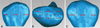
In order to quantitatively evaluate condylar surface changes, pre- and post-operative segmented condylar images were superimposed over three prominent registration points around the condylar neck using the manual registration function in Geomagic Studio® 12 (Fig. 1). After the manual registration, the global registration function provided by Geomagic Studio® 12 was used, and the reconstructed images were fitted automatically. Observer bias was eliminated since the procedures were automatically conducted. Using the Geomagic Studio® 12 tool, we calculated thousands of distances automatically (in millimeters) between the pre- and postoperative points on the registered image, such that the differences between two surfaces at any location could be quantified. These point-to-point distances were defined as condylar surface changes. The maximum distance, average distance, and standard deviation of these thousands of point-to-point distances were automatically calculated by Geomagic Studio® 12 for each condyle. Using these numeric values, we could identify how much the condylar surfaces changed after surgery.
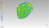
A tool in Geomagic Studio® 12 converted the surface distances into a color map for each registered 3D condylar image. Relative to the preoperative model, the positive (+) values of surface distances were expressed as red coloration (bone formation), the negative (-) values of surface distances were expressed as blue (bone resorption) and values close to zero were expressed as green (no change) (Fig. 2).
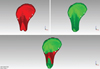
The color maps were used to evaluate the condylar remodeling types. The registered 3D images were cut through one plane, which was located around the condylar neck, and the area below that plane was erased. The plane used in this study was drawn over the thinnest portion of the condylar neck and parallel to a plane having the largest area among a number of areas for cross sections. The cross sections included the condylar long axis, which intersected the medial and lateral pole of the condylar head. An adjusted version of the method of An et al.3 was used to classify remodeling types on the remaining condylar head. The cutting plane used by An et al.3 intersected the medial and lateral pole areas of the condyle, but the location of the plane in this study was adjusted down to near the condylar neck area. The condylar head was divided into six areas (anteromedial, anteromiddle, anterolateral, posteromedial, posteromiddle, and posterolateral areas; Fig. 3) and each area was classified into three types of condylar remodeling (bone formation, no change, and bone resorption). The process of classifying the remodeling types of each condylar area was carried out twice (two weeks apart), and the degree of agreement of the classifications was evaluated to ensure intraobserver reliability.
All of the above procedures were performed to compare the condyle surface changes and remodeling types between the right and left condyles and between the sexes. All procedures were conducted by a single oral and maxillofacial radiologist.
Statistical analysis
All statistical analyses were performed using the Statistical Package for Social Sciences for Windows version 21.0 (IBM Corp., Armonk, NY, USA). Differences were considered to be significant at p<0.05. The mean values of the maximum distances, average distances, and standard deviations of each condyle were calculated, reflecting condylar surface changes.
The condylar remodeling types were evaluated by frequency.
The Wilcoxon signed-rank test was performed to compare the condylar surface changes between the right and left condyles. The marginal homogeneity test, which evaluated the uniformity of the distribution of two related variables, was performed to compare the frequencies of condylar remodeling types between the right and left condyles.
Additionally, the t-test and Mann-Whitney test were conducted to compare the condylar surface changes between the sexes. In order to compare the frequency of condylar remodeling types between the sexes, the chi-square test and Fisher's exact test were performed. Intraobserver reliability was evaluated using Cohen's kappa index.
Results
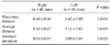
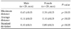
The mean values of the maximum distances, average distances, and standard deviations of condylar surface changes after orthognathic surgery were 0.41±0.06 mm, 0.11±0.03 mm, and 0.09±0.02 mm, respectively (Table 1).
The intraobserver reliability for the classification of condylar remodeling types on the condylar head showed almost perfect agreement (Cohen's kappa index; average, 0.895). Bone resorption occurred more frequently (38.3%) than other types of condylar remodeling. However, bone formation in the anteromedial area and bone resorption in the anterolateral and posterolateral areas were observed to a much greater extent (>50%) than other remodeling types (Table 2).
No statistically significant differences were found in condylar surface changes or the frequency of condylar remodeling types between the right and left condyles (Table 3, Table 4). However, condylar surface changes in males were significantly larger than in females (p<0.05; Table 5). No significant differences were found in the frequencies of condylar remodeling types between the sexes (Table 6).
Discussion
The evaluation of condylar morphology after orthognathic surgery is critical for predicting the clinical prognosis. Progressive condylar resorption has been found to be an irreversible complication and a factor in the development of late skeletal relapse after bilateral sagittal split, Le Fort I, or bimaxillary osteotomy.7 Recently, 3D images have been widely used to assess this condylar morphology. Xi et al.8 presented a reproducible tool for the 3D rendering of the condyle, which enabled the longitudinal follow-up and quantitative analysis of condylar changes. Schilling et al.9 showed that condylar registration was reliable and could be used to quantify subtle bony differences in 3D condylar morphology.
For these reasons, this study used 3D condylar images to assess condylar surface changes and remodeling types. Moreover, to the best of our knowledge, this was the first study to compare the right and left condyles and to compare these parameters according to sex.
In this study, three registration points on the pre- and postoperative 3D condylar images were used for super-imposition. These points were selected from around the condylar neck area, since the condylar neck area exhibited relatively less change than other areas. Moreover, the registration points were needed to align the images, because the pre- and postoperative reconstructed 3D images from one patient were not exactly the same size. After this manual registration, an automatic global registration was then conducted once more in order to increase the accuracy.
In this study, the method proposed by An et al.3 was used to evaluate the frequency of condylar remodeling types, but the location of the cutting plane on the registered 3D condylar images was adjusted down to near the condylar neck area. The cutting plane in the study of An et al.3 intersected the medial and lateral pole areas of the condyle. However, it was hypothesized that the medial and lateral pole areas could change after surgery, and indeed, this change was confirmed by the color map, which presented the condylar remodeling types of the registered 3D condylar images.
Table 1 shows that the extent of condylar surface change after surgery, expressed as the mean of the average point-to-point distance, was 0.11±0.03 mm in this study, which was larger than the mean and standard deviation (0.01± 0.09 mm) reported by An et al.3 and smaller than the average displacement of all surfaces reported by Cevidanes et al.10 (0.77±0.17 mm).
We found that bone resorption occurred more frequently (38.3%) than other types of condylar remodeling (bone formation, 27.7%; no change, 34.0%). However, it cannot be suggested that bone resorption after surgery was the predominant pattern because the frequencies of these three remodeling types were similar. However, bone formation was the predominant pattern in the anteromedial area (55.0%), while bone resorption predominated in the anterolateral (55.0%) and posterolateral (52.5%) areas (Table 2). In other words, condylar remodeling after surgery was most commonly achieved through bone resorption. These results were similar to those of the previous studies by An et al.3 and Park et al.2
Comparisons between the right and left condyles and between the sexes have not been explored in previous studies. In this study, no significant differences were found in the frequency of condylar remodeling types in each area between the sexes or between the right and left condyles (Tables 4 and 6). However, males exhibited significantly greater condylar surface changes than females (Table 5).
Some researchers have proposed that forced condylar positioning in orthognathic surgery could lead to condylar remodeling.1112 Hwang et al.13 suggested that a posteriorly inclined condylar neck should be considered a relevant nonsurgical risk factor in condylar resorption following orthognathic surgery. An et al.3 found that inward condylar rotation after orthognathic surgery was closely related to condylar surface change. Nonetheless, the exact cause of condylar surface change remains controversial.
However, these changes on the condylar surface could, in turn, induce post-operative TMJ symptoms and early or late postoperative skeletal relapse.21415 We found that bone resorption in the lateral area and bone formation in the anteromedial area were especially prominent. Additionally, condylar surface changes after orthognathic surgery occurred; in particular, the condylar surface change in males was greater than in females.
However, this study had some limitations. In particular, each group consisted of a small sample (20 males, 20 females). Additionally, the clinical significance of the values in this study is a potential limitation, since all quantitative values were smaller than the slice thickness (0.75 mm). For these reasons, further studies are needed.


 XML Download