

 PDF
PDF ePub
ePub Citation
 Print
Print


Introduction
Radiographic digital imaging has been widely used in medicine, but it was only in the 1980s that the first intraoral digital image receptors were developed for use in dentistry.1 Recently, digital radiography has become the most modern diagnostic imaging modality in dental practice and, in the near future, it is expected to completely replace conventional radiography.
Scientific studies have demonstrated that digital radiographic imaging is a suitable alternative to improve diagnostic accuracy, minimizing the radiation-related risks and optimizing the outcomes for both patients and professionals. This has facilitated the acceptance of this imaging modality over film-based radiography by dental practitioners worldwide.23456 In many European countries and the United States, the practice of digital radiology is a reality; 24 however, dental practitioners in Brazil experience relatively less accessibility.
The effectiveness of digital dental radiographic systems has been widely reported,710 but little is known about their acceptance by Brazilian dentists. Thus, taking into account that Brazil is a developing country and has a large number of dentists,11 the aims of this study were to investigate the use and acceptance of digital radiographic examinations by Brazilian dental practitioners in daily practice and to evaluate the advances that have occurred over the past 5 years.
Materials and Methods
This study was designed according to guidelines of the local Institutional Research Ethics Committee and was conducted after receiving approval (#039/2011). A total of 273 dental practitioners were enrolled in specialization and refresher courses in any field of dentistry at the Piracicaba Dental School, University of Campinas (UNICAMP), Sao Paulo, Brazil at the time of the study; more specifically, 181 and 92 dental practitioners were enrolled in the years 2011 and 2015, respectively.
A self-administered questionnaire containing 15 questions that had been validated for reproducibility was used to collect the data for the study. Ten specialists in oral and maxillofacial radiology evaluated the questionnaire to verify that the questions were explicit and relevant. Written consent was obtained after verbal and written explanations about the purpose of the study. Initially, a pilot study was conducted with 20% of the total sample to verify the validity and applicability of the proposed methodology. The questionnaire was then distributed to the respondents by the researchers, who remained present to provide additional explanations if needed. The respondents were asked about sociodemographic factors and their knowledge and use of digital radiographic examinations.
The data were tabulated in a Microsoft Office Excel 2013 (Microsoft Corporation, Redmond, WA, USA) spreadsheet. Statistical analyses were performed using BioEstat software version 5.0 (Ayres Company, Pará, Brazil). After descriptive analysis, the chi-square and Fisher exact tests were used to evaluate the associations among variables, with a significance level of 5% (α=0.05).
Results
Sociodemographic factors
Ninety-nine (54.6%) of the 181 potential participants responded to the questionnaire in the year 2011 and 82 (89.1%) of the 92 potential participants did so in 2015, resulting in 181 respondents (66.3% of the 273) in the final sample. Some respondents did not answer all the questions. In such cases, only the usable responses for each particular question were used to calculate the percentages.
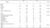
The age range of the respondents was 22 to 48 years (mean, 27.1±4.9) in 2011, and 21 to 60 years (mean, 28.4±7.2) in 2015, and most respondents were from the state of Sao Paulo (61.6% and 56.1% in 2011 and 2015, respectively). In both years, the plurality of respondents were enrolled in endodontics courses (22.2% in 2011 and 26.8% in 2015), and the majority worked in private practice and had graduated within the last 5 years (Table 1).
Knowledge and use of digital radiographic examinations
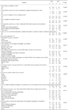
Table 2 summarizes the results about the knowledge and use of digital radiographic examinations by dental practitioners in the years 2011 and 2015. In 2011, most dentists had a computer in their workplace (question 1) and did not digitize conventional (film-based) images (question 4), which was not significantly different from the results obtained in 2015 (p>0.05 for both variables). A significant increase (p=0.0138) in computer use to receive the results of imaging exams (question 3) and, among those who did not have a computer in the workplace, a growing interest in computerizing in the next 5 years (question 2) was observed.
Compared to the year 2011, a significantly greater number of dentists made use of digital radiographic examinations in 2015 (p<0.0001). Moreover, the frequency of dental practitioners who had worked with digital radiographic examinations for more than 3 years increased (p=0.0411).
The preference for digital radiographic examinations increased between the years 2011 and 2015, but not to a statistically significant extent (p>0.05). In the data obtained in 2015, only 9 (12.9%) of the dentists who reported having used digital radiographic examinations preferred the conventional method, and 6 (66.7%) of this latter group reported having used digital radiographic examinations for less than a year (information not presented in Table 2).
A significant increase (p=0.0316) in the use of digital intraoral radiography was observed. The number of dental practitioners using e-mail to receive imaging exams of their patients increased, and the number of those who received printed copies decreased, but this difference was still not significant (p>0.05). The majority of dentists did not use specific software applications to assess digital radiographic examinations. An overall increase occurred in the use of digital tools to assist in the interpretation of the images, such as adjustment of brightness and contrast, zooming, and task-specific enhancement tools.
In the opinion of 121 respondents (96.8% of the 125 who reported ever having used digital radiographic examinations in question 6) from both years, digital radiography provides benefits over conventional radiography (question 13), such as better image quality, ease of storage, and easier communication with other professionals. In 2011 and 2015, the cost was considered to be high, which was felt to be the main disadvantage or limitation to the use of digital radiographic examinations.
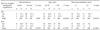
No significant associations (p>0.05) were found between the use of digital radiographic examinations and the following variables: type of practice (public or private), age (20-30 years or older than 30 years) and time since graduation (less than 5 years or 5 years or more), in both the years 2011 and 2015 (Table 3).
Discussion
For this study, dentists from one of the most important dental schools in Brazil responded to a questionnaire consisting of questions regarding dental practitioners' acceptance and use of digital radiographic examinations in the years 2011 and 2015. These respondents were selected because they were professionals who acquired or requested either conventional or digital imaging techniques in their daily practice. In contrast to developed countries, most dental images in Brazil are obtained in specialized radiology clinics and sent to dental offices.
A pilot study was conducted with a questionnaire to verify the validity and applicability of the proposed methodology. According to Shelley et al.,12 conducting a pilot study reduces measurement error due to the possibility of identifying confusing or misleading questions.
The participants in this study were young, with a mean age between 27 and 28 years. Although the majority of the respondents were from the state of Sao Paulo, as expected, since that is where UNICAMP is located, there were representatives from all 5 geographic regions of the country in this study. Statistical analysis showed no significant associations between age and the use of digital radiographic examinations, which is in agreement with data reported in another study.6 In a study conducted in 2010 among with general dental practitioners in Flanders, Belgium, the age of dentists was not evaluated because the researchers did not believe it reflected conditions of practice, since some young dentists work with old equipment and vice-versa.13
Dental practitioners who have recently graduated were expected to be more familiar with digital radiographic examinations, considering the recent development of digital technology. However, no statistically significant difference was found between the time since graduation and the use of digital radiographic examinations. Conversely, a study performed in Indiana, USA, observed that participants with less than 10 years of professional practice were proportionately the smallest group using digital radiographic examinations.4
Despite the large number of computerized dental offices, only 34.3% of dentists reported using a computer to receive imaging exams in 2011, but this number increased considerably to 53.7% in 2015. Importantly, most of the respondents reported a great interest in computerizing their offices in the future. This makes it evident that only a minority will probably not have access to digital radiographic examinations in the near future, which favors the transition process. According to a study conducted by Wenzel and Møystad,5 dental practitioners who made use of digital radiography had more computers in their offices, and a quarter of those who did not use digital radiographic examinations did not have computers in their work environment. Brian and Williamson4 showed that 36.7% of non-users of digital radiographic examinations planned to make it part of their clinical routine within 5 years, which is similar to the results of this study. In a study conducted by Dölekoğlu et al.,6 67% of respondents reported using of digital radiographic examinations.
Film-digitized radiography was mostly used for archiving and/or sharing with other professionals. This confirms a growing trend in digital image use and also confirms the advantages cited by Wenzel and Møystad14 in relation to the ease of communication between practitioners and patients, as well as data archiving and retrieval.
In the years 2011 and 2015, 55.6% and 85.4% of the respondents, respectively, reported making use of some form of digital imaging, with intraoral radiography and computed tomography being the most cited forms. In a study conducted in Norway in the beginning of the year 2000, only 14% of the respondents reported using digital radiography, 5 while in Indiana, USA, this number reached 19.7% in 20074 and, in Turkey, 67% in 2011.6 This sequence of studies done between 2001 and 2011 confirms the important growing trend of acceptance of digital radiology.
In 2015, 71.4% of the respondents indicated that they preferred digital images. Out of those who preferred conventional images, 66.7% had been using digital radiographic examinations for less than a year, which may reflect their lack of knowledge of the benefits of digital radiographic examinations. This suggests that continuing education in dentomaxillofacial radiology is essential, since greater acceptance is expected from those who are more aware of the advantages of digital over conventional radiology.
Most of the respondents in the present study recognized the advantages of digital radiographic examinations, such as image quality, ease of storage, and easier communication between professionals. Image quality and ease of storage were also among the most frequently cited advantages in other studies.56 In the study of Brian and Williamson, 4 the most-cited benefits were saving time (87%) and the elimination of processing-related problems (77%). Additionally, Dölekoğlu et al.6 found radiation dose reduction (79%) to be a major reason for using digital radiographic examinations. The present study also revealed that the main limitations to the use of the digital technology were related to its high cost, which is in agreement with the findings of previous studies.245615
In conclusion, most of the Brazilian dental practitioners who participated in this study made use of digital radiographic examinations. Moreover, the use of digital radiology has increased in Brazil over the past 5 years.
 XML Download