

 PDF
PDF ePub
ePub Citation
 Print
Print


Multiple myeloma is the most common primary bone malignancy with an unknown etiology. It occurs frequently in older age groups and occurs twice as often in men as in women. The most common clinical signs and symptoms of multiple myeloma include bone pain, fatigue, anemia, and infectious diseases, and it is characterized by multiple punched-out radiolucent lesions.1 The clinical manifestations of the disease occur due to an expanding mass of plasma cells in the bone marrow and factors produced by these cells, such as monoclonal immunoglobulin, Bence-Jones proteins, and osteoclast-activating factors.2
Oral and maxillofacial manifestations of multiple myeloma very rarely present as an initial sign.1 Jaw lesions, with an incidence of 8%-15%, may be a primary manifestation in the advanced stages of multiple myeloma.3 A review of several case series and case studies indicated that the maxillary involvement of multiple myeloma has been infrequently reported.4,5,6,7,8 To the best of our knowledge, very few case reports of multiple myeloma with maxillary involvement have so far been published in the literature.
We report a case of multiple myeloma involving the maxilla in a 50-year-old man who experienced swelling and bone pain with mobile teeth in the left maxillary alveolar region. Computed tomography (CT) imaging of the skull and other radiographs showed multiple punched-out radiolucent lesions. A panoramic radiograph revealed an ill-defined osteolytic lesion in the left maxilla, eroding the walls and the floor of the maxillary sinus, which was eventually determined to be a rare presentation of multiple myeloma.
Case Report
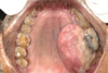
A 50-year-old man presented with a diffuse swelling in the left maxillary alveolar region with mobility of the adjacent teeth. The upper left first and second molars had been extracted 15 days previously due to pain; following the extraction, the lesion became enlarged (Fig. 1). Diffuse swelling in the left maxillary region had obliterated the left nasolabial fold extraorally. A sessile, soft, non-pulsatile, non-tender, non-hemorrhagic intraoral mass extended from the left maxillary second premolar to the tuberosity region, and extended medially up to the midpalatine raphe (Fig. 2). The lymph nodes were not palpable. The clinically considered differential diagnoses were carcinoma of the maxillary sinus and metastatic carcinoma.
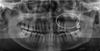
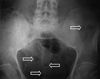
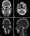
A panoramic radiograph revealed an ill-defined osteolytic lesion in the left posterior maxilla with resorption of the hard palate and the floor of the maxillary sinus (Fig. 3). As the panoramic radiograph did not show any extensions of the lesion, a posterio-anterior view of skull was taken, which showed punched-out radiolucent lesions indicative of multiple myeloma as a radiological diagnosis. In order to further confirm the diagnosis of multiple myeloma, a radiographic survey was carried out. An antero-posterior radiograph of the pelvis (Fig. 4) and a coronal section CT of the skull showed multiple punched-out radiolucent lesions (Fig. 5A). An axial section CT showed a soft tissue density mass measuring 4.2×4.1 cm in the left maxilla, eroding the medial and lateral walls and extending into the nasal cavity and the buccal space (Fig. 5B). The mass eroded the floor of the maxillary sinus and left maxillary alveolus in the coronal section CT (Fig. 5C). A sagittal section CT revealed erosion of the anterior wall of the maxillary sinus with involvement of the premaxillary tissues (Fig. 5D). The radiological differential diagnoses considered were multiple myeloma, carcinoma of the maxillary sinus, and metastatic carcinoma.
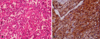
After the radiographic evaluation, an incisional biopsy was taken from the intraoral swelling. A histopathological examination of the specimen revealed atypical plasma cells with large hyperchromatic nuclei and large cytoplasms, of which only few were binucleated (Fig. 6A). The report was suggestive of multiple myeloma. An oral pathologist advised further confirmatory investigations for multiple myeloma. Immunohistochemically, the cells were positive for the CD138 marker (Fig. 6B). A bone marrow biopsy specimen showed 60% plasmacytosis. Serum electrophoresis revealed decreased albumin (2.78 g/dL), increased globulin (5.22 g/dL), a reversed albumin : globulin ratio with increased β1-microglobulin (0.84 g/dL) and β2-microglobulin (2.35 g/dL) levels, and an M-spike in the γ region (1.86 g/dL). Bence-Jones proteins were absent in the urine. Based on all the reports, a final diagnosis of multiple myeloma was made, the patient underwent dexamethasone and thalidomide chemotherapy in a hospital specializing in oncology, and is doing well.
Discussion
Multiple myeloma is the most aggressive primary bone malignancy.9 It accounts for ~1% of all malignancies and >10% of hematologic malignancies.10 The most common initial manifestations of multiple myeloma are bone pain (58%), fatigue (32%), and weight loss (24%).11 Secondary invasion of the skeletal tissue is one of the most important characteristics of the disease.12 The bone marrow reveals a large amount of abnormal plasma cells, which produce M-protein, light chain proteins (κ and λ), and cytokines. Excessive production of M-protein causes hyperviscosity of the blood, which in turn leads to renal dysfunction.13,14
Oral manifestations of multiple myeloma have an incidence of 2%-70%, and rarely present as the first sign of the disease.15,16 Swelling, orofacial pain, mobility of teeth, paresthesia, hemorrhage, fracture, and root resorption are more frequently found in the mandible than the maxilla.4,12 In our case study, bone pain was the first sign, along with mobility of the adjacent teeth in the maxillary left alveolus. Swelling was seen following the extraction of the adjacent teeth, and the patient had no history of hemorrhage or paresthesia.
Jaw involvement has been reported in as many as 30% of multiple myeloma cases, although bony lesions are less common in the maxilla than in the mandible because of the lower amount of hemopoietic marrow in the mandible.4,5,6,7,8 Bruce and Royer3 and Miller et al.17 have reported that 20%-30% of all cases had jaw involvement. Lambertenghi-Deliliers et al.12 showed that none of the 193 cases they examined involved the maxilla; on the contrary, Pisano et al.18 reported four cases of maxillary involvement in 13 patients, and Lae et al.19 in 2003 reported that seven of the 33 cases they examined involved the maxilla. Similarly to the case reports of Pisano et al. and Lae et al., our case demonstrates the maxillary involvement.
Bony lesions in the jaws are directly attributed to the osteoclastic-activating factor, a lymphokine that is responsible for the development of these lesions.16 Radiographically bony lesions reveal multiple well-defined punche-dout radiolucencies without a definitive cortical margin that often contain abnormal plasma cell proliferations.4 These radiolucent lesions occur more frequently in the mandible than the maxilla, particularly in the posterior region, ramus, and condylar process.14 Our case study had multiple punched-out radiolucent lesions in the skull and pelvis, whereas the left maxilla had an ill-defined radiolucent lesion, which was a rare presentation of multiple myeloma.
Early multiple myeloma may not reveal observable changes on plain radiographs. However, advanced imaging techniques can show evidence of active myeloma in approximately 20% of patients with negative radiographs.20 A CT scan can visualize focal alterations of the bone marrow before they can be detected with conventional radiographs.15 Positron emission tomography and positive emission tomography-CT with fluorodeoxyglucose may help to detect new non-suspected lesions when staging the disease, with important implications for treatment and assessing the response to treatment.21
The differential diagnoses of multiple myeloma in children include multiple metastatic lesions and Langerhans' cell disease. In adults, multiple myeloma and metastatic carcinoma are highly probable when several bones are involved. The clinically considered differential diagnoses in our case were carcinoma of the maxillary sinus and metastatic carcinoma. A panoramic radiograph revealed an ill-defined osteolytic lesion in the left maxillary sinus, which further indicated that carcinoma of the maxillary sinus was a relevant differential diagnosis. A further radiological examination showed multiple punched-out radiolucent lesions in the skull and pelvis, which led to a radiological diagnosis of multiple myeloma. Since multiple myeloma is more common than multiple metastatic diseases, it should appear higher in the differential diagnosis. The differential diagnosis of these lesions requires a multidisciplinary approach to diagnosis. Asymptomatic multiple small radiolucencies in the jaws of an apparently healthy adult can represent multiple distinct bone marrow defects.22
The diagnosis of multiple myeloma depends on the identification of abnormal monoclonal plasma cells, a full blood count, a bone marrow biopsy, levels of M-protein in the serum or urine, and a clinical image consistent with multiple myeloma. Serum electrophoresis identifies M-protein in ~93% of patients. Urine electrophoresis may identify M-protein in ~60% of patients. Additionally, ~70 % of myelomas secrete IgG, with κ light chains being more common (63%).23 In the present case, serum electrophoresis showed an IgG monoclonal spike of 1.86 g/dL with a κ light chain. Nevertheless, Bence-Jones proteins were not detected in the urine. Radiographs showed multiple punched-out radiolucent lesions and a CT scan revealed a malignancy in the maxillary sinus. The histopathological and immunohistochemical reports led to the final diagnosis of multiple myeloma, which was supported by bone marrow biopsy and laboratory investigations.
The involvement of the jaw bones is a primary manifestation that often occurs in the advanced stages of multiple myeloma. In conclusion, we have reported a case of multiple myeloma presenting with bone pain and an illdefined osteolytic radiolucency in the maxilla as a first sign, reinforcing the fundamental role of dentomaxillofacial radiologists in the early recognition of oral lesions that reflect underlying systemic disease, thus preventing or reducing morbidity and mortality in such cases.

 XML Download